Which is the most common congenital cardiac defect?
Which of the following is a manifestation of Tetralogy of Fallot?
Reversal of shunt is not possible in natural history of?
Tetralogy of Fallot presents with one of the following:
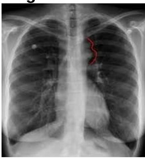
The chest radiograph shown below is from a 25-year-old male patient presenting with hypertension. The image demonstrates bilateral inferior rib notching. What is the most likely diagnosis?

True about Ebstein anomaly is ?
Eisenmenger syndrome-True are All except ?
A 9 year old boy with Fallot's tetralogy, had high grade fever followed by focal seizure 2 days prior to hospital admission. His blood counts were increased and predominantly polymorphs. CT scan of the head showed a focal lesion suggestive of an abscess. Where would be the commonest location of brain abscess in this patient?
Which of the following is the most appropriate true statement regarding ostium primum atrial septal defect (ASD)?
Pressure difference of 5 mm Hg between the two upper limbs occurs in which congenital heart disease?
Explanation: ***Ventricular septal defect*** - **Ventricular septal defect (VSD)** is the most common form of congenital cardiac defect, occurring in approximately 2 to 6 per 1000 live births. - It involves a **hole in the septum** separating the left and right ventricles, allowing oxygenated blood to shunt from the left to the right side of the heart. *Atrial septal defect* - While common, an **atrial septal defect (ASD)** is less frequent than VSD, typically accounting for about 10% of congenital heart diseases. - It involves a **hole in the septum** between the left and right atria, leading to a left-to-right shunt. *TAPVC* - **Total anomalous pulmonary venous connection (TAPVC)** is a rare and severe congenital heart defect where all four pulmonary veins drain abnormally. - It accounts for only about 1% of all congenital cardiac defects. *Transposition of great arteries* - **Transposition of the great arteries (TGA)** is another relatively rare and critical congenital heart defect. - In TGA, the **aorta originates from the right ventricle** and the **pulmonary artery from the left ventricle**, leading to two separate circulatory systems.
Explanation: **VSD** - A **ventricular septal defect (VSD)** is one of the four cardinal defects comprising Tetralogy of Fallot, allowing for communication between the right and left ventricles [1]. - The size of the VSD is typically large, leading to equalization of pressures between the two ventricles [1]. *Left axis deviation* - **Left axis deviation** is not a typical electrocardiographic finding in Tetralogy of Fallot; instead, right axis deviation is more common due to **right ventricular hypertrophy** [1]. - It is more characteristic of conditions like **left anterior fascicular block** or chronic left ventricular overload. *Left ventricular hypertrophy* - **Left ventricular hypertrophy** is not a characteristic feature of Tetralogy of Fallot; rather, **right ventricular hypertrophy** is a diagnostic component due to the obstruction of the right ventricular outflow tract [1]. - While the left ventricle may be affected by volume changes, it does not typically undergo hypertrophy in Tetralogy of Fallot. *All of the options* - This option is incorrect because, as explained, **left axis deviation** and **left ventricular hypertrophy** are not characteristic manifestations of Tetralogy of Fallot. - Only a **VSD** is one of the core defects in this complex congenital heart condition [1].
Explanation: ***TOF*** - In **Tetralogy of Fallot (TOF)**, the shunt is typically right-to-left from birth due to **pulmonary stenosis** and a large **VSD** [1]. - This fixed **right-to-left shunt** means that reversal to a left-to-right shunt does not occur naturally [1]. *PDA* - A **patent ductus arteriosus (PDA)** typically features an initial **left-to-right shunt**. - However, if **pulmonary hypertension** develops, the shunt can reverse to become **right-to-left** (Eisenmenger syndrome), meaning reversal *is* possible. *VSD* - A **ventricular septal defect (VSD)** initially presents with a **left-to-right shunt** [2]. - Prolonged systemic-level pressures in the pulmonary arteries can lead to **pulmonary vascular disease** and eventual shunt reversal to **right-to-left** (Eisenmenger syndrome) [2]. *ASD* - An **atrial septal defect (ASD)** typically causes an initial **left-to-right shunt**. - Over time, significant **pulmonary hypertension** can develop, leading to shunt reversal to **right-to-left** (Eisenmenger syndrome), indicating reversal is possible.
Explanation: ***Central cyanosis with clubbing*** - Tetralogy of Fallot is a **cyanotic congenital heart defect**, meaning it causes inadequate oxygenation of the blood, leading to central cyanosis. - **Chronic hypoxemia** (low oxygen levels) associated with cyanotic heart disease typically results in **clubbing of the fingers and toes** over time. *Normal ECG and Chest x-ray* - An **ECG** in Tetralogy of Fallot typically shows **right ventricular hypertrophy** and an **axis deviation**, which is not normal. - A **chest X-ray** in Tetralogy of Fallot often reveals a characteristic **'boot-shaped' heart** due to right ventricular hypertrophy and decreased pulmonary artery markings. *Cardiomegaly* - While there is **right ventricular hypertrophy**, significant global cardiomegaly (enlargement of the entire heart) is **not a classic or consistent feature** of Tetralogy of Fallot. - The boot-shaped appearance on X-ray reflects the hypertrophied right ventricle, but the overall heart size may not be drastically enlarged. *Left ventricular hypertrophy* - Tetralogy of Fallot is characterized by **right ventricular outflow tract obstruction** and a **large ventricular septal defect**, leading primarily to **right ventricular hypertrophy**. - **Left ventricular hypertrophy** is not a typical finding in Tetralogy of Fallot; in fact, the left ventricle may be smaller due to decreased pulmonary venous return.
Explanation: ***Coarctation of Aorta*** - The chest radiograph shows findings consistent with **rib notching**, which is a classic sign of coarctation of the aorta due to increased collateral circulation through intercostal arteries. - The history of **hypertension** in a male patient, especially if presenting at a younger age or with differential blood pressures between upper and lower extremities, strongly suggests coarctation of the aorta. *Tetralogy of Fallot* - Characterized by a **boot-shaped heart** due to right ventricular hypertrophy and pulmonary outflow obstruction. - Would typically present with **cyanosis** and decreased pulmonary vascular markings, not rib notching or isolated hypertension. *Ebstein's Anomaly* - Involves apical displacement of the **tricuspid valve**, leading to atrialization of the right ventricle and severe tricuspid regurgitation. - Chest X-rays often show **severe cardiomegaly** (huge heart due to right atrial enlargement) and decreased pulmonary vascularity, which are not depicted here. *TAPVC* - Total anomalous pulmonary venous connection (TAPVC) involves all pulmonary veins draining into the systemic circulation. - The classic chest X-ray finding for supracardiac TAPVC is a **"snowman" or "figure of 8" sign** due to dilated anomalous vessels and superior vena cava, which is absent in this image.
Explanation: Right atrial dilatation - Ebstein anomaly is characterized by the **apical displacement** of the tricuspid valve leaflets, which incorporates a portion of the right ventricle into the right atrium, functionally increasing its size. - This **atrialization of the right ventricle** leads to significant **right atrial dilatation** as it has to handle a larger volume [1]. *Right ventricular dilatation* - While there is a functional impairment of the right ventricle due to the displaced tricuspid valve, the **working part of the right ventricle** is often small and hypoplastic, not dilated. - The "atrialized" part of the right ventricle contributes to **right atrial dilatation**, not ventricular dilatation [1]. *Left atrial dilatation* - Ebstein anomaly primarily affects the **right side of the heart**, specifically the **tricuspid valve** and right ventricle. - There is no direct anatomical or physiological mechanism in Ebstein anomaly that would typically cause **left atrial dilatation**. *Left ventricular dilatation* - Similar to left atrial dilatation, Ebstein anomaly is a **right-sided heart defect**. - **Left ventricular morphology and function** are generally preserved in Ebstein anomaly, and dilatation is not a characteristic feature.
Explanation: ***RV & LV walls come back to normal size*** - In **Eisenmenger syndrome**, there is **irreversible pulmonary hypertension** and **ventricular hypertrophy** due to chronic volume and pressure overload. - The right ventricle (RV) undergoes significant hypertrophy and dilatation to pump against increased pulmonary vascular resistance, and the left ventricle (LV) may also be affected due to the shift in the ventricular septum and changes in preload and afterload conditions. Therefore, these chambers **do not revert to normal size**. *Dilatation of central pulmonary artery* - This is a characteristic finding in **pulmonary hypertension**, which is a hallmark of Eisenmenger syndrome. - The central pulmonary arteries **dilate** due to the increased pressure, while more distal arteries constrict. *Peripheral pruning of pulmonary arteries* - In Eisenmenger syndrome, there is **progressive remodeling** of the pulmonary vasculature, leading to **narrowing and obliteration** of the smaller arteries. - This "pruning" effect means the distal pulmonary arteries become less visible on imaging. *Pulmonary veins are not distended* - **Eisenmenger syndrome** is primarily a condition of the **pulmonary arteries**, characterized by increased pressure and resistance. - The pulmonary veins which carry oxygenated blood to the left atrium, typically **do not become distended** as the primary issue is arterial and not a post-capillary or left-sided heart failure problem that would cause venous congestion.
Explanation: **Cerebellum** - In patients with **cyanotic congenital heart disease** like Fallot's tetralogy, brain abscesses are typically supplied by the **posterior circulation**, making the **cerebellum** the most common location. [1] - The **right-to-left shunt** allows bacteria to bypass pulmonary filtration and directly enter systemic circulation, increasing the risk of infection in the brain, predominantly in areas supplied by the vertebral and basilar arteries. [1], [3] *Parietal lobe* - While brain abscesses can occur in the parietal lobe, it is more commonly associated with spread from a **frontal or sphenoid sinusitis** or direct trauma, not typically from cyanotic heart disease. - Abscesses in the parietal lobe are more often seen in **immunocompromised patients** or those with endocarditis causing septic emboli. [2] *Temporal lobe* - Temporal lobe abscesses are frequently a complication of **otitis media** or **mastoiditis**, with infection spreading directly or via venous drainage. - This patient's presentation does not suggest an ear infection as the primary source. *Thalamus* - Thalamic abscesses are rare and usually occur as a result of **hematogenous spread** from distant infections, particularly in immunocompromised individuals. - While possible, they are not the most common location for brain abscesses in patients with cyanotic congenital heart disease.
Explanation: Current guidelines indicate that **ostium primum ASDs** are structural defects involving the **atrial septum** adjacent to the **AV valves** and are not amenable to spontaneous closure [1]. Their location near the **crux of the heart** and association with **cleft mitral valves** makes them more complex and less likely to resolve naturally compared to secundum ASDs. **Ostium primum ASDs** typically cause a **left axis deviation (LAD)** on the ECG due to an associated defect in the **atrioventricular (AV) conduction system**, leading to uncoordinated ventricular depolarization. **Right axis deviation (RAD)** is more commonly seen in **large secundum ASDs** with significant left-to-right shunting and resultant **pulmonary hypertension**. **Holt-Oram syndrome** is primarily associated with **secundum ASDs**, not ostium primum ASDs. Genetic or chromosomal abnormalities, such as Down's syndrome, are also known to cause septal defects [2]. **Ostium primum ASDs** account for about **15-20%** of all ASDs. **Secundum ASDs** are the most common type, typically involving the fossa ovalis, making up approximately **70-75%** of all atrial septal defects [1].
Explanation: ***Supra-valvular aortic stenosis*** - **Supravalvular aortic stenosis** causes a **pressure gradient** across the aortic valve, leading to a significant **pressure difference** between the upper limbs, typically with a **higher pressure** in the right arm. - This is due to the **Coanda effect**, where the high-velocity jet of blood preferentially flows up the **right subclavian artery** as it exits the aorta. *HOCM (Hypertrophic Obstructive Cardiomyopathy)* - HOCM is characterized by hypertrophy of the **left ventricular septum** causing **outflow tract obstruction**, but it does not typically cause a significant **pressure difference** between the upper limbs. - The obstruction primarily affects **ventricular ejection** rather than differential flow to major arteries. *Coarctation of Aorta* - **Coarctation of the aorta** causes a significant **blood pressure difference** between the upper and lower extremities, with higher pressures in the arms [1]. - However, it does not typically cause a marked **pressure difference between the two upper limbs**, unless the coarctation is pre-ductal and affects the subclavian artery circulation asymmetrically, which is less common for a difference of just 5 mmHg. *TOF (Tetralogy of Fallot)* - **Tetralogy of Fallot** is a cyanotic heart disease involving **pulmonary stenosis**, ventricular septal defect, overriding aorta, and right ventricular hypertrophy [2]. - While it causes significant circulatory abnormalities and potential for **hypoxia**, it does not inherently lead to a measurable **pressure difference** between the upper limbs.
Get full access to all questions, explanations, and performance tracking.
Start For Free