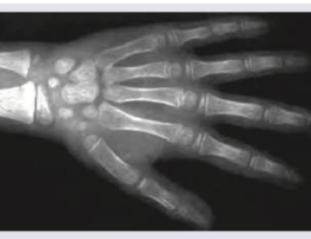
X-ray of wrist joint of an under-trial female convict who claims to be below 18 years was performed. The bone age of the patient as analyzed from the X-ray is: (Recent NEET Pattern 2016-17)

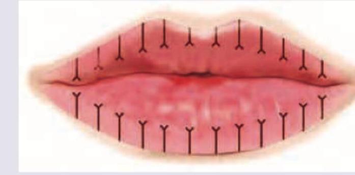
Which of the following lip prints is shown below?

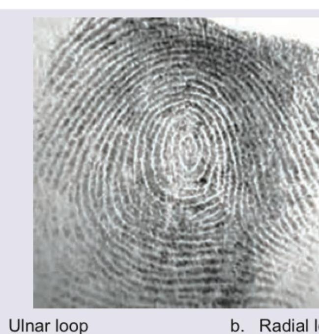
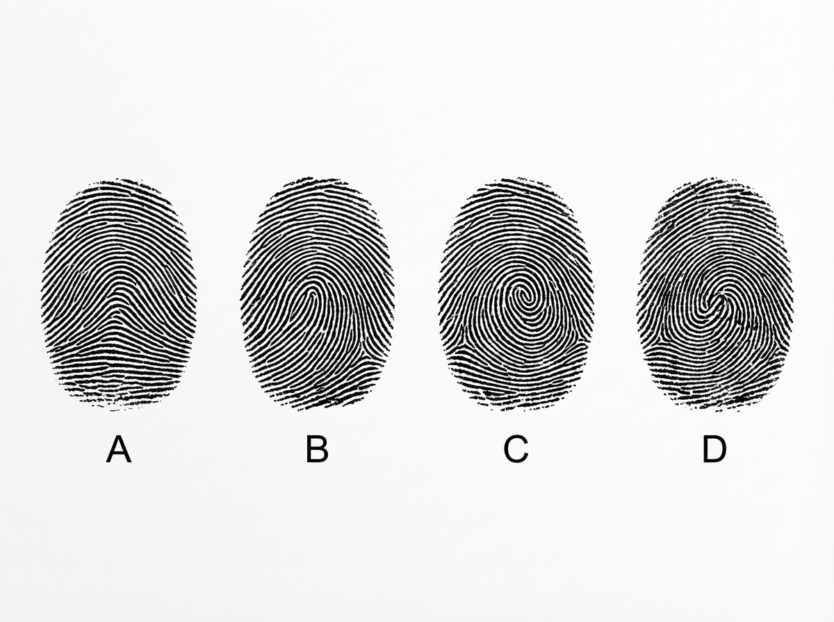
Which of the following fingerprint pattern is shown below?

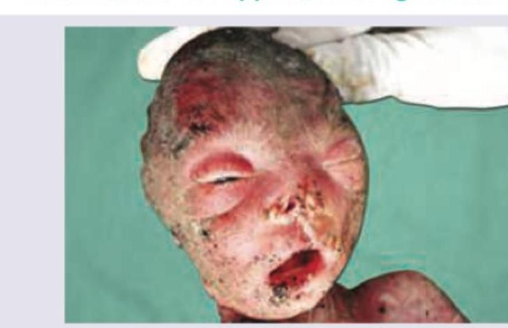
The abandoned body of a fetus was found in a dustbin. What is the approximate age of the fetus?

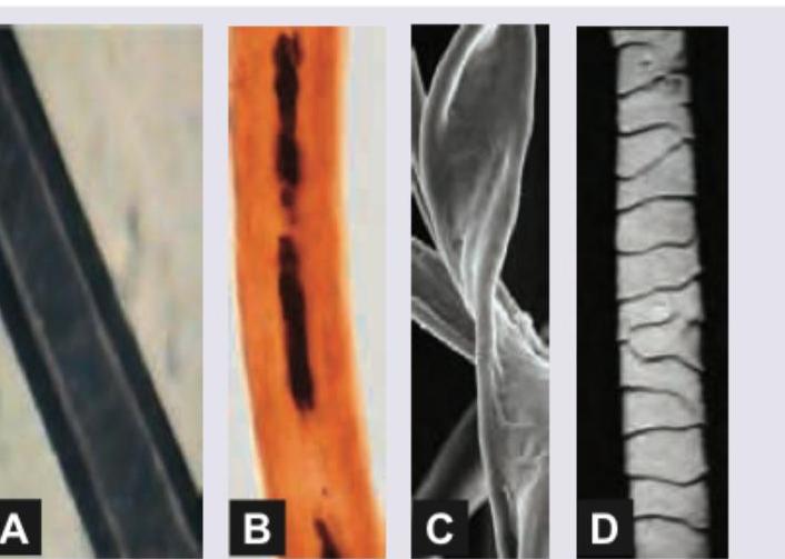
Which of the following about hair/fibers is correct?

Which is the most common type of finger print pattern? (Recent NEET Pattern 2016-17)

Which of the following tests for identifying blood stains is shown in the image below?

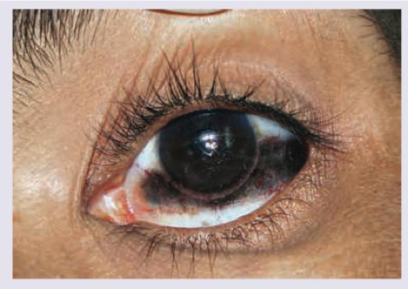
All are true about the image shown except:

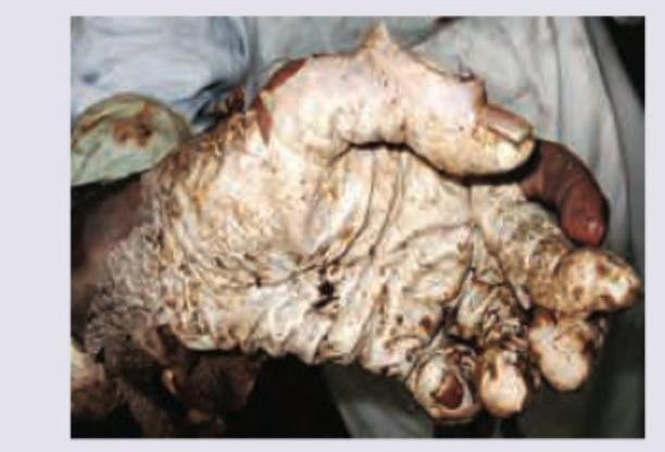
All are true about the condition shown except:

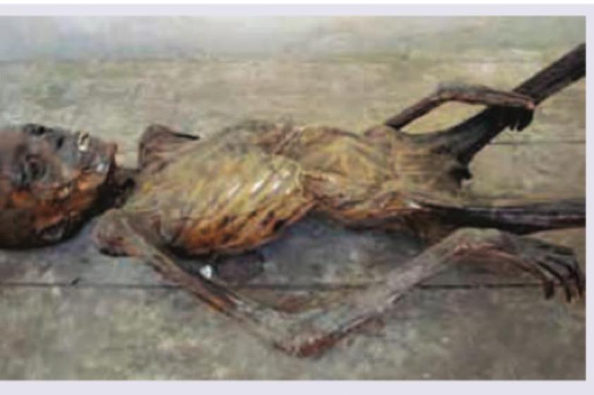
All of following are true about the condition shown in the image except:

Explanation: ***Less than 18 years*** - The X-ray image shows that the **epiphyseal plates** at the distal ends of the radius and ulna, as well as in the phalanges and metacarpals, are still **open or partially fused**. - Complete fusion of these plates, especially the **distal radial epiphysis**, typically occurs around **18-19 years** in females, indicating the individual is likely under 18 years of age. *Less than 16.5 years* - The image shows a relatively mature skeletal structure where many of the **carpal bones** are fully ossified, and some epiphyseal fusion is evident, suggesting an age older than 16.5 years. - While still growing, the extent of development surpasses that typically seen at 16.5 years, where more distinct growth plates would be visible. *Less than 17 years* - Similar to the explanation for 16.5 years, the degree of **ossification** and **epiphyseal closure** observed, particularly in the carpals and partially in the long bones, implies an age beyond 17 years. - The image presents a more advanced skeletal maturity than commonly expected for someone definitively younger than 17 years. *Less than 21 years* - While complete skeletal maturity in some individuals can extend beyond 18 years, the significant **open growth plates** in the distal radius and ulna strongly indicate that the individual is not yet skeletally mature, which is generally achieved by 21 years. - If the individual were close to 21, these plates would be expected to be completely closed and fully fused.
Explanation: ***Type II*** - The image displays lip prints that are **branched**, resembling a tree-like pattern, which is characteristic of Type II lip prints as per Susumu Suzuki's classification. - Type II furrows are often seen in the middle portion of the lips, representing a common pattern of lip wrinkles. *Type I* - Type I lip prints consist of **clear-cut vertical grooves** that run across the entire lip. - These grooves do not show any branching or intersection. *Type III* - Type III lip prints are characterized by **intersecting grooves**, creating a crisscross pattern. - They tend to be more complex than branched patterns, with lines crossing each other. *Type IV* - Type IV lip prints exhibit **reticular patterns**, which are a mesh-like or net-like arrangement of grooves. - This pattern is highly complex and distinct from branched or intersecting grooves.
Explanation: ***Whorl*** - A **whorl pattern** is characterized by ridges that make a complete circuit, often forming a spiral or concentric circles around a central point, as clearly seen in the image. - It typically has **two deltas** and a core, which distinguishes it from loop and arch patterns. *Ulnar loop* - In an ulnar loop, the ridges enter from one side of the finger, form a **loop**, and exit towards the **ulnar side** (little finger side) of the hand. - It has **only one delta** and a core, which is not consistent with the pattern shown. *Radial loop* - In a radial loop, the ridges enter from one side, form a **loop**, and exit towards the **radial side** (thumb side) of the hand. - Similar to the ulnar loop, it has **only one delta** and a core, which is not what is depicted in the image. *Plain arch* - A plain arch is the simplest pattern, characterized by ridges that enter from one side, rise in the center (forming a **wave-like** or **tent-like shape**), and exit on the opposite side. - It completely **lacks deltas**, cores, or loops, which is distinctly different from the given image.
Explanation: ***7 months*** - The image shows a fetus with **fused eyelids**, visible **lanugo** (fine hair all over the body), and developing but still wrinkled skin, which are characteristic features seen at approximately **7 months (28 weeks) of gestation**. - At this age, the fetus is considered **viable** and has a significant chance of survival outside the womb with medical intervention. *4 months* - A fetus at **4 months** (16 weeks) would be much smaller, typically around 10-12 cm in length, and would not display the developed features seen in the image. - While **lanugo** begins to appear, the body would appear less developed and often more translucent compared to the pictured fetus. - The **eyelids remain fused** at this stage. *5 months* - By **5 months** (20 weeks), the fetus is larger (around 15-20 cm), and lanugo becomes more prominent, but the **eyelids are still fused** and it looks less developed compared to the image. - The skin would still appear very thin and translucent with visible blood vessels, which is not as pronounced in the image. *6 months* - At **6 months** (24 weeks), the fetus is gaining weight, and some fat deposition begins, but the skin is often still quite wrinkled and red due to the absence of significant subcutaneous fat. - The **eyelids are still fused or just beginning to open**, and while significant development has occurred, the overall developmental features suggest a slightly older fetus.
Explanation: ***A= Animal hair, B= Human hair, C= Cotton fiber, D= Wool fiber*** - Image A shows **animal hair**, characterized by a **discontinuous or fragmented medulla** (the dark central core visible along the shaft) and distinct scale patterns - Image B depicts **human hair**, which typically has a **continuous, absent, or fragmented medulla** (less prominent than animal hair) with relatively uniform diameter and uneven pigment distribution - Image C shows **cotton fibers**, which are **flat, ribbon-like, and twisted** with a characteristic convoluted, tape-like appearance under microscopy - Image D illustrates **wool fiber**, distinguished by its prominent **overlapping cuticular scales** that create a distinctive scaly surface pattern *A= Cotton fiber, B= Human hair, C= Animal hair, D= Wool fiber* - Image A does **not** show the flat, ribbon-like, twisted morphology characteristic of **cotton fibers**; instead it shows a hair with visible medulla - Image C is **not animal hair** as it lacks the distinct medulla and scale patterns typical of mammalian hair *A= Human hair, B= Animal hair, C= Cotton fiber, D= Wool fiber* - Images A and B are **reversed**: Image A shows the more prominent fragmented medulla typical of **animal hair**, not the finer structure of human hair - Image B shows the characteristics more consistent with **human hair**, not animal hair *A= Animal hair, B= Human hair, C= Wool fiber, D= Cotton fiber* - Images C and D are **reversed**: Image C shows the twisted ribbon-like structure of **cotton**, not the scaly surface of wool - Image D shows the overlapping scales characteristic of **wool fiber**, not the plant-based structure of cotton
Explanation: ***B*** - Pattern B exhibits a **loop** design, where ridges enter from one side of the finger, recurve, and exit from the same side. - **Loops** are the **most common type of fingerprint pattern**, accounting for approximately 60-70% of all fingerprints. *A* - Pattern A shows an **arch** design, characterized by ridges that enter from one side, rise in the middle, and exit on the opposite side without recurving. - **Arches** are the **least common fingerprint pattern**, found in about 5% of individuals. *C* - Pattern C displays a **whorl** design, specifically a central pocket loop or a plain whorl, where ridges form circular or spiral patterns around a central point. - **Whorls** are the second most common pattern, occurring in roughly 25-35% of fingerprints. *D* - Pattern D also represents a **whorl** pattern, similar to C. This can be either a double loop whorl or an accidental whorl depending on the exact configuration of multiple loops or irregular patterns. - While whorls are significant, they are less frequent than loops.
Explanation: ***Teichmann's Test*** - The image displays characteristic **rhombic, dark brown crystals of hemin (ferriprotoporphyrin chloride)**, which are formed in Teichmann's test when **blood** is heated with glacial acetic acid and a halide (e.g., NaCl). - This test is a confirmatory **microchemical test** for the presence of blood, specifically detecting the heme component. *Luminol Test* - The Luminol test is a **presumptive test** that produces a **bluish-white luminescence** when it reacts with the iron in hemoglobin. - It does not involve the formation of crystals visible under a microscope. *Kastle-Meyer Test* - The Kastle-Meyer test is a **presumptive color test** that turns pink in the presence of blood due to the **peroxidase-like activity of hemoglobin** in reducing phenolphthalin. - It does not involve microscopic crystal formation. *Benzidine Test* - The Benzidine test is another **presumptive color test** that turns blue in the presence of blood, also due to the peroxidase-like activity of hemoglobin. - Similar to the Kastle-Meyer test, it is a color reaction and does not result in the formation of crystals as shown in the image.
Explanation: ***Cuticle separates from palm and soles by 48 hours*** - The image depicts the characteristic appearance of a hand after prolonged immersion in water, a phenomenon known as **washerwoman's hands** or **cutis anserina aquosa**, observed in bodies that have been submerged for an extended period. - The separation of the cuticle from the palms and soles typically occurs much later than 48 hours, usually around **2-3 weeks** of submersion, and is referred to as **glove and stocking maceration** or **desquamation**. - This statement is **incorrect** as the timeline is vastly understated. *Occurs due to imbibition of water* - The wrinkling and maceration of the skin, especially on the palms and soles, is due to the **imbibition (absorption) of water** into the stratum corneum. - This process leads to the characteristic corrugated appearance seen in individuals who have spent significant time in water, which is relevant in forensic pathology for estimating postmortem submersion time. *Bleaching of cuticle is evident after 12 hours* - **Bleaching of the cuticle**, where the nails and surrounding skin lighten, is an early sign of prolonged water immersion, usually becoming evident within **12-24 hours** of submersion. - This change is due to water absorption and subsequent physical alteration of the keratin structures. *Can present with cutis anserina (goosebumps)* - **Cutis anserina** (goosebumps or goose flesh) refers to the roughening of the skin caused by contraction of erector pili muscles, typically stimulated by cold water exposure or postmortem changes. - This finding **can be present** in bodies recovered from water, especially in cases of drowning or cold water immersion, as a response to the cold environment. - It is a true associated finding, though distinct from the maceration changes that define washerwoman's hands.
Explanation: ***Finger prints are preserved*** - This is the **INCORRECT** statement about mummification, making it the correct answer to this EXCEPT question - In mummification, the skin undergoes **severe dehydration and shrinkage**, causing the epidermal ridges that form fingerprints to become distorted or lost - The desiccation process makes the skin brittle and causes it to **shrivel and crack**, destroying the fine details necessary for fingerprint identification - While some gross skin features may remain, the **friction ridge details are typically obliterated**, making fingerprint analysis impossible or unreliable *DNA samples can predict gender and race* - DNA can be successfully extracted from mummified remains despite the desiccation process - Modern forensic DNA analysis can accurately determine **biological sex** (through sex chromosome analysis) and **ancestry/racial background** (through genetic markers) - Mummification, while destroying soft tissue architecture, often **preserves DNA better than putrefaction** due to the dry environment *Occurs in absence of water* - **Mummification** is a form of body preservation that occurs in **dry, arid conditions** with minimal moisture - The absence of water inhibits bacterial growth and prevents the usual putrefactive decomposition - Rapid desiccation of tissues leads to preservation of body form through **natural mummification** *Skin splits can mimic injury* - As the skin dries and contracts during mummification, it becomes **brittle and develops cracks and fissures** - These post-mortem **desiccation artifacts** can closely resemble ante-mortem wounds, cuts, or injuries - This poses a significant challenge in forensic analysis, requiring careful examination to distinguish **artifact from true injury**
Explanation: This is an **"EXCEPT"** question - we need to identify the FALSE statement about Tache Noire. ***Correct Answer: Seen after 2-3 days of death*** - This statement is **FALSE** and therefore the correct answer to this "except" question. - Tache Noire forms much **earlier** in the post-mortem period, typically within **2-6 hours** after death if the eyes remain open. - This is a relatively **early post-mortem change** due to desiccation, not a late change occurring after days. *Incorrect: Seen when eye remains open after death* - This is a **TRUE** statement about Tache Noire. - Occurs when **eyelids are not closed** after death, allowing exposure to air and desiccation of the cornea and sclera. *Incorrect: Triangular patch on both side of cornea* - This is a **TRUE** statement. - Tache Noire appears as **brownish-black triangular patches** of desiccated sclera on either side of the cornea. - Typically observed at the **lateral and medial canthi**. *Incorrect: Seen due to phenomenon of Trucking* - This is a **TRUE** statement. - The color change involves **desiccation and oxidation** of hemoglobin in underlying blood vessels. - Associated with **"trucking" or "sludging"** of blood cells in capillaries, contributing to the brownish-black appearance.
Personal Identification Methods
Practice Questions
Anthropometry
Practice Questions
Dactylography (Fingerprinting)
Practice Questions
Dental Identification
Practice Questions
DNA Profiling
Practice Questions
Facial Reconstruction
Practice Questions
Superimposition Techniques
Practice Questions
Hair and Fiber Analysis
Practice Questions
Handwriting Analysis
Practice Questions
Identification of Remains
Practice Questions
Mass Disaster Victim Identification
Practice Questions
Age, Sex and Race Determination
Practice Questions
Get full access to all questions, explanations, and performance tracking.
Start For Free