Identical twins may not have:
What is the most common location for supernumerary teeth?
The sacrum fuses to become a single bone at approximately what age?
Which anatomical feature is most useful for sex determination?
Following the recovery of a skull by the police, the relatives of a missing person want to confirm his identity. Which of the following techniques can be used to figure out a person's identity using the skull and photograph of the person?
A body is found with rigor mortis fully developed in all muscle groups. The ambient temperature is 25°C. Approximately how many hours have elapsed since death?
Which X-ray is used to prove that an individual is more than 18 years of age?
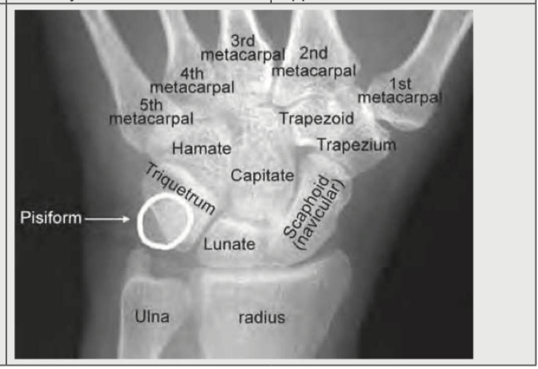
The image shows ossification centers at the lower end of the radius and ulna which are not fused, and the pisiform is present. Based on this, what is the most accurate conclusion about the age?

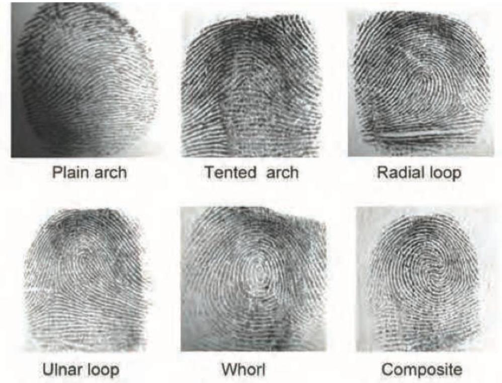
The image shows presence of which pattern? Notice the different patterns shown in the image.

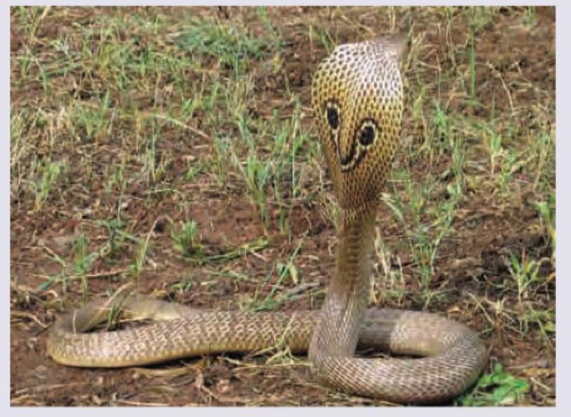
Which snake is shown below?

Explanation: **Explanation:** The correct answer is **B. Same fingerprint pattern.** **Why it is correct:** Identical (monozygotic) twins originate from a single fertilized ovum that splits into two. While they share the same genetic blueprint, **fingerprints (dactylography)** are determined by both genetics and **epigenetic/environmental factors** in utero. During the 10th to 24th week of gestation, the contact of the developing fetus's fingertips with the amniotic fluid, uterine walls, and local growth stresses creates unique ridge patterns. Therefore, even identical twins have distinct, unique fingerprints, making dactylography the "gold standard" for differentiating them in forensic investigations. **Why the other options are incorrect:** * **A. Same DNA fingerprint:** Since monozygotic twins come from the same zygote, their nuclear DNA sequence is identical. Standard DNA profiling cannot distinguish between them (though advanced deep sequencing may find rare mutations). * **C. Same blood group:** Blood groups (ABO, Rh, etc.) are determined purely by inherited genes. Since identical twins share 100% of their DNA, they will always have the same blood group. * **D. Same HLA system:** The Human Leukocyte Antigen (HLA) system is genetically determined. Identical twins are "syngeneic," meaning they have identical HLA types, making them perfect matches for organ transplantation without the risk of rejection. **High-Yield Clinical Pearls for NEET-PG:** * **Dactylography (Galton System):** The most reliable method of identification because no two fingers (even on the same hand) have the same pattern. * **Quetelet’s Rule:** States that the fingerprints of no two persons are ever identical. * **Bertillonage:** An obsolete system of identification based on physical measurements (Anthropometry). * **Poroscopy (Locard’s Method):** Study of the number, size, and distribution of sweat pores on the ridges; used when only fragmentary prints are available.
Explanation: **Explanation:** Supernumerary teeth (hyperdontia) are teeth that appear in addition to the normal dental formula. In forensic medicine and clinical dentistry, identifying these anomalies is crucial for age estimation and individual identification. **Why Option A is Correct:** The **upper incisor region** (specifically the maxillary midline) is the most common site for supernumerary teeth. The most frequent type is the **Mesiodens**, a small, peg-shaped tooth located between the two central maxillary incisors. Statistics show that approximately 90% of all supernumerary teeth occur in the maxilla, with the midline incisor area being the primary location. **Why Other Options are Incorrect:** * **B. Canine:** Supernumerary teeth in the canine region are extremely rare. When they occur, they are usually found in the maxilla rather than the mandible. * **C. Molar:** While "paramolars" (buccal/lingual to molars) and "distomolars" (fourth molars) do occur, they are statistically less frequent than mesiodens. * **D. Lower incisor:** Supernumerary teeth in the mandibular incisor region are significantly less common than those in the maxillary incisor region. **High-Yield Clinical Pearls for NEET-PG:** * **Mesiodens:** The single most common supernumerary tooth (found in the maxillary midline). * **Prevalence:** More common in permanent dentition than deciduous dentition; more common in males (2:1 ratio). * **Associated Syndromes:** Multiple supernumerary teeth are strongly associated with **Cleidocranial Dysplasia** and **Gardner’s Syndrome**. * **Forensic Significance:** Dental anomalies like supernumerary teeth serve as "individualizing characteristics" in forensic identification when comparing antemortem and postmortem dental X-rays.
Explanation: **Explanation:** The sacrum is composed of five separate vertebrae at birth. The process of fusion occurs in stages, beginning from the lower segments and moving upwards. 1. **Why 25 years is correct:** The fusion of the sacral vertebrae typically begins after puberty (around 15–18 years). The lower segments (S3–S5) fuse first, followed by the upper segments. The final fusion of the **S1 and S2 vertebrae**—and the complete consolidation of the sacrum into a single bone—is usually finalized by the age of **25 years**. In forensic age estimation, a completely fused sacrum is a reliable indicator that the individual is at least 25 years old. 2. **Why other options are incorrect:** * **15 years:** At this age, the sacral vertebrae are still largely separate or just beginning the initial stages of primary fusion. * **30 & 40 years:** By these ages, the sacrum is already fully fused. While degenerative changes (like osteophytes) might begin later in life, the primary ossification and fusion are completed much earlier. **High-Yield Clinical Pearls for NEET-PG:** * **Order of Fusion:** Sacral fusion occurs from **below upwards** (S5 to S1). * **The "Gap" Rule:** If a gap is still visible between S1 and S2 on an X-ray, the individual is likely under 25 years of age. * **Sternum Correlation:** The fusion of the body of the sternum (segments) also completes around age 25, making it a useful corroborative marker. * **Ischial Tuberosity:** The epiphysis of the ischial tuberosity also fuses at approximately 20–25 years.
Explanation: **Explanation:** In forensic anthropology and medicolegal practice, the **Pelvis** is the most reliable and accurate skeletal element for sex determination. This is due to the biological necessity of the female pelvis to adapt for childbirth (parturition). These functional reproductive requirements result in distinct morphological differences that are more pronounced than in any other bone in the body. The pelvis provides an accuracy rate of approximately **95%** for sex identification. **Analysis of Options:** * **Pelvis (Correct):** It exhibits high sexual dimorphism. Key features include a wider sub-pubic angle (80-90° in females vs. 50-60° in males), a broader greater sciatic notch, and a circular pelvic brim in females compared to a heart-shaped brim in males. * **Skull (Incorrect):** The skull is the second most reliable feature (approx. 80-90% accuracy). While it shows dimorphism (e.g., prominent supraorbital ridges and mastoid processes in males), these traits are influenced by population and nutrition, making it less definitive than the pelvis. * **Femur & Tibia (Incorrect):** Long bones are used for stature estimation and sexing through "metric analysis" (measuring head diameter or length). However, they are less reliable than the pelvis or skull because their dimensions often overlap between sexes. **High-Yield Clinical Pearls for NEET-PG:** * **Accuracy Hierarchy:** Pelvis (95%) > Skull (80-90%) > Pelvis + Skull (98%) > Long bones (80%). * **Pre-auricular Sulcus:** If deep and well-marked, it is a strong indicator of a female skeleton. * **Chilotic Index:** Calculated using the ilium; it is higher in females. * **Washburn’s Index (Ischio-pubic Index):** The most reliable metric for sexing the pelvis; it is significantly higher in females.
Explanation: ***Superimposition*** - **Superimposition technique** involves electronically or manually overlaying the antemortem photograph onto the recovered skull image to align key craniometric points (e.g., orbits, nasal aperture, chin). - This method is specifically designed to visually compare the distinctive features and contours of the facial structure recorded in the photograph with the underlying skeletal structure of the **skull**, confirming identity. *Anthropometry* - **Anthropometry** is the scientific study of the measurements and proportions of the human body, specifically used in forensic science to estimate age, sex, and stature from skeletal remains. - While useful for broad identification parameters, it relies on quantitative measurements of the skull (like cranial indices) and does not involve the direct visual comparison required when matching a photograph. *DNA fingerprinting* - **DNA fingerprinting** requires comparison of the recovered genetic profile from the bone tissue with a known reference sample (e.g., from suspected relatives or personal items). - Although highly accurate, this technique does not utilize the *photograph* as the primary comparative material; it compares genetic information, not morphological features. *Cheiloscopy* - **Cheiloscopy** is the forensic technique involving the analysis and identification of lip prints (furrows and wrinkles on the lips). - This technique is completely irrelevant for identification based on a **skull** and a general antemortem photograph.
Explanation: ***18-24 hours*** - Rigor mortis is **fully developed** in all muscle groups between **12 and 24 hours** after death under typical ambient temperatures (around 25°C). - At 18-24 hours post-mortem, rigor mortis has progressed through all muscle groups and is at its maximum development. - The process begins in smaller muscles (e.g., face, neck) and progresses to larger muscle groups due to **ATP depletion** in muscle fibers. *6-12 hours* - During this period, rigor mortis is in the **establishment phase**, progressing from smaller to larger muscles. - While rigor may be present in many muscle groups at 12 hours, it typically reaches **full development** in all groups closer to 12-24 hours. - This represents an intermediate stage, not complete rigor in all muscle groups. *36-48 hours* - By this time, rigor mortis is typically beginning to **pass off or disappear (secondary flaccidity)**, starting again in the small muscles. - **Rigor usually lasts** for 24-48 hours total, but begins to resolve after about 24-36 hours. - This represents the resolution phase, not the fully developed stage. *2-4 hours* - Rigor mortis usually begins to become noticeable in the jaw and neck muscles during the **first 2 to 4 hours** after death. - This stage is the **onset or early establishment phase**, not full development. - Only the smallest muscle groups show rigidity at this stage.
Explanation: ***Medial end of clavicle and sternum*** - The **medial (sternal) end of the clavicle** is the last center of ossification to fuse, typically fusing between 18 and 25 years of age, making it the most reliable skeletal site for confirming legal majority (**age >18 years**). - An X-ray view of this junction determines if the clavicular epiphysis is completely fused to the diaphysis, verifying physical maturity beyond the critical age of 18. ***Appearance of inner end of clavicle, knee joint, and wrist joint*** - The inclusion of the **wrist joint** and **knee joint** makes this option inaccurate, as their major epiphyseal plates (e.g., distal radius/ulna, distal femur/proximal tibia) generally fuse by 16–17 years. - These earlier fusions are useful for estimating age approaching 16 or 17 but are unreliable for determining if an individual has crossed the threshold of **18 years**. ***Fusion of ischial tuberosity and lower end of humerus*** - Fusion of the epiphysis of the **ischial tuberosity** typically occurs around 15 to 16 years of age (sometimes up to 17), making it fuse too early to confirm age greater than 18. - The **lower end of the humerus** often completes fusion significantly earlier (e.g., secondary centers around 13–16 years), rendering it unsuitable for the confirmation of legal majority. ***Medial end of clavicle and skull*** - While the medial end of the clavicle is correct, combining it with the **skull** is inappropriate, as the study of **cranial sutures** is highly variable and unreliable for pinpointing the age of 18. - Cranial suture fusion is generally used for broad age estimation in older adults, and its inclusion in combination renders this option medically and forensically inaccurate for proving the age of majority.
Explanation: **14-16 years** - The presence of the **pisiform** and unfused epiphyses at the **distal radius and ulna** is consistent with an age range of 14-16 years. The pisiform typically begins to ossify around 10-12 years in females and 12-14 years in males, while the distal radial and ulnar epiphyses usually fuse later, completing closure around 16-18 years. - In females, the distal radius begins ossifying around **12-14 years** and fuses around **16-17 years**. In males, it starts ossifying around 14 years and fuses around 17-18 years. The ulna follows a similar pattern, typically fusing a bit later than the radius. *<12 years* - While the pisiform may begin to ossify in some individuals around 10-12 years, the **distal radial and ulnar epiphyses are generally still widely open** and not yet nearing fusion at this age. - Many carpal bones would still be in earlier stages of development, and the overall skeletal maturity would be less advanced than depicted. *12-14 years* - During this period, the **pisiform is usually ossified**, but the **fusion of the distal radius and ulna has not typically begun**. The image shows the epiphyses of the radius and ulna are present but not yet fused, suggesting a slightly older age than 12-14 years, especially considering the advanced development of other carpal bones. - The epiphyses appear well-formed and distinct, indicating a stage where fusion is approaching but not yet initiated, which is usually after 14 years. *<17.5 years* - While this option is technically correct in that 14-16 years is less than 17.5 years, it is too broad and not the **most accurate conclusion**. The specific features like the absence of fusion in the distal radius and ulna help narrow down the age range more precisely. - After 17.5 years, especially in males, the **distal radial and ulnar epiphyses** would typically show signs of complete or near-complete fusion.
Explanation: ***Tented arch*** - A **tented arch** fingerprint pattern is characterized by ridges that rise sharply in the center, forming a "tent-like" or "peak-like" structure. - Unlike a **plain arch**, the ridges do not flow smoothly from one side to the other; instead, they converge at a distinct angle in the middle. *Plain arch* - A **plain arch** pattern involves ridges that enter from one side of the impression and flow out the other side with a slight rise or wave in the center. - It lacks the sharp, angular rise seen in a tented arch. *Radial loop* - A **radial loop** is characterized by one or more ridges entering upon either side, recurring by curving around, and exiting upon the same side as they entered, flowing towards the **radius bone** (thumb side) of the hand. - This pattern has a clear **delta** and a **core**, which are absent in arch patterns. *Ulnar loop* - An **ulnar loop** is similar to a radial loop, but the ridges flow towards the **ulna bone** (little finger side) of the hand. - Like radial loops, it features a **delta** and a **core**, indicating a looping pattern, unlike the simple flow of arches. *Whorl* - A **whorl** fingerprint pattern typically has at least one ridge that makes a complete circuit, often forming a spiral or concentric circle. - It is defined by having **two or more deltas**, which distinguishes it clearly from loops and arches. *Composite* - A **composite** pattern is a more complex fingerprint pattern that combines characteristics of two or more fundamental patterns, such as a loop and a whorl or two loops. - This typically results in a combination of **deltas and cores** that do not fit into a single primary classification.
Explanation: **Cobra** - The image clearly shows a cobra with its distinctive **hood spread** and the characteristic **spectacle mark** on its neck. - The hood is formed by the extension of its ribs, a common defense mechanism specific to cobras. *Common krait* - Common kraits typically have a slender body with **glossy black or bluish-black scales** and distinct **white or yellowish crossbands**. - They do not spread a hood or have a spectacle mark. *Saw scaled viper* - Saw-scaled vipers are generally small, stout-bodied snakes with a characteristic **zigzag pattern** on their back. - They are known for their **sizzling sound** produced by rubbing their scales together, not a hood display. *Russell viper* - Russell's vipers are large, venomous snakes with a pattern of **dark brown elliptical or circular spots** outlined in black along their body. - They do not possess a hood but are known for their loud hiss.
Personal Identification Methods
Practice Questions
Anthropometry
Practice Questions
Dactylography (Fingerprinting)
Practice Questions
Dental Identification
Practice Questions
DNA Profiling
Practice Questions
Facial Reconstruction
Practice Questions
Superimposition Techniques
Practice Questions
Hair and Fiber Analysis
Practice Questions
Handwriting Analysis
Practice Questions
Identification of Remains
Practice Questions
Mass Disaster Victim Identification
Practice Questions
Age, Sex and Race Determination
Practice Questions
Get full access to all questions, explanations, and performance tracking.
Start For Free