What is the multiplying factor for estimating stature from the humerus in males?
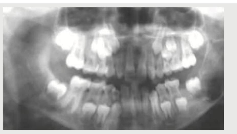
What is the minimum age of this person?

In which month does the ossification center of the calcaneus appear?
Faded tattoo marks can be visualized by?
Faint letter marks can be made visible by:
Which bone specimen is best for sex determination?
All of the following are types of fingerprint patterns except:
Exhumation is done following court order. Only skull bone was found with basiocciput fused with basisphenoid. What is the approximate age?
Palatoprints for identification of the person are performed by taking prints from which area of the hard palate?
What does the Chilotie line help in determining?
Explanation: ### Explanation **1. The Medical Concept (Why Option B is Correct)** Estimation of stature from long bones is a cornerstone of forensic identification, primarily utilizing **Karl Pearson’s formula** or **Pan’s formula**. In the Indian context, Pan’s formula is frequently cited for its simplicity. The "Multiplying Factor" (MF) is a constant derived from the ratio of total body height to the length of a specific long bone. For the **humerus in males**, the multiplying factor is **5.3 to 6.0**. In most standardized forensic textbooks (like Reddy’s), **6** is the accepted high-yield value for the humerus. **2. Analysis of Incorrect Options** * **Option A (12):** This value is too high. No long bone has a multiplying factor this large; it would imply the humerus is only 1/12th of the body height. * **Option C (9):** This is not a standard factor for any major long bone. * **Option D (10):** This is the multiplying factor for the **Clavicle** (approx. 10–11). **3. High-Yield Clinical Pearls for NEET-PG** To excel in stature estimation questions, memorize these standard Multiplying Factors (MF) for males: * **Humerus:** 6.0 * **Radius:** 6.5 * **Ulna:** 6.0 * **Femur:** 3.7 (The most accurate bone for stature estimation) * **Tibia:** 4.5 * **Fibula:** 4.5 **Key Rule:** If a range is given, the femur is the most reliable bone for height estimation because it contributes most directly to the vertical height of the individual. Always remember that stature decreases by approximately **0.06 cm per year after the age of 30**.
Explanation: ***6 years*** - The eruption of the **first permanent molar** (6-year molar) establishes a definitive minimum age of 6 years in forensic age estimation. - This **dental milestone** is considered one of the most reliable indicators for determining that a person has reached at least 6 years of age. *Less than 12 years* - This is too broad and non-specific for forensic age determination using **dental eruption patterns**. - While it may include the correct age range, it fails to provide the **precise minimum age** established by specific dental milestones. *5 years* - At 5 years, the **first permanent molars** have not yet erupted, making this age insufficient as a minimum. - **Primary dentition** is still predominantly present at this age, lacking the key permanent tooth markers for age estimation. *18 years* - This represents the age of **third molar** (wisdom tooth) eruption, which is irrelevant to establishing a minimum age of 6. - Using 18 years as a minimum would **overestimate** the actual age significantly when first permanent molars are the reference point.
Explanation: **Explanation:** The appearance of ossification centers is a critical parameter in Forensic Medicine for determining the age of a fetus or a newborn. The **calcaneus** is the first bone of the foot to begin ossification. 1. **Why 5th month is correct:** The primary ossification center for the calcaneus appears during the **5th month of intrauterine life (IUL)**. This is a high-yield milestone used in forensic anthropology and radiology to assess fetal maturity. 2. **Analysis of incorrect options:** * **3rd month:** At this stage, ossification begins in the shafts of long bones (like the femur and humerus) and the skull, but not in the tarsal bones. * **7th month:** This is when the ossification center for the **talus** typically appears. * **9th month:** This period is marked by the appearance of the **distal end of the femur** (36 weeks) and the **proximal end of the tibia** (38-40 weeks), which are classic indicators of a full-term fetus. **High-Yield Clinical Pearls for NEET-PG:** * **Order of Tarsal Ossification:** Calcaneus (5th month IUL) → Talus (7th month IUL) → Cuboid (at birth/9th month IUL). * **Rule of Thumb:** The presence of the distal femoral epiphysis (Casper’s sign) and the proximal tibial epiphysis indicates the fetus is viable and has reached full term. * **Sternum:** The manubrium and 1st piece of the body ossify at the 5th-6th month IUL. * **Memorable Sequence:** 5th month (Calcaneus) → 7th month (Talus) → 9th month (Cuboid).
Explanation: **Explanation:** **1. Why Ultraviolet (UV) Rays are Correct:** Tattoo marks are created by depositing pigment into the dermis. Over time, these marks may fade due to phagocytosis by macrophages or skin regeneration. Even when a tattoo is no longer visible to the naked eye (faded or erased), residual pigment particles often remain in the deeper layers of the dermis or the regional lymph nodes. Under **Ultraviolet (UV) light**, these residual pigments or the resulting scar tissue exhibit **fluorescence**, making the pattern discernible. This is a standard forensic technique for identifying individuals with obliterated or aged tattoos. **2. Analysis of Incorrect Options:** * **B. Spectrophotometer:** This instrument is used to measure the intensity of light as a function of wavelength. While it can identify specific chemical substances or poisons, it is not a visualization tool for anatomical patterns like tattoos. * **C. Ordinary Light:** Faded tattoos are, by definition, difficult or impossible to see under the visible spectrum (ordinary light) because the pigment density is too low to provide contrast against the skin. * **D. X-ray:** X-rays are used to visualize radiopaque structures like bones or metallic foreign bodies. Most tattoo inks are not sufficiently radiopaque to be visualized on a standard radiograph. **3. High-Yield Clinical Pearls for NEET-PG:** * **Lymph Node Pigmentation:** If a tattoo is completely removed, the pigment can often still be found in the **proximal regional lymph nodes** (e.g., axillary nodes for an arm tattoo). * **Infrared Photography:** Besides UV light, **Infrared (IR) photography** is another highly effective method for visualizing faded or charred tattoo marks. * **Medicolegal Importance:** Tattoos are a "secondary" means of identification. They are particularly useful in decomposed or mutilated bodies where facial features are lost. * **Vitreous Humor:** In cases of decomposition, remember that the vitreous humor is the last fluid to degrade and is used for post-mortem biochemistry (e.g., Potassium levels).
Explanation: ### Explanation **Correct Option: A. Infrared photography** Infrared (IR) photography is the gold standard for visualizing faint, faded, or obliterated marks (such as tattoos, ink, or letter marks) on the skin or documents. This is based on the principle of **differential absorption and reflection**. Even when a mark appears invisible to the naked eye, the residual pigment or altered skin texture absorbs IR radiation differently than the surrounding healthy tissue. Since IR rays have longer wavelengths, they can penetrate the superficial layers of the skin, reflecting off the underlying pigment to create a high-contrast image on IR-sensitive film. **Why other options are incorrect:** * **B. Spectrophotometer:** This instrument is used to measure the intensity of light as a function of its wavelength. While useful in toxicology for identifying chemical substances or hemoglobin derivatives, it cannot produce a visual image of a physical mark. * **C. Ordinary light:** Faint marks are often below the threshold of visual perception under white light because the contrast between the mark and the background skin is too low. * **D. X-ray:** X-rays are used to visualize dense structures like bone or metallic foreign bodies. They are not sensitive enough to detect superficial pigment changes or faint surface lettering. **High-Yield Clinical Pearls for NEET-PG:** * **Tattoo Identification:** IR photography is specifically used to identify tattoos that have been erased, scarred over, or are present on decomposed/putrefied skin. * **Ultraviolet (UV) Light:** While IR is best for faint marks, UV light (Wood’s lamp) is typically used to detect **semen stains** (fluorescence) or certain skin infections. * **Reflected UV Photography:** Can be used to visualize "latent" bite marks or healed injuries that are no longer visible. * **VSC (Video Spectral Comparator):** A modern forensic tool that uses various light spectra (including IR) to detect document forgeries and alterations.
Explanation: **Explanation:** Sex determination is a critical step in forensic identification. The **Pelvis** is the most reliable bone for this purpose because it exhibits the most significant sexual dimorphism. These differences are biological adaptations in females to facilitate childbirth (parturition), making the female pelvis wider, shallower, and more spacious compared to the narrower, heavier, and more robust male pelvis. According to Krogman, the accuracy of sex determination using the pelvis alone is approximately **95%**. **Analysis of Options:** * **Pelvis (Correct):** Offers the highest accuracy (95%) due to functional reproductive requirements. Key features include the wider sub-pubic angle, presence of the pre-auricular sulcus, and the rectangular shape of the pubis. * **Skull (Option C):** This is the second most reliable bone for sexing, with an accuracy of about **90%**. Male skulls are generally larger with prominent supraorbital ridges and mastoid processes. * **Femur (Option A):** While long bones can be used (accuracy ~80%), they rely on measurements like the head diameter and bicondylar width, which are less definitive than pelvic morphology. * **Mandible (Option D):** Useful when other bones are missing, but less reliable than the pelvis or skull. Male mandibles are typically larger with a square chin and more everted angles. **High-Yield Clinical Pearls for NEET-PG:** * **Accuracy Hierarchy:** Pelvis (95%) > Skull (90%) > Pelvis + Skull (98%) > Entire Skeleton (100%). * **Washburn’s Index (Ischio-pubic Index):** Used for sexing the pelvis; it is higher in females. * **Pre-auricular Sulcus:** If deep and well-defined, it is strongly indicative of a female (parous). * **Sciatic Notch:** A wide, shallow notch is characteristic of a female; a narrow, deep notch indicates a male.
Explanation: **Explanation:** The identification of individuals through fingerprints (Dactylography/Galton System) is based on the permanent and unique patterns of dermal ridges. According to the **Galton-Henry classification**, there are four primary types of fingerprint patterns. **Why "Circle" is the correct answer:** There is no standard fingerprint pattern known as a "Circle." While some patterns like Whorls may appear circular, they are formally classified under specific terminology. Therefore, "Circle" is the odd one out. **Analysis of other options:** * **Loops (60-70%):** The most common pattern. Ridges enter from one side, curve back, and exit on the same side. They are further divided into Ulnar and Radial loops. * **Whorls (25-35%):** Ridges form concentric circles or spirals. (Note: Option C, **Composite**, is a sub-type of the Whorl group in some classifications or a separate category representing a combination of two or more patterns). * **Arches (5%):** The rarest pattern. Ridges enter from one side and exit on the opposite side without turning back. **High-Yield NEET-PG Pearls:** 1. **Dactylography:** Also known as the "Galton System." It is the most reliable method of identification (error rate is 1 in 64 billion). 2. **Permanence:** Fingerprints appear at the **4th month of intrauterine life** and remain unchanged until death (and even after, until the skin decomposes). 3. **Poroscopy (Locard’s Method):** The study of sweat pore patterns on the ridges; useful when only fragmentary prints are available. 4. **Bertillonage:** An obsolete system based on physical measurements (Anthropometry), now replaced by dactylography. 5. **Legal Aspect:** Under Section 73 of the Indian Evidence Act, a court can direct any person to provide fingerprints for comparison.
Explanation: ### Explanation **1. Why Option A is Correct:** The fusion of the **basiocciput** with the **basisphenoid** (forming the spheno-occipital synchondrosis) is one of the most reliable skeletal markers for age estimation in late adolescence and early adulthood. * In males, this fusion typically occurs between **18 and 21 years**. * In females, it may occur slightly earlier (around 17–20 years). Since the skull bone found shows this fusion, the most accurate age range among the options is **18–21 years**. **2. Why Other Options are Incorrect:** * **Option B (40–50 years):** By this age, cranial sutures like the sagittal and coronal sutures would show significant closure (ectocranial and endocranial), and the spheno-occipital synchondrosis would have been fused for decades. * **Option C (70–80 years):** This represents an elderly skull, characterized by thinning of the table, loss of teeth (alveolar resorption), and complete obliteration of most cranial sutures. * **Option D (6–7 years):** At this age, the basiocciput and basisphenoid are still separated by a distinct cartilaginous plate. Fusion has not yet commenced. **3. High-Yield Clinical Pearls for NEET-PG:** * **Spheno-occipital Synchondrosis:** Also known as the "Basilar Suture." It is a primary cartilaginous joint. * **Order of Suture Closure (Endocranial):** Sagittal (25y) → Coronal (30y) → Lambdoid (35y). * **Metopic Suture:** Usually closes by **2 years** of age. If it persists, it is called metopism. * **Exhumation Rule:** In India, there is no time limit for exhumation (unlike some Western countries). It requires a written order from a **Magistrate** (Executive, Judicial, or Sub-divisional). * **Gustafson’s Method:** Used for age estimation from teeth in adults (reliable after age 21).
Explanation: **Explanation:** **Palatoprinting**, also known as **Palatoscopy** or **Rugoscopy**, is the study of the patterns of the **palatal rugae** (Plicae palatinae transversae) for individual identification. These are the irregular, asymmetric mucosal ridges located on the **anterior third** of the hard palate, specifically behind the incisive papilla. 1. **Why Anterior is Correct:** The palatal rugae are anatomically confined to the **anterior** part of the hard palate. They are highly individualistic, stable throughout life (resisting decomposition and thermal trauma due to protection by the tongue and cheeks), and unique even in twins. This makes the anterior palate the only viable site for rugoscopy. 2. **Why Other Options are Incorrect:** * **Middle and Posterior:** These areas of the hard palate are relatively smooth and lack the distinct ridge patterns (rugae) necessary for identification. * **Lateral:** While rugae extend laterally from the median raphe, the primary anatomical landmark and concentration of these ridges are defined by their position in the anterior segment. **High-Yield Facts for NEET-PG:** * **Classification:** The most commonly used system for rugae classification is the **Trobo system** or **Lysell classification**. * **Stability:** Rugae patterns remain unchanged from the 3rd month of intrauterine life until death, though they may change position slightly if teeth are lost. * **Forensic Significance:** Palatoscopy is particularly useful in identifying victims of **burns** or **mass disasters** where fingerprints may be destroyed, as the palate is protected by the surrounding orofacial structures. * **Cheiloscopy:** A related concept involving **lip prints** (Quetelet’s classification), also used for identification.
Explanation: **Explanation:** The **Chilotie line** (also known as the Chilotic line) is a morphometric parameter used in forensic anthropology to determine the **sex** of an individual from the human pelvis (specifically the sacrum and ilium). **1. Why Sex is the Correct Answer:** The Chilotie line is an imaginary line extending from the iliopectineal eminence to the nearest point on the auricular surface of the ilium. It is divided into two segments: the **pelvic part** (internal) and the **sacral part** (external). * In **females**, the pelvic part is longer than the sacral part (Chilotie Index > 100). * In **males**, the sacral part is longer than the pelvic part (Chilotie Index < 100). This sexual dimorphism exists because the female pelvis is evolutionarily adapted for childbirth, requiring a wider pelvic inlet. **2. Why Other Options are Incorrect:** * **Race:** Racial determination (Ethnicity) typically relies on cranial features (e.g., Cephalic Index) or the morphology of the nasal aperture and zygomatic bones. * **Age:** Age estimation in skeletal remains is determined by the eruption of teeth, ossification centers, and the closure of cranial sutures (e.g., Gustafson’s method for teeth or the McKern-Stewart criteria for the pubic symphysis). * **Stature:** Stature is estimated using the length of long bones (Femur, Humerus) via **Pearson’s formula** or **Trotter and Gleser’s formula**. **High-Yield Clinical Pearls for NEET-PG:** * **Sciatic Notch:** The most reliable indicator of sex in the pelvis. Females have a wider, shallower notch (>60°), while males have a narrow, deep notch (<50°). * **Pre-auricular Sulcus:** More common and deeper in females (especially those who have borne children). * **Washburn’s Index (Ischio-pubic Index):** Another pelvic metric for sex determination; it is significantly higher in females. * **Rule of Thumb:** If the entire skeleton is available, sex can be determined with 100% accuracy; the pelvis alone offers ~95% accuracy.
Personal Identification Methods
Practice Questions
Anthropometry
Practice Questions
Dactylography (Fingerprinting)
Practice Questions
Dental Identification
Practice Questions
DNA Profiling
Practice Questions
Facial Reconstruction
Practice Questions
Superimposition Techniques
Practice Questions
Hair and Fiber Analysis
Practice Questions
Handwriting Analysis
Practice Questions
Identification of Remains
Practice Questions
Mass Disaster Victim Identification
Practice Questions
Age, Sex and Race Determination
Practice Questions
Get full access to all questions, explanations, and performance tracking.
Start For Free