An infant previously diagnosed with a large muscular VSD comes to the office with complaints from the mother of fatigue and poor feeding over the past month. You note the child has not gained weight since the previous visit 2 months ago. The child is apathetic, tachypneic, and has wheezes and crackles on lung auscultation. What is the most likely cardiac diagnosis based on this patient's presentation?
Autopsy finding after 24 hours in a case of death due to myocardial infarction is
What causes sudden decreased end tidal CO2 in GA?
A man who is chronic alcoholic will develop which type of cardiomyopathy?
Commonest cause of heart failure in infancy is:
What is defined as a negative autopsy?
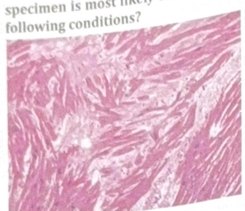
An athlete collapsed and expired while playing school basketball. Histology of the cardiac specimen is most likely to indicate which of the following conditions?

A healthy middle-aged man who became emotionally upset during an argument with his brother suddenly developed chest pain and collapsed. He was declared dead upon arrival at the hospital. What is the most likely diagnosis?
Which of the following statements about sudden infant death syndrome (SIDS) is false?
What does Casper's dictum indicate?
Explanation: ***Congestive heart failure*** - The infant's symptoms of **fatigue**, **poor feeding**, **no weight gain**, **apathy**, **tachypnea**, and **wheezes/crackles** are classic signs of **congestive heart failure** in an infant. - A **large muscular VSD** can lead to significant left-to-right shunting, causing **pulmonary overcirculation** and symptoms of heart failure. *Congenital heart block* - This condition involves an abnormality in the heart's electrical conduction system, leading to a **slow heart rate (bradycardia)**. - While it can cause fatigue, it typically doesn't present with respiratory symptoms like **tachypnea** and **rales** unless profound bradycardia leads to heart failure. *Prolonged QT syndrome* - This is an **electrical disorder** that can cause **arrhythmias** and sudden cardiac death, often presenting with syncope or seizures. - It does not typically manifest with the signs of **pulmonary congestion** (wheezes, crackles) or feeding difficulties seen in this infant. *Hypertrophic cardiomyopathy* - This condition involves thickening of the heart muscle, leading to **outflow obstruction** and diastolic dysfunction. - While it can cause symptoms of poor feeding and fatigue, the prominent respiratory symptoms like **tachypnea** and **crackles** are more indicative of pulmonary venous congestion secondary to a large shunt.
Explanation: ***Coagulative necrosis*** - Coagulative necrosis is the predominant histological finding after **myocardial infarction**, typically occurring within the first 12 hours [1]. - It results in preserved tissue architecture with **cellular outlines** remaining visible, indicating ischemic tissue damage [1,2]. *Liquefactive necrosis* - Commonly associated with **bacterial infections** or brain infarction, it leads to the transformation of tissue into liquid pus, which is not characteristic of myocardial infarction. - It occurs later and is not typically observed in heart tissue within 12 hours post-infarction. *Fat necrosis* - Primarily occurs due to damage to **adipose tissue**, as seen in cases of pancreatitis or trauma, and is not relevant to myocardial injury. - It is characterized by the release of **lipases** and fatty acids, a response not seen in myocardial infarction. *Caseous necrosis* - Often associated with **tuberculosis** or fungal infections, presenting as cheese-like necrotic tissue, it is not a feature of myocardial infarction. - This type of necrosis appears much later and reflects chronic granulomatous inflammation rather than acute ischemic damage. **References:** [1] Kumar V, Abbas AK, et al.. Robbins and Cotran Pathologic Basis of Disease. 9th ed. The Heart, p. 552. [2] Kumar V, Abbas AK, et al.. Robbins and Cotran Pathologic Basis of Disease. 9th ed. The Heart, pp. 552-554.
Explanation: ***Cardiac arrest*** - In **cardiac arrest**, there is a sudden cessation of effective **cardiac output**, which leads to a dramatic reduction in pulmonary blood flow. - As a result, **CO2 is not transported to the lungs** for exhalation, causing an abrupt and severe drop in **end-tidal CO2**. *Pulmonary embolism* - A **pulmonary embolism** causes an acute obstruction of pulmonary arterial blood flow, leading to an **increase in alveolar dead space**. - While it can decrease **end-tidal CO2** due to reduced perfusion, the drop is often less sudden and complete than in cardiac arrest, and the primary mechanism is **ventilation-perfusion mismatch**. *Pulmonary hypertension* - **Pulmonary hypertension** involves chronically elevated pressures in the pulmonary arteries, which can lead to **right ventricular dysfunction** and reduced cardiac output over time. - It typically causes a more gradual and chronic reduction in **end-tidal CO2** due to impaired gas exchange, rather than a sudden, precipitous drop. *Malignant hyperthermia* - **Malignant hyperthermia** is characterized by a rapid and severe increase in **metabolic rate** and CO2 production. - This condition typically leads to a **sudden increase in end-tidal CO2** as the body produces more CO2 than can be eliminated, rather than a decrease.
Explanation: ***Dilated cardiomyopathy*** - Chronic alcohol abuse is a major cause of **dilated cardiomyopathy**, where the heart's pumping chambers (ventricles) become enlarged and weakened, leading to reduced cardiac output [1]. - This condition often called **alcoholic cardiomyopathy**, is characterized by **ventricular dilation** and **systolic dysfunction**. *Hypertrophic cardiomyopathy* - This condition involves thickening of the heart muscle, often genetic, and is not directly caused by **chronic alcoholism**. - While alcohol can worsen pre-existing heart conditions, it does not typically lead to primary **hypertrophic cardiomyopathy**. *Myocarditis* - **Myocarditis** is an inflammation of the heart muscle, usually caused by viral infections or autoimmune processes. - Although heavy alcohol use can weaken the immune system, it is not a direct cause of viral or primary inflammatory myocarditis. *Pericarditis* - **Pericarditis** is the inflammation of the pericardium, the sac surrounding the heart, most commonly due to viral infections or autoimmune conditions. - While alcohol abuse can have various systemic effects, it is not a recognized direct cause of **pericarditis**.
Explanation: **Congenital heart disease** - **Congenital heart defects** are the leading cause of heart failure in infancy due to structural abnormalities that impair normal cardiac function and blood flow. - Conditions like **ventricular septal defects (VSDs)**, **atrial septal defects (ASDs)**, and **patent ductus arteriosus (PDA)** can lead to volume overload or pressure overload, resulting in heart failure. *Cardiomyopathy* - While a cause of heart failure, **cardiomyopathy** is less common than congenital heart disease in infancy. - It involves primary myocardial dysfunction, which can be genetic, metabolic, or idiopathic. *Rheumatic fever* - **Rheumatic fever** is a post-streptococcal inflammation that can affect the heart, but it is rare in infancy and more typically seen in older children and adolescents. - Its incidence has also significantly decreased in developed countries due to improved hygiene and antibiotic use. *Myocarditis* - **Myocarditis**, often virally induced, can cause heart failure in infants but is less frequent than congenital heart defects. - It involves inflammation of the heart muscle, leading to impaired contractility.
Explanation: ***No cause of death is found after both gross and histopathological examination.*** - A **negative autopsy** is declared when comprehensive examination, including both macroscopic (gross) and microscopic (histopathological) assessment, fails to identify a definitive **cause of death**. - This outcome can be due to various reasons, such as death from **functional disturbances** (e.g., arrhythmias, metabolic imbalances) or processes not evident morphologically. *Cause is apparent on gross examination but not on histopathological examination.* - This scenario describes situations where a cause might be evident visually (e.g., a large hemorrhage) but further microscopic investigation is still needed for confirmation or detailed understanding. - It does not align with a "negative" autopsy, as a cause has already been *grossly identified*. *Gross findings are minimal.* - While minimal gross findings might precede a negative autopsy, this statement alone is insufficient to define it. - A negative autopsy specifically requires the absence of a cause of death even after subsequent **histopathological examination**. *Cause is apparent on gross examination but not found due to constraints on the part of the doctor.* - This option refers to a failure in diagnostic process due to external factors or limitations by the examiner, not an inherent lack of discernible cause. - It suggests a missed diagnosis, not that a cause could not be found through comprehensive investigation.
Explanation: ***Hypertrophic cardiomyopathy (HCM)*** - The image shows **myocardial disarray and hypertrophy**, characterized by haphazardly arranged and abnormally branched cardiac muscle cells with large, irregular nuclei, which is a classic histologic finding in HCM [1]. - HCM is the most common cause of **sudden cardiac death in young athletes**, often during exertion, due to ventricular arrhythmias arising from the disarrayed myocardium [1]. *Dilated cardiomyopathy (DCM)* - Histology for DCM typically shows **myocyte atrophy**, thinning of the ventricular walls, and interstitial fibrosis, not the marked disarray and hypertrophy seen here [3]. - DCM leads to **progressive cardiac enlargement and systolic dysfunction**, and while it can cause sudden death, it is less common in athletes than HCM [4]. *Restrictive cardiomyopathy (RCM)* - RCM is characterized by **stiff, non-compliant ventricles** with impaired diastolic filling, often due to conditions like amyloidosis or sarcoidosis, showing interstitial infiltration or fibrosis. - The image does not show evidence of significant **interstitial infiltration or severe fibrosis** characteristic of RCM; instead, it highlights myocyte pathology. *Arrhythmogenic right ventricular dysplasia (ARVD)* - ARVD is characterized by the **replacement of right ventricular myocardium with fibrofatty tissue**, which would be evident histologically as fat and fibrous infiltration [2]. - While ARVD can cause sudden death in athletes, the displayed image primarily shows **myocyte hypertrophy and disarray**, not extensive fibrofatty replacement [2]. **References:** [1] Kumar V, Abbas AK, et al.. Robbins and Cotran Pathologic Basis of Disease. 9th ed. The Heart, pp. 577-578. [2] Kumar V, Abbas AK, et al.. Robbins and Cotran Pathologic Basis of Disease. 9th ed. The Heart, pp. 576-577. [3] Kumar V, Abbas AK, et al.. Robbins and Cotran Pathologic Basis of Disease. 9th ed. The Heart, p. 576. [4] Kumar V, Abbas AK, et al.. Robbins and Cotran Pathologic Basis of Disease. 9th ed. The Heart, pp. 559-560.
Explanation: ***Takotsubo cardiomyopathy*** - Presents with sudden **chest pain** and collapse following **severe emotional stress**, mimicking a myocardial infarction but often without significant coronary artery disease. - It involves dramatic and transient left ventricular systolic dysfunction, often described as **apical ballooning**, that can lead to acute heart failure and sudden death. *Dilated cardiomyopathy* - Typically presents with **progressive heart failure symptoms** such as dyspnea and fatigue, not sudden collapse after acute emotional stress. - It is characterized by **ventricular dilation** and impaired systolic function, developing over time from various causes. *Arrhythmogenic right ventricle dysplasia* - This condition primarily affects the **right ventricle**, leading to fibrofatty replacement of myocardial tissue and increasing the risk of **ventricular arrhythmias** and sudden cardiac death, especially during exertion. - The presentation of sudden collapse after emotional stress, without prior athletic activity, makes it less likely than Takotsubo cardiomyopathy. *Chronic ischemic cardiomyopathy* - This condition results from **long-standing coronary artery disease**, leading to **myocardial remodeling**, reduced systolic function, and chronic heart failure. - While it can cause sudden cardiac death due to arrhythmias, the acute onset after emotional upset in an otherwise healthy individual is less typical for a chronic process [1].
Explanation: ***Cause is prolonged breast feeding*** - This statement is **false** because **breastfeeding** is actually considered a **protective factor** against SIDS, not a cause. - The longer an infant is breastfed, the lower the risk of SIDS. - Studies consistently show that breastfed infants have a **36-50% reduced risk** of SIDS. *Peak incidence is between 2-4 months of age* - This statement is **true** because SIDS most commonly occurs between **2-4 months of age**. - Over 90% of SIDS cases occur within the **first 6 months of life**. - The peak incidence is at **2-3 months** of age. *Seen in premature babies* - This statement is **true** because **prematurity** is a well-established **risk factor** for SIDS. - Premature infants have underdeveloped neurological and respiratory control systems. - Low birth weight and prematurity increase SIDS risk **2-3 fold**. *Occurs only in male children* - This statement is **false** because **SIDS affects both male and female infants**. - While there is a slight male predominance (approximately **60% male vs 40% female**), SIDS is **not exclusive to males**.
Explanation: ***Rate of putrefaction*** - **Casper's dictum** states that the rate of **putrefaction** in air is approximately equal to the rate of putrefaction in water for eight times longer, and in earth for sixteen times longer. - This principle is used in **forensic pathology** to estimate the **post-mortem interval** or **time since death** based on environmental conditions. *Identification of a deceased individual* - **Identification** involves methods like fingerprinting, dental records, **DNA analysis**, or unique physical characteristics. - While essential in forensic investigations, it is not the primary focus of Casper's dictum. *Estimation of time since death* - Although Casper's dictum helps in estimating time since death, it specifically addresses the **comparative rates of decomposition** in different environments. - Time since death estimation also involves other factors like **rigor mortis**, **algor mortis**, and **livor mortis**. *Assessment of child abuse indicators* - This involves recognizing specific patterns of injuries, fractures, or neglect, and is a critical aspect of **forensic pediatrics**. - Child abuse assessment is unrelated to the principles of decomposition described by Casper's dictum.
Get full access to all questions, explanations, and performance tracking.
Start For Free