CT numbers of water and bone are respectively:
What is the forensic method of identification that utilizes lip prints?
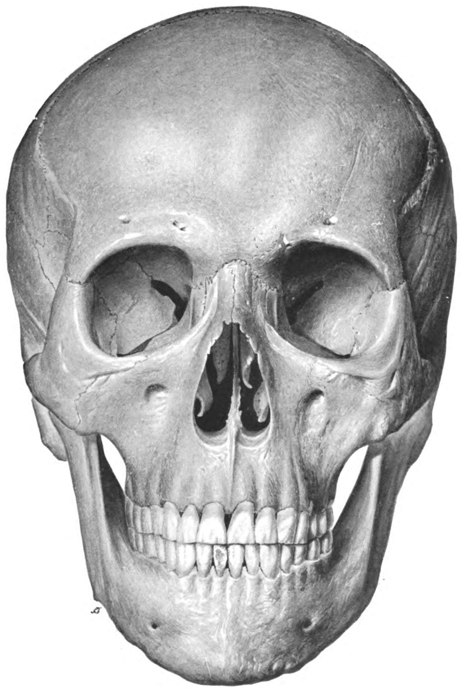
True statement about the skull shown below:

Ghost teeth are seen in which of the following?
What type of epiphysis is represented by the epiphysis at the tip of the coracoid process?
Subpubic angle in females is
Pell and Gregory classification includes all of the following except:
An abdominal CT shows 'champagne glass' appearance of pelvic bones. Which additional finding would best support Paget's disease?
Which test is most sensitive for detecting semen in forensic investigations?
Which of the following biomedical wastes can be incinerated?
Explanation: ***0, + 1000*** - The **CT number (Hounsfield Unit)** for **water** is defined as **0**, serving as a reference point for all other tissues in CT imaging. - **Bone**, particularly **cortical bone**, has a high density and thus corresponds to a CT number of approximately **+1000 HU**. *100,0* - This option incorrectly assigns a CT number of **100 to water**, which is fundamentally incorrect as water is defined as **0 HU**. - It also assigns **0** to **bone**, which is the CT number for water, not bone. *+1000,-100* - This option correctly identifies **+1000 HU** for dense bone but incorrectly assigns **-100 HU to water**, which is the CT number typically associated with fat, not water. *0,-1000* - While **0 HU** is correct for water, **-1000 HU** is the CT number for **air**, not bone. - Bone has a high positive CT number due to its high density, whereas air has a very low negative CT number.
Explanation: ***Cheiloscopy*** - **Cheiloscopy** is the scientific study of lip prints for human identification, based on the unique patterns of furrows on the human lips. - These patterns are considered individual and permanent, making them useful in forensic investigations. *Dactylography* - **Dactylography** is the study of fingerprints, which involves analyzing the unique patterns of ridges and furrows on the fingertips for identification. - It is one of the most widely used and reliable methods for personal identification in forensic science, but does not involve lip prints, *Poroscopy* - **Poroscopy** is a forensic technique that involves the examination of the pores on the ridges of fingerprints. - It is used to individualize fingerprints when there is insufficient ridge detail, but it focuses on pores, not lip prints. *Trichology* - **Trichology** is the scientific study of hair and scalp. - In forensics, it involves analyzing hair samples to determine characteristics such as origin, race, and presence of toxins, but not lip prints.
Explanation: ***Prominent supraorbital ridges, prominent glabella, square chin, acute fronto-nasal angle - Male*** - The image displays features typical of a **male skull**, including a pronounced **supraorbital ridge** (brow ridge), a prominent **glabella** (area between the eyebrows), a more **square and robust chin**, and an **acute fronto-nasal angle**. - These characteristics reflect the usually heavier and denser bone structure in males, adapted for stronger muscle attachments. *Round shaped orbit, prominent frontal eminences - Female* - **Female skulls generally have rounder orbits** and more prominent frontal eminences, giving the forehead a fuller, more rounded appearance. - The skull in the image does not exhibit these characteristics; the orbits are squarer, and the frontal eminences are not particularly prominent. *Prominent supraorbital ridges, prominent glabella, square chin, acute fronto-nasal angle - Female* - This statement incorrectly attributes features typically found in **male skulls** to a female skull. - Female skulls are generally smoother and less rugged, lacking the prominently developed ridges and angles seen in the image. *Round shaped orbit, prominent frontal eminences - Male* - This statement incorrectly attributes features typical of **female skulls** to a male skull. - Male skulls usually have squarer orbits and less prominent frontal eminences, with a more sloping forehead compared to females.
Explanation: ***Regional odontodysplasia*** - **Ghost teeth** is a characteristic radiographic appearance seen in **regional odontodysplasia** due to severe hypomineralization and thin enamel and dentin. - This condition involves both the enamel and dentin, resulting in teeth that appear faint and poorly formed on radiographs. *Dens in dente* - This condition refers to a tooth anomaly where the **enamel organ invaginates into the dental papilla** before calcification. - Radiographically, it appears as a **tooth within a tooth**, not as faint or ghost-like structures. *Dentin dysplasia* - This is a hereditary developmental disturbance of dentin formation, categorized into two types, Type I and Type II. - Type I presents with **short or absent roots** and pulp chamber obliteration, while Type II shows **thistle-shaped pulp chambers** and pulp stones, neither of which are described as "ghost teeth." *None of the options* - This option is incorrect because **regional odontodysplasia** is indeed associated with the characteristic presentation of "ghost teeth."
Explanation: ***Atavistic*** - An **atavistic epiphysis** is a phylogenetic remnant representing a part that was a **separate bone in lower vertebrates** but has become fused in humans during evolution. - The **tip of the coracoid process** is classified as an atavistic epiphysis because the coracoid was a separate bone in reptiles and birds, and the apical epiphysis at its tip represents this evolutionary remnant. - This epiphysis appears around **puberty (15-18 years)** and fuses by the **mid-twenties** [1]. [1] *Traction epiphysis* - A **traction epiphysis** is located at sites of **major muscle or tendon attachment** and does not contribute to longitudinal bone growth. - While the coracoid process does have a traction epiphysis at its **base** (for attachments of short head of biceps and coracobrachialis), the question specifically asks about the **tip/apex** of the coracoid process. - The **tip** primarily represents a phylogenetic remnant rather than serving as the primary attachment point, making it atavistic rather than traction in classification. *Pressure epiphysis* - A **pressure epiphysis** is found at the **articular ends of long bones**, transmits weight-bearing forces, and contributes significantly to **longitudinal bone growth** [1]. - The coracoid process tip is a non-articular projection that does not bear weight or transmit pressure across joints. - It does not contribute to longitudinal growth of the scapula [1]. *None of the options* - This option is incorrect because the tip of the coracoid process definitively fits the classification of an **atavistic epiphysis** based on its evolutionary origin and developmental characteristics.
Explanation: ***90 degrees*** - The **subpubic angle** in females typically ranges from **80-90 degrees**, making **90 degrees** the most accurate answer among the given options. - This wider angle is a distinguishing feature of the **female pelvis**, reflecting adaptations for childbirth and is significantly wider than the male angle (50-60 degrees). - Standard anatomy references cite the female subpubic angle as approximately **90 degrees** at the upper limit of normal. *120 degrees* - While the female pelvis has a wider angle than males, **120 degrees** exceeds the normal anatomical range. - This value is an overestimation and not representative of the typical female subpubic angle. *180 degrees* - A subpubic angle of **180 degrees** would imply a completely flat, straight line between the pubic rami, which is anatomically impossible. - This value does not represent any normal anatomical configuration in the human pelvis. *70 degrees* - An angle of **70 degrees** is characteristic of the **male subpubic angle**, which is narrower (typically 50-60 degrees, but can be up to 70 degrees). - This narrower angle is not conducive to childbirth and distinguishes the male from the female pelvis.
Explanation: ***Root size*** - The Pell and Gregory classification for impacted mandibular third molars assesses the **spatial relationship** between the impacted tooth and surrounding structures - It specifically evaluates: (1) **Class I-III** based on the relationship to the ramus and second molar, and (2) **Position A-C** based on depth relative to the occlusal plane - **Root size and morphology** are NOT parameters in this classification system, making this the correct answer *Angulation of 3rd molar* - While angulation is important in surgical planning, it is classified by **Winter's classification** (mesioangular, distoangular, vertical, horizontal), not by Pell and Gregory - However, Pell and Gregory does assess the **position** of the tooth, which is different from its angulation *Height of mandible* - The Pell and Gregory classification indirectly relates to mandibular dimensions through its assessment of available **anteroposterior space** - **Class I:** Sufficient space between distal of 2nd molar and anterior border of ramus - **Class II:** Space is less than the mesiodistal width of the 3rd molar crown - **Class III:** 3rd molar is completely within the ramus *Relationship to adjacent teeth* - The Pell and Gregory classification specifically includes the **spatial relationship** of the impacted 3rd molar to the 2nd molar and the ascending ramus - This relationship to adjacent structures is a fundamental component of the classification
Explanation: ***Cotton wool skull*** - The **champagne glass appearance** of the pelvic bones is a characteristic radiological finding in **Paget's disease**, indicating bone remodeling with cortical thickening and expansion of the iliac bones. - The presence of a **cotton wool skull**, characterized by patchy areas of sclerosis and lucency on skull radiographs, is another highly specific and common finding in advanced Paget's disease, further supporting this diagnosis. *Bamboo spine* - **Bamboo spine** is a characteristic radiographic finding in **ankylosing spondylitis**, representing calcification of the spinal ligaments and fusion of vertebrae. - This finding is not associated with Paget's disease of bone. *Ivory vertebra* - An **ivory vertebra** refers to a homogeneously dense and sclerotic vertebral body, which is a classic radiographic sign seen in **Paget's disease**, **metastatic prostate cancer**, and occasionally in **lymphoma**. - While ivory vertebra can occur in Paget's disease, the **cotton wool skull** in conjunction with the champagne glass pelvis provides a more specific diagnostic pattern, as cotton wool appearance is almost pathognomonic for Paget's disease. *Bone islands* - **Bone islands** (enostoses) are common, benign focal areas of cortical bone within the cancellous bone, usually seen as uniformly dense, well-circumscribed lesions on imaging. - They are typically asymptomatic and unrelated to Paget's disease, which involves widespread disorganized bone remodeling.
Explanation: ***Acid phosphatase test*** - This test is highly sensitive for detecting **prostatic acid phosphatase (PAP)**, a key enzyme found in high concentrations in semen. - A positive result, indicated by a **rapid color change**, suggests the presence of seminal fluid, even in small quantities. *Luminol test* - The luminol test is used to detect **bloodstains** at a crime scene, causing them to luminescence, but it is not specific for semen. - While it can react to other substances and potentially give **false positives** for semen, its primary use is for blood detection. *Barberio test* - The Barberio test is a **confirmatory microcrystal test** for spermine, a component of semen. - It involves the formation of specific crystals; however, it is less sensitive for initial screening than the acid phosphatase test and requires the presence of spermine. *Florence test* - The Florence test is a **confirmatory microcrystal test** for choline, another component of semen. - Similar to the Barberio test, it is a corroborative test that requires the formation of specific crystals and is not used for initial, highly sensitive screening.
Explanation: ***Human anatomical wastes*** - **Human anatomical wastes**, such as **tissues**, **organs**, and **body parts** obtained from surgical procedures or autopsies, are typically incinerated. - **Incineration** is an effective method for sterilizing and safely disposing of such bio-hazardous materials, reducing them to ash and destroying pathogens. *Pressurized gas containers* - **Pressurized gas containers**, like oxygen tanks or aerosol cans, contain **flammable** or **explosive gases** and can rupture under extreme heat. - Incineration poses a significant **safety risk** due to potential explosions and the release of harmful gases. *Radiographic wastes* - **Radiographic wastes** include items such as **used X-ray films** and **spent fixer/developer solutions**, which contain heavy metals like silver. - Incineration can release **toxic fumes** and **pollutants** into the atmosphere and is not the recommended disposal method for these materials. *PVC* - **Polyvinyl chloride (PVC)**, when incinerated, releases **toxic fumes** including **dioxins** and **furans**, which are harmful to human health and the environment. - **Non-chlorinated plastics** are preferred for incineration to minimize the release of these highly toxic compounds.
Get full access to all questions, explanations, and performance tracking.
Start For Free