Which of the following is a type of observational study that analyzes population-level data?
A 40-year-old man presents with daytime sleepiness and impaired concentration and memory. On examination his BMI is 41 kg/m2, BP is 160/100 mm Hg. His awake ABG analysis is given: PaO2=66 mm Hg, PaCO2=50 mm Hg, HCO3=28 mEq/L. What is the most likely diagnosis?
Severe Obstructive sleep apnea is defined as AHI of greater than
Berger waves (alpha waves) of EEG have a rhythm of how many Hz?
Most sensitive method of monitoring cardiovascular ischemia in the perioperative period is -
Which of the following is not a diagnostic criterion for SIRS?
A child presents with night blindness, delayed dark adaptation. Which investigation is to be done further to confirm the diagnosis?
In current obstetrics practice, what is the best test for monitoring sensitized Rh negative mother?
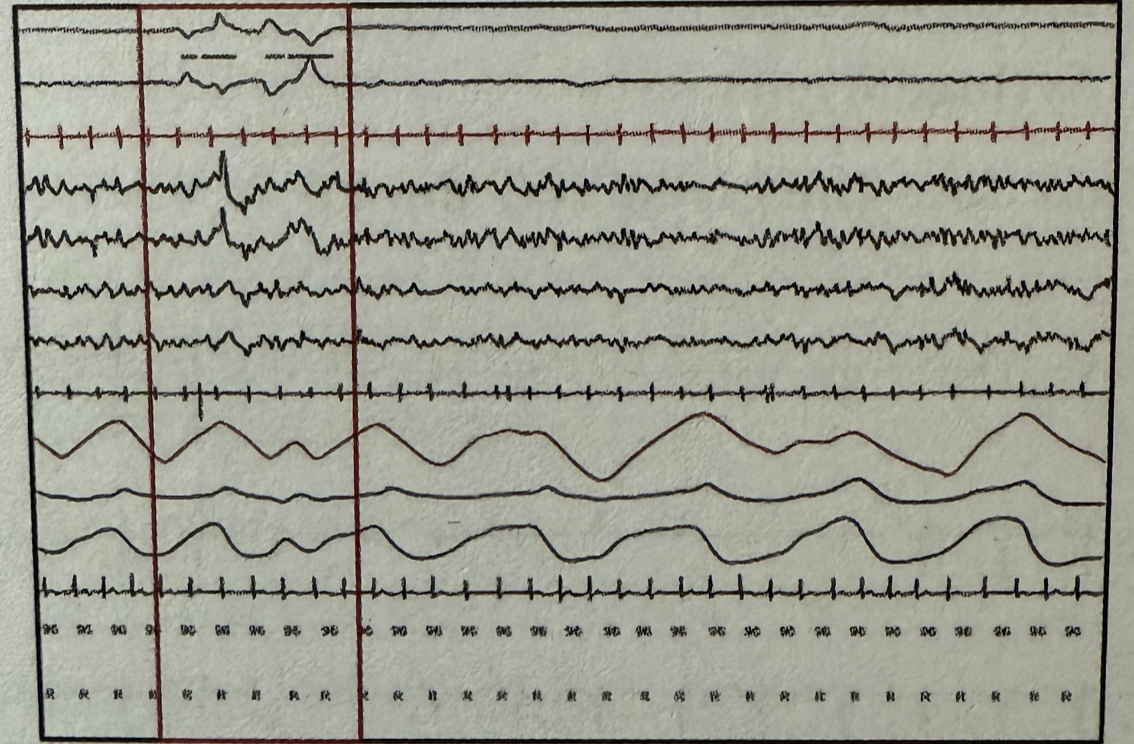
During polysomnography, which stage of sleep is represented by the marked areas when observing the following wave patterns? EOG (Electrooculography) EEG (Electroencephalography) EMG (Electromyography)

All of the following are correct about the image shown except:
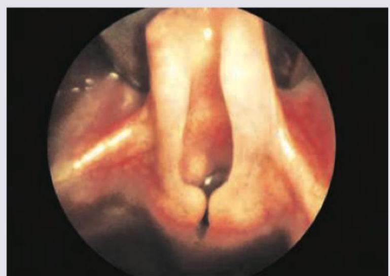
Explanation: ***Ecological study*** - This type of study examines the relationship between an exposure and an outcome at the **population level** rather than the individual level. - It often uses aggregated data, such as incidence rates of disease in different geographic areas, to identify associations. *Case-control study* - This is an **individual-level observational study** that compares individuals with a disease (cases) to individuals without the disease (controls) and looks back retrospectively at their exposures. - It is used to investigate potential risk factors for a disease but does not analyze population-level data directly. *Randomized controlled trial* - This is an **experimental study design** where participants are randomly assigned to an intervention group or a control group. - It is considered the gold standard for establishing causality but does not analyze observational population-level data. *Longitudinal study* - This is an **individual-level observational study** that follows the same group of individuals over a period of time, collecting data at multiple points. - While it observes changes over time, it typically focuses on individual-level trends and outcomes, not aggregated population data.
Explanation: ***Obstructive Sleep Apnea (Correct Answer)*** - Classic triad: **morbid obesity (BMI 41 kg/m²)**, **excessive daytime somnolence**, and **systemic hypertension (160/100 mmHg)** — hallmarks of OSA - **ABG findings** (PaO2=66 mmHg, PaCO2=50 mmHg, HCO3=28 mEq/L) indicate **chronic nocturnal hypoxemia and hypercapnia** with compensatory **metabolic alkalosis** from repeated apneic episodes - **Cognitive impairment** (impaired concentration and memory) results from **sleep fragmentation** and intermittent nocturnal hypoxia - Obesity promotes **pharyngeal fat deposition** → upper airway narrowing and collapse during sleep → recurrent obstructive events *Narcolepsy* - Causes excessive daytime sleepiness but is **not associated with obesity, hypertension, or ABG abnormalities** - Hallmarks include **cataplexy**, sleep paralysis, and hypnagogic/hypnopompic hallucinations — none present here - Caused by **orexin (hypocretin) deficiency**; associated with **HLA-DQB1*06:02**; ABG is normal *Obesity Hypoventilation Syndrome (OHS / Pickwickian Syndrome)* - Defined as **awake PaCO2 >45 mmHg + BMI >30 kg/m²** with exclusion of other causes of hypoventilation - OHS frequently coexists as an **overlap with and consequence of severe OSA** rather than being the primary diagnosis - In this setting, **OSA is the most prevalent and primary diagnosis**; OHS is specifically considered when awake hypoventilation persists despite adequate OSA treatment *Central Sleep Apnea* - Results from **failure of central respiratory drive** (brainstem), not upper airway obstruction - Associated with **congestive heart failure, opioid use, high-altitude exposure, or neurological disease** — none present here - Not characteristically associated with morbid obesity; clinical and ABG picture here favors an **obstructive** rather than central pattern
Explanation: ***30 events/hour*** - A **severe form of obstructive sleep apnea (OSA)** is diagnosed when the Apnea-Hypopnea Index (AHI) is greater than or equal to **30 events per hour** [1]. - The AHI represents the average number of **apnea and hypopnea events** per hour of sleep [1]. *15 events/hour* - An AHI of **15 to 30 events/hour** typically defines **moderate sleep apnea**, not severe. - This level indicates a significant number of sleep disturbances, but less than what is categorized as severe. *25 events/hour* - An AHI of **25 events/hour** falls within the **moderate range** of OSA severity (15-30 events/hour). - It does not meet the criteria for severe OSA, which requires a higher AHI. *20 events/hour* - An AHI of **20 events/hour** also falls into the **moderate category** of OSA. - This value is above the threshold for mild OSA (5-15 events/hour) but below the threshold for severe OSA.
Explanation: ***8-13 Hz*** - **Berger waves**, also known as **alpha waves**, are defined by their frequency range of **8 to 13 Hz** in the electroencephalogram (EEG). - These waves are typically observed when a person is in a relaxed, awake state with their eyes closed. *0-4 Hz* - This frequency range corresponds to **delta waves**, which are characteristic of deep sleep and certain brain pathologies. - Delta waves are much slower and have higher amplitude compared to alpha waves. *4-7 Hz* - This frequency range is associated with **theta waves**, commonly seen during light sleep, drowsiness, and some meditative states. - Theta waves are slower than alpha waves and indicate a state of reduced alertness. *13-30 Hz* - This frequency range represents **beta waves**, which are associated with active thinking, problem-solving, and alertness with open eyes. - Beta waves are faster and typically have lower amplitude than alpha waves.
Explanation: ***TEE*** - **Transesophageal echocardiography (TEE)** is the most sensitive method for detecting perioperative myocardial ischemia because it can visualize **regional wall motion abnormalities** and changes in **ventricular function** much earlier than ECG. - **Ischemia** directly impairs the contractility of the affected myocardium, leading to subtle changes in wall motion that TEE can identify. *NIBP* - **Non-invasive blood pressure (NIBP)** monitoring can detect **hemodynamic changes** (like hypotension or hypertension) that may precede or accompany ischemia. - However, these changes are **non-specific** and occur relatively late, making NIBP a less sensitive indicator of early ischemia. *ECG* - **Electrocardiography (ECG)** monitors the electrical activity of the heart and can detect **ST-segment changes** indicative of ischemia. - While useful, ECG changes may appear later than wall motion abnormalities, and **silent ischemia** can be missed if the leads are not optimally placed or if the ischemia does not produce significant electrical changes. *Pulse oximeter* - A **pulse oximeter** measures **oxygen saturation** in the peripheral blood. - It is primarily used to assess **respiratory function** and tissue oxygenation, and it does not directly monitor myocardial ischemia or cardiac function.
Explanation: ### Hypotension - **Hypotension** is a criterion for **sepsis** and **septic shock**, but not for **SIRS** itself. - **SIRS** criteria are based on inflammatory responses, while hypotension indicates a more severe systemic compromise. *Tachycardia* - **Tachycardia**, defined as a **heart rate >90 beats per minute**, is a diagnostic criterion for **SIRS** [1]. - It reflects the body's physiological stress response to a systemic inflammatory state [1]. *Tachypnoea* - **Tachypnoea**, indicated by a **respiratory rate >20 breaths per minute** or a **PaCO2 <32 mmHg**, is a diagnostic criterion for **SIRS** [1]. - This symptom shows the body's effort to compensate for metabolic acidosis or increased oxygen demand. *Leucocytosis* - **Leucocytosis**, defined as a **white blood cell count >12,000/mm³** or **<4,000/mm³**, or the presence of **>10% immature neutrophils (bands)**, is a diagnostic criterion for **SIRS** [1]. - This indicates a significant systemic inflammatory response in the blood [1].
Explanation: ***ERG*** - **Electroretinography (ERG)** measures the electrical responses of various retinal cells, including **rods** and **cones**, to light stimuli. - In conditions like **retinitis pigmentosa** which cause night blindness and delayed dark adaptation, ERG will show characteristic abnormal or extinguished responses, confirming retinal dysfunction. *Retinoscopy* - **Retinoscopy** is an objective method to assess the refractive error of the eye by observing the light reflex from the retina. - It does not directly evaluate the functional integrity of photoreceptors or diagnose conditions causing **night blindness**. *Dark adaptometry* - **Dark adaptometry** measures the time it takes for the eye to adapt to dim light after exposure to bright light, quantifying the function of **rod photoreceptors**. - While it can *detect* delayed dark adaptation, it is a functional test that assesses the symptom, not the underlying cause provided by ERG. *EOG* - **Electrooculography (EOG)** measures the potential difference between the cornea and the retina, primarily assessing the function of the **retinal pigment epithelium (RPE)**. - While useful for conditions like **Best's disease**, it is less direct for evaluating generalized rod dysfunction causing night blindness compared to ERG.
Explanation: ***Middle cerebral artery Doppler wave forms*** - This is currently the most widely accepted and **non-invasive** method for monitoring **fetal anemia** in Rh-sensitized pregnancies. - An increase in the **peak systolic velocity (PSV)** in the middle cerebral artery indicates that the fetus is increasing cardiac output to compensate for a reduced oxygen-carrying capacity due to anemia. *Biophysical profile* - The biophysical profile assesses various fetal parameters like **movement**, **tone**, **breathing**, and **amniotic fluid volume**, which are often altered late in the course of severe fetal anemia. - It is a **less sensitive** indicator of early or moderate fetal anemia compared to MCA Doppler. *Amniotic fluid spectrophotometry* - This method measures the **bilirubin levels** in amniotic fluid, which correlates with the severity of hemolysis. - It is an **invasive procedure** (amniocentesis) and has largely been replaced by non-invasive MCA Doppler due to associated risks and better predictive value of Doppler. *Fetal blood sampling* - Fetal blood sampling (cordocentesis) provides a direct measurement of **fetal hemoglobin** and other blood parameters. - While definitive, it is a **highly invasive procedure** with significant risks, reserved primarily for confirmation of severe anemia or for direct transfusion, not for routine monitoring.
Explanation: ***NREM I sleep*** - This stage is characterized by a transition from wakefulness to sleep, identifiable by the appearance of **slow eye movements** in the EOG and a reduction in EEG frequency with the presence of **theta waves**. - The EMG shows a decrease in muscle tone but without the complete atonia seen in REM sleep. *REM sleep* - **Rapid eye movements** are characteristic in the EOG, and the EEG shows **low-amplitude, mixed-frequency waves** similar to wakefulness. - The EMG would display profound muscle atonia, which is not evident in the provided tracing. *NREM II sleep* - This stage is marked by the presence of **sleep spindles** and **K-complexes** in the EEG, which are absent in the marked area. - Eye movements are generally absent, and muscle activity continues to be low. *NREM III sleep* - This is the deepest stage of sleep, characterized by **high-amplitude, slow-delta waves** (20-50% of the epoch) in the EEG. - Eye movements are typically absent, and muscle tone is very low but not completely absent.
Explanation: ***High pitched expiratory stridor*** - The image depicts an **omega-shaped epiglottis** and collapsed aryepiglottic folds, consistent with **laryngomalacia**. - Laryngomalacia typically presents with **inspiratory stridor**, not expiratory, resulting from airway collapse during inspiration. - **This is the EXCEPT answer** - high-pitched expiratory stridor is NOT a feature of laryngomalacia. *Omega shaped epiglottis* - The image clearly shows an **omega-shaped epiglottis**, a characteristic feature of **laryngomalacia**. - This anatomical variation contributes to the collapse of supraglottic structures during inspiration. *Cry is normal* - In laryngomalacia, the **vocal cords** themselves are not affected, so the **cry typically remains normal**. - The abnormal sounds (stridor) arise from the supraglottic structures, not the vocal cord function during crying. *10% cases need surgery due to development of OSA or Cor Pulmonale* - While most cases of laryngomalacia are self-limiting, approximately **10% of infants may require surgical intervention** (supraglottoplasty). - This is usually due to severe symptoms like **obstructive sleep apnea (OSA)**, failure to thrive, or the rare development of **cor pulmonale**.
Get full access to all questions, explanations, and performance tracking.
Start For Free