What is the most common structural location within the bone for conventional chondrosarcoma?
All of the following statements about synovial sarcoma are true, except -
A 68-year-old man has many months history of progressive hearing loss, unsteady gait, tinnitus, and facial pain. An MRI scan reveals a tumor at the cerebellopontine angle. Which of the following cranial nerves is this tumor most likely to affect?
Which brain tumor is the most radiosensitive?
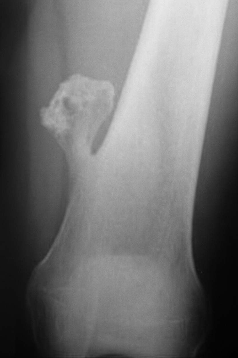
A 14 year old male presents with mushroom like tumor in the distal femur for past 2 years. Which of the following features suggest malignant transformation?

Which of the following is an epiphyseal tumor?
Nasopharyngeal chordoma arises from:-
Chordoma arises from:
A lady comes to OPD after fall from scooty. Her vitals are stable. She is having continuous, clear watery discharge from nose after 2 days. This is most likely a feature of?
FISCH classification is used for-
Explanation: ***Intramedullary*** - This is the **most common location** for conventional chondrosarcoma, accounting for approximately **75-85%** of cases [1]. - These tumors arise within the **medullary cavity** (central or intramedullary location) and grow expansively within the bone [1]. - They typically show lytic destruction with internal ring-and-arc or popcorn calcifications on imaging. - Conventional chondrosarcoma is synonymous with central or intramedullary chondrosarcoma [1]. *Periosteal* - Periosteal (surface) chondrosarcoma is a **rare subtype** accounting for only **1-2%** of chondrosarcomas. - These arise from the periosteum on the bone surface, not within the medullary cavity. - They have a better prognosis compared to conventional (intramedullary) chondrosarcoma. *Juxtacortical* - This refers to tumors arising at or near the **bone surface** (cortical region) [1]. - Peripheral/juxtacortical chondrosarcoma accounts for approximately **10-15%** of cases and is much less common than the central type. - Secondary chondrosarcoma arising from osteochondroma is an example of peripheral chondrosarcoma [1]. *Epiphyseal* - Chondrosarcoma **rarely arises in the epiphysis** of long bones. - Conventional chondrosarcoma typically involves the **metaphysis and diaphysis**, with predilection for flat bones (pelvis, ribs) and long bones (femur, humerus) [2]. - Epiphyseal location would be highly unusual for this tumor. **References:** [1] Kumar V, Abbas AK, et al.. Robbins and Cotran Pathologic Basis of Disease. 9th ed. Bones, Joints, and Soft Tissue Tumors, pp. 1202-1204. [2] Cross SS. Underwood's Pathology: A Clinical Approach. 6th ed. Common Clinical Problems From Osteoarticular And Connective Tissue Disease, pp. 673-674.
Explanation: ***Originate from synovial lining*** - This statement is **false**. While named "synovial sarcoma" due to its histological resemblance to synovium, it **does not originate from synovial cells** [1]. - Its origin is believed to be from **primitive mesenchymal cells** that can differentiate along various cell lines, not directly from the synovial lining of joints. *Usually seen in patients less than 50 years of age* - This statement is **true**. Synovial sarcoma predominantly affects **adolescents and young adults**, with a median age of diagnosis typically in the 3rd or 4th decade of life [1]. - It is one of the more common soft tissue sarcomas in this younger age group. *Occur more often at extraarticular sites* - This statement is **true**. Despite its name, synovial sarcoma most frequently occurs in **extra-articular locations**, often near large joints but not within the joint capsule itself [1]. - Common sites include the deep soft tissues of the extremities, especially the **thigh and knee**, and less often the trunk or head and neck. *Knee and foot are common sites involved* - This statement is **true**. The **knee** (particularly the thigh region around the knee) and **foot/ankle** are indeed among the most frequent locations for synovial sarcoma [1]. - These tumors often present as a deep-seated mass in these areas. **References:** [1] Kumar V, Abbas AK, et al.. Robbins and Cotran Pathologic Basis of Disease. 9th ed. Bones, Joints, and Soft Tissue Tumors, pp. 1225-1226.
Explanation: ***eighth cranial nerve*** - The **eighth cranial nerve (vestibulocochlear nerve)** is located in the **cerebellopontine angle** and is responsible for **hearing and balance**. [1], [2] - Symptoms like **progressive hearing loss, tinnitus, and unsteady gait (vertigo)** are classic signs of compression or damage to this nerve, often caused by an **acoustic neuroma (vestibular schwannoma)** in this region. [2], [3] - **CN VIII is the FIRST and MOST COMMONLY affected nerve** in cerebellopontine angle tumors, making it the correct answer. - The **facial pain** mentioned suggests compression of the **trigeminal nerve (CN V)** by a large tumor, which can occur as the tumor expands, but CN VIII remains the primary nerve affected. *sixth cranial nerve* - The **sixth cranial nerve (abducens nerve)** innervates the **lateral rectus muscle**, responsible for **abduction of the eye**. - Damage would typically result in **diplopia** and an inability to move the eye laterally, which is not described. - This nerve is **rarely affected** by CPA tumors due to its anatomical location. *tenth cranial nerve* - The **tenth cranial nerve (vagus nerve)** controls **pharyngeal and laryngeal muscles**, as well as **parasympathetic innervation to many organs**. - Damage would typically cause **dysphagia**, **hoarseness**, or autonomic dysfunction, none of which are presented. - The vagus nerve is **not typically affected** by CPA tumors. *fourth cranial nerve* - The **fourth cranial nerve (trochlear nerve)** innervates the **superior oblique muscle**, aiding in **eye movement**. - Damage would primarily lead to **vertical diplopia**, particularly when looking down and in, which is not mentioned as a symptom. - This nerve is **not affected** by CPA tumors due to its location.
Explanation: ***Medulloblastoma*** - **Medulloblastoma** is highly **radiosensitive** due to its rapid cell proliferation and immature cellular characteristics, making radiation therapy a cornerstone of treatment. - This tumor commonly originates in the **cerebellum** and is one of the most common malignant brain tumors in children. *Ependymoma* - **Ependymomas** are generally only moderately **radiosensitive**; while radiation is used, it is often delivered in higher doses directly to the tumor bed. - These tumors arise from **ependymal cells** lining the ventricles and spinal cord. *Glioblastoma multiforme* - **Glioblastoma multiforme (GBM)** is known for its marked **radioresistance**, requiring high doses of radiation often in combination with chemotherapy, and still having a poor prognosis. - It is the most aggressive and common type of primary **brain tumor in adults**, characterized by rapid growth and extensive infiltration. *Astrocytoma* - The **radiosensitivity** of astrocytomas varies significantly by grade; **low-grade astrocytomas** are relatively radioresistant, while **anaplastic astrocytomas** have intermediate radiosensitivity. - These tumors originate from **astrocytes**, a type of glial cell, and can occur in various parts of the brain and spinal cord.
Explanation: ***Cartilage thickness >2 cm*** - A **cartilage cap thickness greater than 2 cm** in an osteochondroma in an adult (or >3 cm in children) is a strong indicator of **malignant transformation** into a secondary peripheral **chondrosarcoma**. [2], [3] - **Key imaging finding:** Cartilage cap measured on MRI or CT scan - Other features suggesting malignant transformation include continued growth after skeletal maturity, new or increasing pain, cortical destruction, and new soft tissue mass. [2], [3] *Presence of cartilage cap* - All osteochondromas have a cartilage cap by definition - this is a normal feature, not a sign of malignancy. [1] - The **thickness** of the cap, not its presence, is what matters. *Location in metaphysis* - Osteochondromas typically arise from the metaphysis near the growth plate - this is a normal location. [3] - Location alone does not indicate malignant transformation. *Size less than 1 cm* - Small size suggests a benign, stable lesion. - Malignant transformation is suggested by **increasing size** and growth after skeletal maturity, not small size. [2] **References:** [1] Kumar V, Abbas AK, et al.. Robbins and Cotran Pathologic Basis of Disease. 9th ed. Bones, Joints, and Soft Tissue Tumors, p. 1202. [2] Cross SS. Underwood's Pathology: A Clinical Approach. 6th ed. Common Clinical Problems From Osteoarticular And Connective Tissue Disease, pp. 672-673. [3] Kumar V, Abbas AK, et al.. Robbins and Cotran Pathologic Basis of Disease. 9th ed. Bones, Joints, and Soft Tissue Tumors, pp. 1202-1204.
Explanation: ***Chondroblastoma*** - **Chondroblastoma** is a rare, benign cartilaginous tumor that typically originates in the **epiphysis** of long bones before the closure of growth plates. - It is histologically characterized by chondroblast-like cells, multinucleated giant cells, and chondroid matrix, and radiographically appears as a lytic lesion with a sclerotic rim in the epiphysis. *Osteosarcoma* - **Osteosarcoma** is the most common primary malignant bone tumor and typically originates in the **metaphysis** of long bones, particularly around the knee. - It invades the surrounding bone and soft tissues, often presenting with a **Codman triangle** or **sunburst pattern** on imaging studies. *Osteoid osteoma* - **Osteoid osteoma** is a benign bone-forming tumor primarily found in the **cortex** of long bones, although it can occur in other locations, presenting with nocturnal pain relieved by NSAIDs. - It is characterized by a central radiolucent nidus surrounded by reactive sclerotic bone. *Adamantinoma* - **Adamantinoma** is an extremely rare, low-grade malignant tumor that almost exclusively occurs in the **tibia diaphysis**. - It is thought to originate from epithelial cells and presents as a lytic lesion within the cortical bone, often with a polycystic appearance.
Explanation: Correct Option: Notochord - **Chordomas** are rare malignant tumors that arise from embryonic remnants of the **notochord** [1] - The notochord is a flexible rod-like structure that forms the primitive axial skeleton during embryonic development - Chordomas typically occur along the **midline** at sites where notochordal remnants persist, most commonly at the **skull base (clivus/nasopharynx)** and **sacrococcygeal region** [1] Incorrect Option: Luschka's bursa - **Luschka's bursa** (also known as the pharyngeal bursa) is a small indentation or pocket in the nasopharynx posterior to the pharyngeal tonsil - While located in the nasopharynx, it is a **normal anatomical structure**, not an embryological remnant that gives rise to tumors - Not associated with chordoma development Incorrect Option: Pharyngeal bursa - **Pharyngeal bursa** is another term for **Luschka's bursa** (they are synonymous) - It is a normal anatomical structure in the nasopharynx - Not related to the embryonic remnants that give rise to chordomas Incorrect Option: Rathke's pouch - **Rathke's pouch** is an embryonic invagination of the stomodeum (primitive oral cavity) that gives rise to the **anterior pituitary gland** - Tumors arising from remnants of Rathke's pouch are **craniopharyngiomas**, which are distinct from chordomas - Craniopharyngiomas are typically suprasellar, while chordomas are more commonly found at the clivus or sacrum
Explanation: ***Notochord*** - Chordoma is a **rare, slow-growing malignant bone tumor** that originates from **persistent notochordal remnants**. - The notochord is a flexible rod-like structure that serves as the primary axial support during embryonic development, eventually becoming the **nucleus pulposus** of the intervertebral discs. *Pharyngeal bursa* - The pharyngeal bursa is a normal anatomical variant, a small recess in the **posterior wall of the nasopharynx**. - It is not associated with the development of chordomas. *Rathke's pouch* - Rathke's pouch is an **ectodermal invagination** from the roof of the primitive mouth (stomodeum) that gives rise to the **anterior pituitary gland**. - Tumors arising from remnants of Rathke's pouch are typically **craniopharyngiomas**, not chordomas. *Luschka's bursa* - This term is sometimes used to refer to a **pharyngeal bursa**, as mentioned in the first incorrect option. - It is not the origin of chordomas.
Explanation: ***CSF rhinorrhoea*** - **Clear watery discharge** appearing **two days after head trauma** (fall from scooty) is highly suggestive of **cerebrospinal fluid (CSF) rhinorrhoea**. - This occurs due to a breach in the **skull base**, allowing CSF to leak from the subarachnoid space into the nasal cavity. *Acute respiratory infection* - An acute respiratory infection typically presents with symptoms like **fever, cough**, and **nasal discharge** that is often thicker and discolored, not clear and watery. - The onset of discharge two days after trauma without other signs of infection also makes this less likely. *Rhinitis* - Rhinitis involves inflammation of the nasal mucosa, leading to watery discharge, sneezing, and congestion. - However, the traumatic etiology and the specific timing of the discharge make **CSF leak** a more pertinent diagnosis than simple rhinitis. *Middle cranial fossa fracture* - While a **middle cranial fossa fracture** can cause CSF leakage, the discharge from the nose (rhinorrhoea) typically originates from an **anterior cranial fossa fracture**. - A middle cranial fossa fracture is more commonly associated with **otorrhoea** (CSF leakage from the ear) if the temporal bone is involved.
Explanation: ***Glomus tumor*** - The **FISCH classification** is a surgical staging system used to classify **glomus tumors** based on their extent and involvement of surrounding structures. - This classification helps guide surgical management and predict procedural outcomes for these highly vascular tumors. *Juvenile nasopharyngeal angiofibroma* - **Radkowski's classification**, or **Andrews' classification**, are commonly used for staging **juvenile nasopharyngeal angiofibroma**, not FISCH. - These classifications categorize tumors based on their extension into the nasal cavity, paranasal sinuses, orbit, or intracranial space. *Nasopharyngeal ca* - The staging of **nasopharyngeal carcinoma** is typically based on the **AJCC (American Joint Committee on Cancer) TNM classification system**. - This system assesses the **tumor (T)** size and local extension, **node (N)** involvement, and **metastasis (M)**. *Vestibular schwannoma* - **Vestibular schwannomas** are usually staged using systems that describe their size and extension into the **cerebellopontine angle** and brainstem, such as the **Koos grade**. - The FISCH classification is specifically for **glomus tumors** of the temporal bone and is not applicable to vestibular schwannomas.
Get full access to all questions, explanations, and performance tracking.
Start For Free