Intrathecal fluorescein with endoscopic visualization is useful in diagnosis of?
A lady comes to OPD after fall from scooty. Her vitals are stable. She is having continuous, clear watery discharge from nose after 2 days. This is most likely a feature of?
Most difficult site to remove cholesteatoma from the sinus tympani is related to:
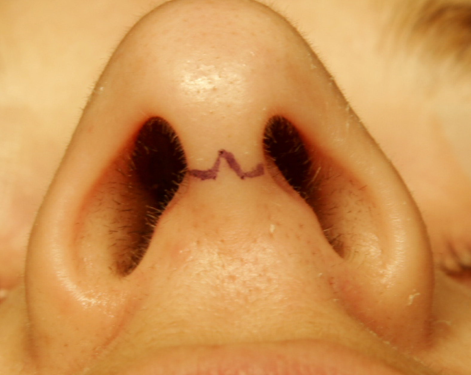
A nasal surgery was carried out with the incision shown in the image. What was the procedure likely carried out?

Which of the following is not a complication of maxillary sinus lavage and insufflation?
Which of the following is a common topical use of the medicine shown in the image?

Statement 1 - A 59-year-old patient presents with flaccid bullae. Histopathology shows a suprabasal acantholytic split. Statement 2 - The row of tombstones appearance is diagnostic of Pemphigus vulgaris.
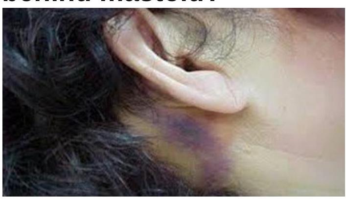
What does a bluish-purple discoloration behind the mastoid indicate?

FISCH classification is used for-
Dohlman surgery in Zenker's diverticulum is:-
Explanation: ***Diagnosis of CSF Rhinorrhoea*** - **Intrathecal fluorescein** is instilled into the cerebrospinal fluid, and its presence in the nasal cavity via endoscopy confirms a **CSF leak**. - This method provides direct visualization of the leak site, which is crucial for surgical planning. *Rhinitis Medicamentosa* - This condition is caused by overuse of **topical decongestants** and characterized by nasal congestion, not a CSF leak. - Diagnosis is typically based on patient history and clinical examination rather than specialized imaging or dye studies. *Multiple ethmoidal polyps* - **Ethmoidal polyps** are benign growths in the ethmoid sinuses, causing nasal obstruction and anosmia. - Diagnosis is made via nasal endoscopy and CT scan, and fluorescein staining is not indicated. *Deviated nasal septum* - A **deviated nasal septum** is a structural abnormality causing unilateral or bilateral nasal obstruction. - Diagnosis is clinical and confirmed by anterior rhinoscopy or nasal endoscopy, with no role for intrathecal fluorescein.
Explanation: ***CSF rhinorrhoea*** - **Clear watery discharge** appearing **two days after head trauma** (fall from scooty) is highly suggestive of **cerebrospinal fluid (CSF) rhinorrhoea**. - This occurs due to a breach in the **skull base**, allowing CSF to leak from the subarachnoid space into the nasal cavity. *Acute respiratory infection* - An acute respiratory infection typically presents with symptoms like **fever, cough**, and **nasal discharge** that is often thicker and discolored, not clear and watery. - The onset of discharge two days after trauma without other signs of infection also makes this less likely. *Rhinitis* - Rhinitis involves inflammation of the nasal mucosa, leading to watery discharge, sneezing, and congestion. - However, the traumatic etiology and the specific timing of the discharge make **CSF leak** a more pertinent diagnosis than simple rhinitis. *Middle cranial fossa fracture* - While a **middle cranial fossa fracture** can cause CSF leakage, the discharge from the nose (rhinorrhoea) typically originates from an **anterior cranial fossa fracture**. - A middle cranial fossa fracture is more commonly associated with **otorrhoea** (CSF leakage from the ear) if the temporal bone is involved.
Explanation: **Posterior facial ridge** - The **posterior facial ridge** forms a critical anatomical barrier, often **obscuring the sinus tympani** and making cholesteatoma removal difficult due to limited access and visibility. - Its presence necessitates careful surgical technique to avoid **facial nerve injury** while ensuring complete eradication of disease. *Anterior facial ridge* - The **anterior facial ridge** is less directly involved in defining the access to the sinus tympani, thus not posing as significant a surgical challenge for cholesteatoma removal in this specific area. - Its anatomical position typically allows for better visualization and maneuverability compared to the posterior ridge. *Epitympanum* - The **epitympanum**, while a common site for cholesteatoma, is generally more accessible surgically than the sinus tympani because of its location superior to the tympanic membrane. - Its removal often involves mastoidectomy or atticotomy, which provides direct views. *Hypotympanum* - The **hypotympanum** is located inferior to the tympanic membrane and is typically more open and accessible, allowing for easier visualization and removal of cholesteatoma. - It does not present the same deep, narrow, and obstructed surgical field as the sinus tympani, especially in relation to the facial nerve.
Explanation: ***Open rhinoplasty*** - The image displays a **transcolumellar incision** (typically inverted V or W-shaped), which is the hallmark approach for **open rhinoplasty**. - This incision allows for direct visualization of the underlying nasal cartilages and bones, enabling precise reshaping of the nose. *Submucosal resection (SMR)* - SMR is a procedure to correct a **deviated nasal septum** by removing cartilage or bone from beneath the mucoperichondrial flaps. - It involves an **intranasal incision**, usually along the septal mucosa, not an external transcolumellar incision. *FESS (Functional Endoscopic Sinus Surgery)* - FESS is a minimally invasive procedure used to treat **chronic sinusitis** and other sinus conditions. - It is performed entirely **endoscopically through the nostrils**, with no external incisions on the nasal columella. *Caldwell-Luc's procedure* - This procedure accesses the **maxillary sinus** through an incision in the upper gum beneath the lip. - It is used for drainage of the maxillary sinus or removal of foreign bodies/tumors, and does not involve an external nasal incision.
Explanation: ***Facial nerve injury*** - The **facial nerve (CN VII)** passes through the parotid gland and temporal bone, far from the maxillary sinus. - There is no anatomical proximity or procedural mechanism during maxillary sinus lavage and insufflation that would put the facial nerve at risk of injury. *Air embolism* - **Insufflation of air** into the maxillary sinus, especially under pressure, can lead to air entering the bloodstream if a blood vessel is inadvertently punctured. - This can result in a serious and potentially fatal **air embolism**, particularly if the air reaches the cerebral circulation. *Orbital injury* - The **medial wall of the maxillary sinus** is in close proximity to the orbit, separated by thin bone. - During lavage, excessive force or incorrect angulation of instruments can perforate this thin bone, leading to **orbital complications** such as periorbital hematoma or injury to orbital contents. *Epistaxis* - During the procedure, the **mucosa of the nasal cavity** or the sinus itself can be traumatized by the instruments used for lavage. - This local trauma to the rich blood supply of these areas can easily cause **nasal bleeding (epistaxis)**.
Explanation: ***Post-adenoidectomy to control bleeding*** - The image shows **Neo-Synephrine (phenylephrine)**, a potent **alpha-adrenergic agonist** that causes **vasoconstriction**. - Its vasoconstrictive properties make it useful topically to reduce **bleeding** during and after surgical procedures like **adenoidectomy**. *Rhino cerebral mucormycosis* - This is a serious fungal infection requiring systemic antifungal therapy, often **amphotericin B**. Topical phenylephrine has no role in treating the infection itself. - While bleeding might be a symptom of mucormycosis, phenylephrine would only offer temporary symptomatic relief, not address the underlying fungal pathology. *Inlay type I myringoplasty* - Myringoplasty is a surgical procedure to repair a perforated eardrum. Topical phenylephrine is not indicated for this procedure. - The primary goal of this surgery is to reconstruct the **tympanic membrane**, and phenylephrine would not contribute to tissue healing or graft integration. *Subglottic stenosis* - This condition involves narrowing of the airway below the vocal cords, often requiring surgical intervention or corticosteroids. - Phenylephrine is a decongestant and vasoconstrictor, and as such, it does not have a therapeutic role in resolving the **fibrotic narrowing** characteristic of subglottic stenosis.
Explanation: ***Correct: Statements 1 & 2 are correct, 2 is not explaining 1*** **Analysis of Statement 1:** - A 59-year-old patient with **flaccid bullae** and **suprabasal acantholytic split** on histopathology is the classic presentation of **Pemphigus vulgaris** - The flaccid (easily ruptured) nature of bullae distinguishes it from tense bullae seen in bullous pemphigoid - The suprabasal location of the split (just above the basal layer) with acantholysis (loss of cell-to-cell adhesion) is pathognomonic - **Statement 1 is CORRECT** ✓ **Analysis of Statement 2:** - The **"row of tombstones" or "tombstone appearance"** is indeed a diagnostic histopathological feature of Pemphigus vulgaris - This appearance results from basal keratinocytes remaining attached to the basement membrane while suprabasal cells separate due to acantholysis - The intact basal cells standing upright resemble a row of tombstones - **Statement 2 is CORRECT** ✓ **Does Statement 2 explain Statement 1?** - Statement 2 describes a **histopathological appearance** (tombstone pattern) that is a **consequence** of the suprabasal split - However, it does NOT explain the **underlying cause** of the flaccid bullae or the suprabasal split - The true explanation involves **IgG autoantibodies against desmoglein 3 (and desmoglein 1)**, which attack intercellular adhesion structures (desmosomes), causing **acantholysis** - Therefore, **Statement 2 does NOT explain Statement 1** ✗ *Incorrect: Statement 2 is the correct explanation for Statement 1* - While both statements describe features of Pemphigus vulgaris, the tombstone appearance is a descriptive finding, not an explanatory mechanism *Incorrect: Statements 1 and 2 are incorrect* - Both statements are medically accurate descriptions of Pemphigus vulgaris features *Incorrect: Statement 1 is incorrect* - Statement 1 correctly describes the cardinal clinical and histopathological features of Pemphigus vulgaris
Explanation: ***Battle sign*** - A **bluish-purple discoloration behind the mastoid** (postauricular ecchymosis) is a classic sign of a **basilar skull fracture**, particularly involving the middle cranial fossa. - This bruising is caused by the extravasation of blood from the fracture site into the soft tissues over the mastoid process. *Bezold abscess* - A Bezold abscess is a rare complication of **mastoiditis**, where infection erodes through the mastoid tip and spreads to the soft tissues of the neck. - It presents as a **painful swelling in the neck** and is typically not associated with a bluish-purple discoloration *behind* the mastoid unless there is significant necrotic tissue or a secondary hematoma, which is not the primary feature. *Both A and B* - These conditions represent distinct pathologies, one related to **trauma (Battle sign)** and the other to **infection (Bezold abscess)**. - While both involve the mastoid region, their underlying causes and typical presentations are different. *None of the options* - The image directly displays the characteristic bruising of a Battle sign, making this option incorrect. - The appearance is highly indicative of a specific medical condition.
Explanation: ***Glomus tumor*** - The **FISCH classification** is a surgical staging system used to classify **glomus tumors** based on their extent and involvement of surrounding structures. - This classification helps guide surgical management and predict procedural outcomes for these highly vascular tumors. *Juvenile nasopharyngeal angiofibroma* - **Radkowski's classification**, or **Andrews' classification**, are commonly used for staging **juvenile nasopharyngeal angiofibroma**, not FISCH. - These classifications categorize tumors based on their extension into the nasal cavity, paranasal sinuses, orbit, or intracranial space. *Nasopharyngeal ca* - The staging of **nasopharyngeal carcinoma** is typically based on the **AJCC (American Joint Committee on Cancer) TNM classification system**. - This system assesses the **tumor (T)** size and local extension, **node (N)** involvement, and **metastasis (M)**. *Vestibular schwannoma* - **Vestibular schwannomas** are usually staged using systems that describe their size and extension into the **cerebellopontine angle** and brainstem, such as the **Koos grade**. - The FISCH classification is specifically for **glomus tumors** of the temporal bone and is not applicable to vestibular schwannomas.
Explanation: ***Endoscopic electrocautery technique*** - The **Dohlman procedure** (Dohlman-Mattsson procedure, 1960) is an **endoscopic electrosurgical technique** that uses **diathermy/electrocautery** to divide the cricopharyngeal muscle (the septum between the esophagus and the diverticulum). - This method creates a common cavity between the esophagus and the diverticulum, allowing food to pass freely and preventing pooling. - It is one of the **classic endoscopic approaches** for treating Zenker's diverticulum and remains widely used. *Endoscopic suturing of pouch* - Endoscopic suturing is not the primary technique for the Dohlman procedure. - The goal is to **divide the septum**, not to suture or reduce the pouch itself. *Laser division of pouch* - **Laser division** of the cricopharyngeal muscle is another endoscopic approach, often called **endoscopic laser diverticulostomy**. - While effective, this is a **different technique** from the Dohlman procedure, which specifically uses electrocautery. *Endoscopic stapling of septum* - **Endoscopic stapling** (using an endoscopic stapler to divide the septum) is associated with the **Collard-Peracchia technique** or endoscopic stapling diverticulostomy. - While this is a modern and effective approach, it is **not the Dohlman procedure**, which historically and traditionally refers to the electrocautery technique.
Get full access to all questions, explanations, and performance tracking.
Start For Free