What is the most common presentation of glottis carcinoma?
Which of the following statements is true regarding congenital subglottic stenosis?
What is the most common mode of treatment for laryngomalacia?
What is the term for a young man whose voice has not broken?
What is the most common benign tumor of the larynx in a child between 2-5 years of age?
Which of the following is indicative of a foreign body in the tracheobronchial tree in a child?
In direct laryngoscopy, which of the following structures can be visualized?
What is the most common congenital abnormality of the larynx?
The Heimlich maneuver is used for what condition?
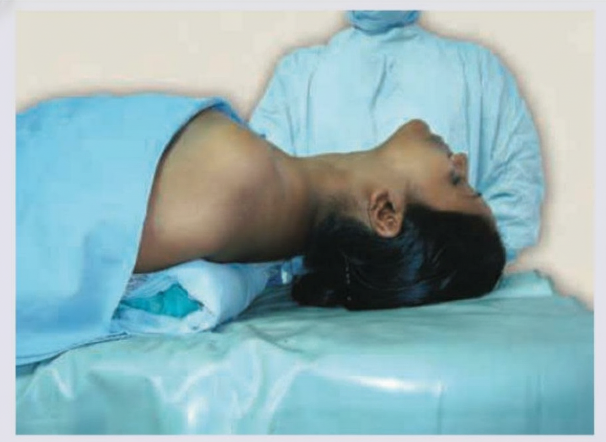
Tonsillectomy is being performed in the given patient. What position is being used?

Explanation: **Explanation:** **Why "Change in Voice" is Correct:** The glottis (vocal cords) is the most common site for laryngeal carcinoma. Because the vocal cords are responsible for phonation, even a tiny mucosal irregularity or growth disrupts the vibratory pattern and glottic closure. This leads to **hoarseness (change in voice)** as the earliest and most consistent symptom. Since the glottic region has **sparse lymphatic drainage**, the tumor remains localized for a long duration, making hoarseness a crucial early warning sign that often leads to early diagnosis and a high cure rate. **Analysis of Incorrect Options:** * **B. Dysphagia:** This is more characteristic of **supraglottic** or **hypopharyngeal** tumors. In glottic cancer, dysphagia occurs only in advanced stages when the tumor involves the extrinsic muscles or the esophagus. * **C. Pain:** Pain (often presenting as referred otalgia via the Vagus nerve) is a late feature indicating deep infiltration, ulceration, or involvement of the laryngeal framework. * **D. Involvement of lymph nodes:** This is **rare** in early glottic carcinoma due to the lack of lymphatics in the true vocal cords (Reinke’s space). In contrast, supraglottic cancers present early with lymph node metastasis because that area is rich in lymphatics. **High-Yield Clinical Pearls for NEET-PG:** * **Most common site of Laryngeal Cancer:** Glottis (60-65%). * **Best Prognosis:** Glottic cancer (due to early symptoms and poor lymphatics). * **Staging:** Any hoarseness persisting for more than **3 weeks** in an adult smoker must be evaluated via indirect laryngoscopy to rule out malignancy. * **Most common pathology:** Squamous Cell Carcinoma (SCC).
Explanation: **Explanation:** Congenital subglottic stenosis (SGS) is the third most common congenital anomaly of the larynx and the most common cause of laryngeal stenosis requiring tracheostomy in infants. It occurs due to the failure of the laryngeal lumen to recanalize during embryonic development. **Breakdown of Options:** * **Options A & B (Diagnostic Criteria):** The subglottis is the narrowest part of the pediatric airway. Diagnosis is based on the diameter of the cricoid ring. In a **full-term neonate**, a diameter **<4 mm** is considered stenotic. In a **premature neonate**, the threshold is **<3 mm**. These measurements are typically confirmed using age-appropriate endotracheal tubes or bronchoscopy. * **Option C (Clinical Presentation):** Unlike laryngomalacia (where the voice may be muffled) or vocal cord palsy (where the cry is weak/breathy), the vocal cords in SGS are typically normal. Therefore, the **cry is usually normal**, but the child presents with **biphasic stridor** and respiratory distress. Since all statements are clinically accurate, **Option D** is the correct answer. **High-Yield Clinical Pearls for NEET-PG:** * **Grading:** The **Cotton-Myer Classification** is used to grade the severity based on the percentage of luminal obstruction (Grade I to IV). * **Most Common Site:** The stenosis most commonly involves the **cricoid cartilage** (the only complete cartilaginous ring). * **Associated Syndrome:** Frequently associated with **Down Syndrome** (Trisomy 21), where the subglottis is naturally smaller. * **Management:** Mild cases (Grade I/II) are managed conservatively; severe cases (Grade III/IV) may require **Laryngotracheal Reconstruction (LTR)** or **Cricotracheal Resection (CTR)**.
Explanation: **Explanation:** **Laryngomalacia** is the most common congenital anomaly of the larynx and the most frequent cause of stridor in infants. It is characterized by an inward collapse of the supraglottic structures (epiglottis, arytenoids) during inspiration, leading to inspiratory stridor. **Why Reassurance is the Correct Answer:** In approximately **90% of cases**, laryngomalacia is a self-limiting condition. The stridor typically appears at 2 weeks of age, peaks at 6–9 months, and resolves spontaneously by 18–24 months as the laryngeal cartilage matures and strengthens. Therefore, the primary management strategy for a thriving infant with mild symptoms is **reassurance** and parental education regarding the natural history of the disease. **Analysis of Incorrect Options:** * **Medical Management:** While conservative measures like upright positioning after feeds or treating comorbid GERD (with PPIs) are used, they are adjuncts to the primary approach of watchful waiting/reassurance. * **Surgical Intervention:** This is reserved only for **severe cases** (approx. 10%) presenting with "danger signs" such as cyanosis, failure to thrive, cor pulmonale, or severe obstructive sleep apnea. The surgery of choice is **Supraglottoplasty**. * **Observation:** While clinically similar to reassurance, in the context of NEET-PG, "Reassurance" is the preferred terminology for a benign, self-resolving condition where no active intervention is required. **Clinical Pearls for NEET-PG:** * **Characteristic Finding:** "Omega-shaped" epiglottis on flexible laryngoscopy. * **Stridor Profile:** Inspiratory stridor that worsens when the infant is supine, crying, or feeding, and improves when prone. * **Associated Condition:** Gastroesophageal Reflux Disease (GERD) is present in up to 80% of these patients and can exacerbate symptoms.
Explanation: **Explanation:** **Puberphonia** (also known as Mutational Falsetto) is a functional voice disorder characterized by the failure of the male voice to transition from the high-pitched prepubertal voice to the low-pitched adult voice during puberty. Despite the larynx being anatomically and physiologically normal, the individual continues to use a high-pitched voice by keeping the vocal cords tense and the larynx elevated. It is often associated with psychological factors or a failure to adapt to the rapid laryngeal changes during adolescence. **Analysis of Incorrect Options:** * **Functional Aphonia:** This is a conversion disorder where the patient speaks only in a whisper despite having a normal larynx. It is usually triggered by emotional stress and is more common in females. * **Plica Ventricularis (Ventricular Dysphonia):** This occurs when the false vocal cords (ventricular folds) are used for phonation instead of the true vocal cords. It results in a rough, low-pitched, and strained voice. * **Androphonia:** This refers to a female possessing a low-pitched, masculine voice. It is often caused by virilization of the larynx due to hormonal imbalances or androgen therapy. **High-Yield Facts for NEET-PG:** * **Treatment of Choice:** Voice therapy (**Gutzmann’s Pressure Test** is both a diagnostic and therapeutic maneuver where downward pressure is applied to the thyroid cartilage to lower the pitch). * **Surgical Management:** If voice therapy fails, **Type III Thyroplasty** (Relaxation Thyroplasty) is performed to shorten and relax the vocal cords, thereby lowering the pitch. * **Laryngeal Findings:** On examination, the larynx is often held high in the neck, and the vocal cords may show a "mutational chink" (posterior gap) during phonation.
Explanation: **Explanation** **1. Why Juvenile Laryngeal Papilloma (JLP) is Correct:** Juvenile Laryngeal Papilloma, a subset of **Recurrent Respiratory Papillomatosis (RRP)**, is the most common benign neoplasm of the larynx in children. It is caused by **Human Papillomavirus (HPV) types 6 and 11**, typically acquired during birth via an infected birth canal. It most frequently presents between **2 and 5 years of age** with symptoms of progressive hoarseness, stridor, and respiratory distress. Histologically, it consists of finger-like projections of non-keratinized stratified squamous epithelium with a central vascular core. **2. Why the Other Options are Incorrect:** * **A. Chondroma:** These are rare, slow-growing cartilaginous tumors that usually affect the cricoid cartilage in adults (typically 40–60 years). * **B. Infantile Hemangioma:** While common in infants, subglottic hemangiomas usually present earlier (within the first 6 months of life) and are characterized by a biphasic growth pattern (proliferation followed by involution). * **C. Scleroma (Rhinoscleroma):** This is a chronic granulomatous infection caused by *Klebsiella rhinoscleromatis*. It primarily affects the nose and subglottis and is characterized by Mikulicz cells, rather than being a true benign tumor of childhood. **3. NEET-PG Clinical Pearls:** * **Most common site:** True vocal cords (due to the transition zone of epithelium). * **Treatment of choice:** CO2 laser excision or microdebrider excision. Avoid tracheostomy if possible to prevent "seeding" of the papilloma distally. * **Adjuvant therapy:** Cidofovir (antiviral) is used in aggressive cases. * **Triad for JLP:** Hoarseness + Stridor + HPV 6/11.
Explanation: **Explanation:** Foreign body (FB) aspiration is a common pediatric emergency, typically occurring in the "exploratory" age group (1–3 years). The diagnosis is primarily clinical, supported by radiological findings. 1. **Clinical Presentation (Option A):** The **"Penetration Syndrome"** is the classic initial phase of FB aspiration. It is characterized by a sudden episode of choking, gagging, and paroxysmal coughing while the child was eating or playing. This history is the most important diagnostic clue, even if the child is currently asymptomatic. 2. **Radiological Findings (Options B & C):** Since most foreign bodies are radiolucent (e.g., peanuts), we look for secondary signs on a chest X-ray: * **Hyperinflation (Obstructive Emphysema):** This occurs due to a **"Ball-valve" effect**, where air enters during inspiration but cannot escape during expiration. This is the most common radiological finding. * **Atelectasis (Collapse):** This occurs due to a **"Stop-valve" effect**, where the airway is completely occluded, leading to the absorption of distal air. **Why "All of the above" is correct:** All three options represent different stages or mechanisms of the same pathology. A positive history combined with either hyperinflation or collapse on X-ray strongly indicates a tracheobronchial foreign body. **High-Yield Clinical Pearls for NEET-PG:** * **Most common site:** Right main bronchus (due to it being wider, shorter, and more vertical). * **Most common FB:** Vegetative matter (Peanuts). * **Gold Standard Investigation:** Rigid Bronchoscopy (both diagnostic and therapeutic). * **Holzknecht Sign:** Mediastinal shift towards the affected side during inspiration (seen in obstructive emphysema). * **Classic Triad:** Wheeze, cough, and diminished breath sounds (though only present in ~60% of cases).
Explanation: Direct laryngoscopy is a procedure used to visualize the larynx and surrounding structures in a straight line of sight, typically performed under general anesthesia. **Explanation of the Correct Answer:** The correct answer is **D (All of the above)** because direct laryngoscopy provides a comprehensive view of the endolarynx and the base of the tongue. * **Lingual surface of the epiglottis:** During the procedure, the blade of the laryngoscope (like a Macintosh blade) is placed in the vallecula, which directly exposes the lingual (anterior) surface of the epiglottis. * **Arytenoid cartilages:** These are key landmarks located at the posterior limit of the glottis. Visualizing the arytenoids and the interarytenoid notch is essential for identifying the laryngeal inlet. * **Cricothyroid membrane:** While this is an external structure, it can be visualized from the *internal* aspect as the area immediately below the vocal folds (subglottis) leading toward the trachea. In the context of "visualization" during surgical laryngoscopy, the entire laryngeal framework, including the internal boundaries of the cricothyroid space, is accessible. **Clinical Pearls for NEET-PG:** * **Positioning:** The "Sniffing position" (flexion of the lower cervical spine and extension of the atlanto-occipital joint) is required to align the oral, pharyngeal, and laryngeal axes. * **Indications:** It is the gold standard for biopsy of laryngeal masses, removal of foreign bodies, and staging of laryngeal endoscopes. * **Key Landmark:** The **Vallecula** is the space between the base of the tongue and the epiglottis; it is the primary site for blade placement in indirect/direct laryngoscopy to elevate the epiglottis. * **Complications:** The most common complication is dental injury (upper incisors).
Explanation: **Explanation:** **Laryngomalacia** is the correct answer as it is the most common congenital anomaly of the larynx, accounting for approximately 60–70% of all congenital laryngeal stridor. It is characterized by an inward collapse of the supraglottic structures (epiglottis, aryepiglottic folds, and arytenoids) during inspiration due to excessive flaccidity of the laryngeal tissues. **Why the other options are incorrect:** * **Laryngeal Web:** This results from the failure of the laryngeal lumen to recanalize during the 10th week of gestation. While significant, it is much rarer than laryngomalacia. * **Subglottic Stenosis:** This is the second most common cause of stridor in infants and the most common congenital abnormality requiring tracheostomy, but it is less frequent than laryngomalacia. * **Subglottic Haemangioma:** This is a benign vascular neoplasm. It typically presents with biphasic stridor at 3–6 months of age (not at birth) and is far less common than structural anomalies. **High-Yield Clinical Pearls for NEET-PG:** 1. **Clinical Presentation:** The hallmark is **inspiratory stridor** that increases with crying, feeding, or lying in the supine position, and improves when the infant is prone. 2. **Diagnosis:** The gold standard is **Flexible Fiberoptic Laryngoscopy**, which shows an "Omega-shaped" (Ω) epiglottis and short aryepiglottic folds. 3. **Management:** Most cases (90%) are self-limiting and resolve spontaneously by 18–24 months. Severe cases (causing failure to thrive or cyanosis) are treated with **supraglottoplasty**.
Explanation: **Explanation:** The **Heimlich maneuver** (abdominal thrusts) is a critical emergency procedure used to relieve **Upper Airway Obstruction** caused by a foreign body. The physiological basis of this maneuver is the **artificial cough**. By applying sudden upward pressure to the abdomen (between the navel and the ribcage), the diaphragm is elevated, which increases intrathoracic pressure and forces air out of the lungs. This sudden bolus of air acts as a force to dislodge and expel the obstructing object from the larynx or trachea. **Analysis of Incorrect Options:** * **Option B (BPPV):** This is managed by canalith repositioning maneuvers, most commonly the **Epley maneuver** or Semont maneuver, to move otoconia back into the utricle. * **Option C (Eustachian tube patency):** This is tested using the **Valsalva maneuver** (forced expiration against a closed nose and mouth) or the **Toynbee maneuver** (swallowing with the nose pinched). * **Option D (Tympanic membrane integrity):** This is assessed via otoscopy or **Siegle’s pneumatic otoscopy**, which checks for the mobility of the drum. **High-Yield Clinical Pearls for NEET-PG:** * **Indications:** Use only when the patient shows signs of severe obstruction (silent cough, cyanosis, or the "universal choking sign"—clutching the neck). * **Contraindication:** Do not perform on infants under 1 year; use **back blows and chest thrusts** instead to avoid abdominal organ injury. * **Modified Heimlich:** In pregnant or morbidly obese patients, **chest thrusts** are performed instead of abdominal thrusts. * **Complication:** Rupture of abdominal viscera or rib fractures can occur if performed incorrectly.
Explanation: ***Rose's position*** - **Supine position** with **neck hyperextended** and head tilted back, providing optimal **surgical access** to the oropharynx and tonsillar fossa. - Facilitates **drainage of blood and secretions** away from the airway, reducing risk of **aspiration** during tonsillectomy. *Trendelenburg position* - **Head-down tilt** with legs elevated above the heart level, primarily used for **pelvic** and **lower abdominal surgeries**. - Would cause **blood pooling** in the head and neck area, impairing visualization and increasing **aspiration risk** during throat procedures. *Barking dog position* - **Prone position** with neck flexed and chin tucked, resembling a dog's posture, not suitable for **oral cavity access**. - This position would **obstruct the surgical field** and make **airway management** extremely difficult during tonsillectomy. *Boyce position* - **Lateral decubitus** position with the patient lying on their side, commonly used for **kidney** and **thoracic surgeries**. - Does not provide adequate **visualization** of the tonsillar area and would make **bilateral access** to both tonsils challenging.
Embryology of the Ear, Nose, and Throat
Practice Questions
Anatomy of the Ear
Practice Questions
Anatomy of the Nose and Paranasal Sinuses
Practice Questions
Anatomy of the Oral Cavity and Pharynx
Practice Questions
Anatomy of the Larynx
Practice Questions
Physiology of Hearing
Practice Questions
Physiology of Balance
Practice Questions
Physiology of Smell and Taste
Practice Questions
Physiology of Speech and Swallowing
Practice Questions
Clinical Examination in ENT
Practice Questions
Diagnostic Investigations in ENT
Practice Questions
Surgical Principles in Otolaryngology
Practice Questions
Get full access to all questions, explanations, and performance tracking.
Start For Free