Which nerve is called the nerve of Wrisberg?
From which embryonic arch does the stapes develop?
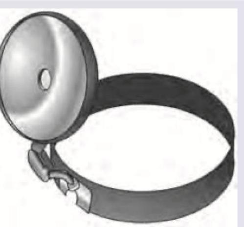
Which of the following is correct about the mirror in the instrument shown?

All are tuning fork test except-
Explanation: ### Explanation The **Nerve of Wrisberg** is the **sensory root of the facial nerve (CN VII)**. The facial nerve emerges from the brainstem as two distinct roots: a large medial motor root and a smaller lateral root, which is the nerve of Wrisberg (nervus intermedius). **Why Option C is Correct:** The nerve of Wrisberg carries three types of fibers: 1. **Special Visceral Afferent (SVA):** Taste sensations from the anterior two-thirds of the tongue (via the chorda tympani). 2. **General Visceral Efferent (GVE):** Parasympathetic secretomotor fibers to the lacrimal, submandibular, and sublingual glands. 3. **General Somatic Afferent (GSA):** Sensory fibers from the external auditory canal and parts of the auricle. **Analysis of Incorrect Options:** * **Option A:** The **motor root** is the larger division of the facial nerve supplying the muscles of facial expression. It is not referred to as the nerve of Wrisberg. * **Option B:** The tympanic branch of the glossopharyngeal nerve (CN IX) is known as **Jacobson’s nerve**. It forms the tympanic plexus and eventually becomes the lesser petrosal nerve. * **Option D:** The **greater auricular nerve** (C2, C3) is a branch of the cervical plexus providing sensation to the skin over the parotid gland and the lower part of the auricle. **Clinical Pearls for NEET-PG:** * **Nomenclature Confusion:** Do not confuse the *Nerve of Wrisberg* (Sensory root of CN VII) with the *Ligament of Wrisberg* (Posterior meniscofemoral ligament in the knee) or the *Cartilage of Wrisberg* (Cuneiform cartilage of the larynx). * **Geniculate Ganglion:** This is where the cell bodies of the sensory fibers of the nerve of Wrisberg are located. * **Ramsay Hunt Syndrome:** Herpes Zoster Oticus involves the nerve of Wrisberg, leading to vesicles in the ear canal and taste loss.
Explanation: The development of the middle ear ossicles is a high-yield topic in ENT embryology. The correct answer is the **Second Branchial Arch (Reichert’s cartilage)**. ### **Explanation of the Correct Answer** The ossicular chain originates from the first and second branchial arches. The **Second Arch (Reichert’s cartilage)** gives rise to the majority of the **stapes** (specifically the head, neck, and crura), the **styloid process**, the **stylohyoid ligament**, and the **lesser cornu of the hyoid bone**. It is important to note that while the stapes superstructure comes from the second arch, the **footplate** and the **annular ligament** have a dual origin, developing partly from the second arch and partly from the **otic capsule** (neuroepithelium). ### **Why Other Options are Incorrect** * **First Arch (Meckel’s cartilage):** This arch gives rise to the **malleus** (except the anterior process) and the **incus** (except the long process). It also forms the mandible and muscles of mastication. * **Third Arch:** This arch forms the **greater cornu** and the lower part of the body of the **hyoid bone**, as well as the stylopharyngeus muscle. * **Fourth Arch:** This arch contributes to the formation of the **laryngeal cartilages** (thyroid and cuneiform) and the muscles of the pharynx and soft palate. ### **High-Yield Clinical Pearls for NEET-PG** * **Nerve Supply Rule:** The nerve of the arch supplies the structures derived from it. Since the stapes is a 2nd arch structure, the **Stapedius muscle** is supplied by the **Facial nerve** (nerve of the 2nd arch). * **Mnemonic:** **M**alleus and **I**ncus = **1**st Arch; **S**tapes = **2**nd Arch. * **Otosclerosis:** This clinical condition primarily involves the stapedial footplate (derived from the otic capsule), leading to conductive hearing loss. * **Congenital Anomalies:** Failure of the second arch development can lead to stapes fixation or malformation, often seen in syndromes like Treacher Collins.
Explanation: ***Focal length of 89 mm, diameter of 2.5 cm*** - The instrument shown is a **head mirror**, used in ENT examinations. It is a **concave mirror** with a **focal length of 89 mm** (approximately 3.5 inches). - The mirror has a **diameter of 2.5 cm**, which is the standard specification for head mirrors used in clinical ENT practice. *Focal length of 25 cm, diameter of 98 mm* - A focal length of **25 cm** is much longer than the standard for a head mirror, which requires a shorter focal length of **89 mm** to focus light effectively at the typical working distance during examination. - A diameter of **98 mm** (almost 10 cm) is also significantly larger than the standard head mirror diameter. *Focal length of 25 cm, diameter of 89 mm* - A **focal length of 25 cm** is incorrect for a head mirror. The correct focal length is **89 mm** (which appears here as the diameter, creating confusion). - This option reverses the measurements - 89 mm is the focal length, not the diameter. *Focal length of 89 mm, diameter of 2.5 inches* - While the **focal length of 89 mm** is correct, the **diameter of 2.5 inches** (approximately 6.35 cm) is larger than the standard specification. - The standard head mirror has a diameter of **2.5 cm**, not 2.5 inches, making this option incorrect due to the wrong unit of measurement.
Explanation: ***Pure tone audiometry*** - This is an **objective test** that uses an audiometer to measure hearing thresholds at various frequencies, determining the degree and type of hearing loss. - It does not involve the use of a **tuning fork** but rather headphones or bone oscillators connected to specialized equipment. *Rinne's test* - This is a **tuning fork test** that compares **air conduction** and **bone conduction** in one ear. - A tuning fork is first placed on the mastoid process and then held near the ear canal to determine which sound is heard longer. *Schwabach test* - The Schwabach test is a **tuning fork test** that compares the **bone conduction** of the patient with that of the examiner. - It assesses the duration of sound perception via bone conduction for both the patient and a normal hearing individual. *Weber's test* - This is a **tuning fork test** that assesses **lateralization of sound** when the tuning fork is placed on the middle of the forehead. - It helps distinguish between conductive and sensorineural hearing loss by indicating which ear hears the sound louder or if it's heard equally in both ears.
Embryology of the Ear, Nose, and Throat
Practice Questions
Anatomy of the Ear
Practice Questions
Anatomy of the Nose and Paranasal Sinuses
Practice Questions
Anatomy of the Oral Cavity and Pharynx
Practice Questions
Anatomy of the Larynx
Practice Questions
Physiology of Hearing
Practice Questions
Physiology of Balance
Practice Questions
Physiology of Smell and Taste
Practice Questions
Physiology of Speech and Swallowing
Practice Questions
Clinical Examination in ENT
Practice Questions
Diagnostic Investigations in ENT
Practice Questions
Surgical Principles in Otolaryngology
Practice Questions
Get full access to all questions, explanations, and performance tracking.
Start For Free