All of the following structures are adult-sized at birth EXCEPT?
Which of the following statements about Infraglottic carcinoma of the larynx is true?
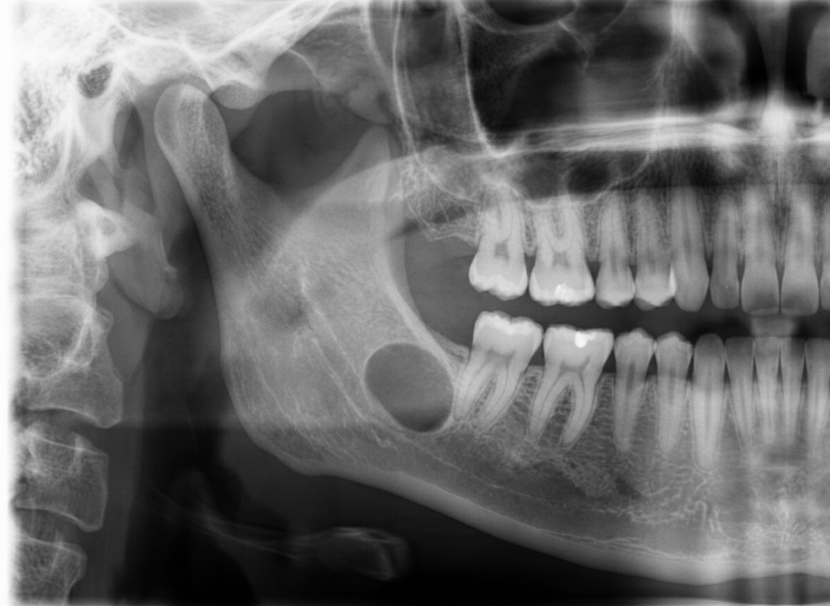
A 45-year-old male presented for a routine dental checkup. All teeth in the lower right quadrant were vital. A panoramic radiograph was taken. Which of the following can be the most probable diagnosis?

Phonation in esophageal speech in a patient who has undergone laryngectomy is produced by which structure?
What are the features of laryngeal carcinoma?
What is the recommended treatment for early vocal nodules?
Mouth opening is considered to be restricted when the distance is
Bryce's sign is seen in which of the following conditions?
What is the primary treatment for a mobile vocal cord tumor?
Eagle syndrome is also known as:
Explanation: The correct answer is **D. Maxillary antrum.** ### **Explanation** In the human body, most structures grow proportionally with age. However, the ear is a unique anatomical exception where several structures reach their full adult dimensions during fetal life or are present at adult size at birth. 1. **Maxillary Antrum (Correct Answer):** The maxillary sinus (antrum) is the first sinus to develop (around the 3rd month of fetal life) but it is **not** adult-sized at birth. At birth, it is merely a small slit-like cavity. It undergoes two main periods of rapid growth (0–3 years and 7–12 years) and only reaches full adult size after the eruption of all permanent teeth (around 15–18 years). 2. **Mastoid Antrum:** This is a large air cell located behind the middle ear. It is unique because it is already adult-sized at birth. However, the mastoid **process** itself is absent at birth and only begins to develop at the end of the 1st year due to the pull of the sternocleidomastoid muscle. 3. **Ear Ossicles:** The Malleus, Incus, and Stapes reach their full adult size and degree of ossification by the time of birth. They do not grow further during a person’s lifetime. 4. **Tympanic Cavity:** The middle ear cleft (tympanic cavity) is essentially adult-sized at birth, though it is initially filled with embryonic mesenchyme that clears as the infant begins to breathe and swallow. ### **High-Yield Clinical Pearls for NEET-PG** * **Other Adult-sized structures at birth:** Inner ear (Labyrinth), Cochlea, and the Tympanic membrane (though the latter is more horizontal in infants). * **The Mastoid Process:** Develops at **1 year** of age. This is why the Facial Nerve is superficial at birth; an incision behind the ear in an infant can easily sever the nerve. * **Eustachian Tube:** In infants, it is shorter (18mm), wider, and more horizontal (10° angle) compared to adults (36mm, 45° angle), making children more prone to Otitis Media.
Explanation: ### Explanation **Correct Answer: D. It commonly spreads to mediastinal lymph nodes.** **1. Why Option D is Correct:** The subglottis (infraglottic region) has a sparse lymphatic network compared to the supraglottis. However, when malignancy occurs here, the primary lymphatic drainage follows the pathway through the **cricothyroid membrane** to the **pre-laryngeal (Delphian)** and **pre-tracheal** nodes. From there, the drainage continues inferiorly into the **paratracheal** and **superior mediastinal lymph nodes**. This deep extension into the mediastinum is a characteristic feature of subglottic spread and often necessitates a mediastinal lymph node dissection during surgery. **2. Why Other Options are Incorrect:** * **Options A & B:** Glottic carcinoma (vocal cords) is the **most common** (approx. 60-65%), followed by Supraglottic carcinoma (approx. 30-35%). Subglottic (Infraglottic) carcinoma is the **rarest**, accounting for less than 5% of all laryngeal cancers. * **Option C:** Submental lymph nodes (Level Ia) primarily drain the floor of the mouth, tip of the tongue, and lower lip. They are not the primary drainage site for laryngeal structures. **3. High-Yield Clinical Pearls for NEET-PG:** * **Prognosis:** Subglottic cancers often present late with stridor or dyspnea because the region is clinically "silent" until the airway is compromised. They have a poorer prognosis compared to glottic cancers. * **Lymphatic Watershed:** The glottis (vocal cords) has virtually **no lymphatic drainage**, which is why early glottic cancer (T1) rarely metastasizes to the neck. * **Delphian Node:** The pre-laryngeal node is a classic "sentinel" node for subglottic and thyroid malignancies. Its involvement often indicates a higher risk of recurrence.
Explanation: ***Stafne bone cyst*** - **Stafne bone cyst** is an **asymptomatic radiolucent lesion** typically found **below the inferior alveolar canal** in the **posterior mandible** of middle-aged males during routine radiographic examination. - The **vital teeth** in the affected quadrant strongly support this diagnosis, as Stafne cysts are **developmental defects** containing **salivary gland tissue** and do not affect tooth vitality. *Dentigerous cyst* - **Dentigerous cysts** are associated with the **crown of an unerupted tooth**, typically **third molars** or **canines**. - They present as **unilocular radiolucencies** surrounding the **crown** of an impacted tooth, which is not described in this case. *Radicular cyst* - **Radicular cysts** arise from **chronic periapical inflammation** and are associated with **non-vital teeth** with **pulpal necrosis**. - Since all teeth in the lower right quadrant are **vital**, this diagnosis is ruled out as radicular cysts require **endodontic infection**. *Calcifying odontogenic cyst* - **Calcifying odontogenic cyst** (Gorlin cyst) typically presents as a **mixed radiolucent-radiopaque lesion** with **calcifications**. - It commonly occurs in the **anterior maxilla** and shows **calcific deposits** on radiographs, unlike the presumed **radiolucent lesion** in the posterior mandible.
Explanation: **Explanation:** In a patient who has undergone a total laryngectomy, the primary source of sound (the larynx) is removed. To regain speech, patients can learn **Esophageal Speech**. **Why the Pharyngo-esophageal (PE) segment is correct:** In esophageal speech, the patient swallows air into the upper esophagus and then expels it in a controlled manner. As the air is forced back up, it causes the mucosal folds of the **Pharyngo-esophageal (PE) segment** (primarily the **Cricopharyngeus muscle**) to vibrate. This vibration acts as the "new vocal cords" (neoglottis), producing a low-pitched sound that is then articulated into speech by the tongue and lips. **Analysis of Incorrect Options:** * **A. Buccal cavity:** While the mouth and buccal cavity are essential for *articulation* (forming words), they do not act as the vibratory source for phonation. * **B. Pharynx:** Although the PE segment is located at the junction of the pharynx and esophagus, the "pharynx" as a whole is too broad an anatomical term. The specific vibratory sphincter is the PE segment. * **C. Trachea:** In a total laryngectomy, the trachea is diverted to a permanent stoma in the neck. It is completely disconnected from the digestive tract/mouth; therefore, tracheal air cannot be used for esophageal speech. **High-Yield Clinical Pearls for NEET-PG:** * **Primary Vibrator:** The Cricopharyngeus muscle is the most important component of the PE segment. * **Voice Quality:** Esophageal speech is low-pitched, hoarse, and has a short phrase length (limited by esophageal air capacity). * **Gold Standard:** While esophageal speech is an option, **Tracheo-esophageal Puncture (TEP)** with a prosthesis (e.g., Blom-Singer valve) is currently the most common and successful method of post-laryngectomy rehabilitation. * **Electrolarynx:** An external battery-operated device used by patients who cannot master esophageal or TEP speech.
Explanation: Laryngeal carcinoma is the most common head and neck malignancy, with Squamous Cell Carcinoma (SCC) accounting for over 95% of cases. **Explanation of Options:** * **The glottis is the most common site (Option A):** In the Indian subcontinent and globally, the glottic region (vocal cords) is the most frequent site for laryngeal cancer (approx. 60-65%), followed by the supraglottis. * **Lesions at the edge of the vocal cord (Option B):** Glottic tumors typically originate on the free edge or the upper surface of the anterior two-thirds of the true vocal cords. * **Hoarseness as an early sign (Option C):** Because glottic tumors involve the vibrating edge of the vocal cords, even a tiny lesion causes air turbulence and irregular vibration, leading to early hoarseness. This often leads to early diagnosis and a better prognosis compared to supraglottic or subglottic tumors, which remain asymptomatic longer. **Clinical Pearls for NEET-PG:** 1. **Lymphatic Spread:** The glottis has sparse lymphatic drainage; therefore, glottic SCC rarely presents with early lymph node metastasis (N0 neck). In contrast, the supraglottis has a rich lymphatic network, often presenting with bilateral neck nodes. 2. **Staging:** T1a involves one vocal cord; T1b involves both cords but with normal mobility. T3 is defined by **vocal cord fixation**. 3. **Risk Factors:** Smoking is the primary risk factor, followed by alcohol (synergistic effect). HPV (types 16, 18) is also associated. 4. **Treatment:** Early-stage (T1, T2) is treated with radiotherapy or laser excision with excellent voice preservation. Advanced stages require surgery (laryngectomy) and/or chemoradiotherapy.
Explanation: **Explanation:** Vocal nodules (Singer’s or Teacher’s nodules) are benign, bilateral, symmetrical swellings located at the junction of the **anterior 1/3rd and posterior 2/3rds** of the vocal folds. They are caused by chronic mechanical trauma due to vocal abuse or misuse. **Why Voice Therapy is Correct:** The primary pathophysiology of early vocal nodules is localized edema and congestion (soft nodules). Since the underlying cause is functional (misuse of the voice), the first-line management is **conservative**. Voice therapy focuses on re-educating the patient on proper breath support and vocal hygiene, which allows the early inflammatory changes to resolve spontaneously. **Analysis of Incorrect Options:** * **A & B (Excision/Laser Ablation):** Surgical intervention (Microlaryngeal Surgery) is reserved only for **chronic, fibrotic (hard) nodules** that have failed a trial of voice therapy. Early surgical intervention in soft nodules is contraindicated as it may lead to unnecessary scarring of the Reinke’s space. * **D (Tissue sampling):** Vocal nodules have a characteristic clinical appearance and history. Routine biopsy is not indicated unless there is clinical suspicion of malignancy (e.g., unilateral lesion, irregular surface, or persistent hoarseness in a smoker). **High-Yield Clinical Pearls for NEET-PG:** * **Location:** Junction of anterior 1/3 and posterior 2/3 (point of maximum vibration). * **Demographics:** Most common in male children and adult females. * **Stroboscopy:** Shows an "hourglass" glottic closure pattern. * **Management Rule:** Always start with **Voice Therapy**; surgery is the last resort.
Explanation: **Explanation:** In clinical practice and otolaryngology, the inter-incisor distance (the distance between the upper and lower central incisors at maximal mouth opening) is the standard measure for assessing mandibular mobility. **1. Why Option A is Correct:** The normal range for mouth opening in an average adult is typically between **40 mm and 50 mm** (roughly equivalent to the width of three fingers). Trismus or restricted mouth opening is clinically defined when this distance falls **below 40 mm**. A measurement of less than 20 mm is considered severe restriction, often necessitating specialized anesthetic techniques like fiberoptic intubation. **2. Why Other Options are Incorrect:** * **Options B, C, and D (50 mm, 53 mm, 58 mm):** These values represent the upper limits of normal or hypermobile ranges. A mouth opening of 50 mm or more is considered perfectly healthy and unrestricted. Using these as thresholds for "restriction" would lead to over-diagnosis, as the majority of the healthy population would be classified as restricted. **Clinical Pearls for NEET-PG:** * **The Three-Finger Test:** A quick bedside clinical assessment; if a patient cannot fit three of their own fingers vertically between their incisors, mouth opening is likely restricted. * **Common Causes:** In the context of ENT, restricted mouth opening is frequently associated with **Oral Submucous Fibrosis (OSMF)**, peritonsillar abscess (Quinsy), temporomandibular joint (TMJ) ankylosis, or pterygoid muscle involvement in head and neck cancers. * **Mallampati Classification:** While mouth opening is a component, remember that Mallampati specifically assesses the visibility of the faucial pillars, soft palate, and uvula to predict difficult intubation.
Explanation: **Explanation:** **Bryce’s Sign** is a clinical diagnostic sign pathognomonic for a **Laryngocele** (specifically the external or combined types). A laryngocele is an abnormal cystic dilatation of the saccule of the laryngeal ventricle, filled with air. Bryce’s sign is elicited by applying manual pressure to the external neck swelling; this results in an audible **hissing sound** as the air is forced out of the sac back into the laryngeal lumen. Additionally, the swelling may decrease in size upon compression. **Analysis of Options:** * **Laryngocele (Correct):** As described, the sign relies on the communication between the air-filled sac and the airway. * **Post-cricoid Carcinoma:** This condition is typically associated with **Trotter’s triad** or the loss of laryngeal crepitus (Moure’s sign) due to post-cricoid fullness, but not Bryce’s sign. * **Angiofibroma:** Juvenile Nasopharyngeal Angiofibroma (JNA) presents with epistaxis and nasal obstruction. Characteristic signs include the **Holman-Miller sign** (anterior bowing of the posterior wall of the maxillary antrum on CT). * **Chronic Tonsillitis:** This presents with halitosis and tonsillar hypertrophy. A relevant sign here is **Irwin Moore’s sign** (pressure on the anterior pillar expresses cheesy material from the crypts). **High-Yield Clinical Pearls for NEET-PG:** * **Laryngocele Association:** Frequently associated with glassblowers and trumpet players due to increased intralaryngeal pressure. * **Radiology:** A "dark" air-filled sac on X-ray/CT that enlarges during a **Valsalva maneuver**. * **Malignancy Link:** In adults, always rule out an underlying squamous cell carcinoma of the ventricle obstructing the saccule. * **Boyce's Sign:** (Do not confuse with Bryce’s) This is the gurgling sound heard on pressure over a Pharyngeal Pouch (Zenker’s Diverticulum).
Explanation: **Explanation:** The primary goal in treating early-stage glottic cancer (where the vocal cord remains mobile) is to achieve a cure while preserving the best possible voice quality. **1. Why Radiotherapy is Correct:** In early-stage laryngeal tumors (T1 and T2), where the **vocal cord is mobile**, both Radiotherapy (RT) and conservative surgery (like CO2 laser excision) offer similar cure rates (approx. 85-95%). However, **Radiotherapy** is traditionally preferred as the primary modality because it preserves the structural integrity of the vocal cord, resulting in a **superior functional voice outcome** compared to surgical resection. **2. Why Other Options are Incorrect:** * **Surgery:** While "Micro-laryngeal surgery" or "Laser cordectomy" are valid options for T1 lesions, "Surgery" as a general term often implies more invasive procedures. In the context of a mobile cord, RT is the classic textbook answer for maximizing voice preservation. * **Chemotherapy:** Chemotherapy is not used as a primary or standalone treatment for early glottic cancer. It is reserved for advanced stages (T3, T4) as part of "Organ Preservation Protocols" (Concurrent Chemoradiotherapy) or for palliative care. **Clinical Pearls for NEET-PG:** * **T1a:** Tumor limited to one vocal cord (mobile). * **T1b:** Tumor involves both vocal cords (mobile). * **T2:** Tumor extends to supraglottis or subglottis with **impaired** cord mobility (but not fixed). * **T3:** Tumor limited to the larynx with **vocal cord fixation**. * **High-Yield:** The most common site of laryngeal cancer is the **Glottis**, but it has the best prognosis due to a sparse lymphatic supply (low risk of nodal metastasis) and early presentation (hoarseness).
Explanation: **Explanation:** **Eagle Syndrome**, also known as **Styalgia** or Styloid Process Syndrome, occurs due to an **elongated styloid process** (greater than 30 mm) or calcification of the stylohyoid ligament. This anatomical variation causes irritation of cranial nerves (V, VII, IX, X) or compression of the carotid arteries. Patients typically present with a dull, nagging facial pain, foreign body sensation in the throat (globus), and odynophagia, often triggered by head rotation or swallowing. **Analysis of Options:** * **A. Styalgia (Correct):** This is the synonymous term for Eagle Syndrome, derived from "Styloid" and "Algia" (pain). * **B. Ludwig’s Angina:** This is a rapidly spreading cellulitis of the submandibular, sublingual, and submental spaces, usually of dental origin. It is a life-threatening emergency due to potential airway obstruction. * **C. Pharyngeal Bursitis (Thornwaldt’s Cyst):** This refers to a chronic inflammation of the nasopharyngeal bursa located in the midline of the posterior nasopharyngeal wall. * **D. Amaurosis:** This refers to partial or total loss of vision without an apparent lesion in the eye itself (e.g., Amaurosis fugax). **High-Yield Clinical Pearls for NEET-PG:** * **Diagnosis:** Confirmed by palpating the styloid process in the tonsillar fossa (reproduces pain) and imaging (Orthopantomogram/OPG or CT scan). * **Treatment:** The definitive treatment is **Styloidectomy** (surgical shortening of the process), which can be performed via a trans-oral or external cervical approach. * **History:** Often follows a history of **tonsillectomy**, as scar tissue can rub against the elongated process.
Embryology of the Ear, Nose, and Throat
Practice Questions
Anatomy of the Ear
Practice Questions
Anatomy of the Nose and Paranasal Sinuses
Practice Questions
Anatomy of the Oral Cavity and Pharynx
Practice Questions
Anatomy of the Larynx
Practice Questions
Physiology of Hearing
Practice Questions
Physiology of Balance
Practice Questions
Physiology of Smell and Taste
Practice Questions
Physiology of Speech and Swallowing
Practice Questions
Clinical Examination in ENT
Practice Questions
Diagnostic Investigations in ENT
Practice Questions
Surgical Principles in Otolaryngology
Practice Questions
Get full access to all questions, explanations, and performance tracking.
Start For Free