What is the primary function of the otolith organs?
The vestibulocochlear nerve (VIII cranial nerve) carries afferent information for:
Positive Romberg test with eyes closed detects a defect in -
Which of the following stimuli is detected by the vestibular macula?
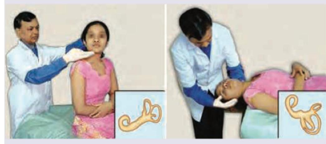
Which of the following test is being performed on the patient?

Most medial nucleus of cerebellum is:
Stimulation of posterior semicircular canal produces -
Which of the following structures is responsible for detecting rotational acceleration?
Which of the following will occur in a girl who suddenly stops spinning after several seconds of spinning to the left?
All are true about vestibular neuritis EXCEPT:
Explanation: ***Detecting the position of the head in space*** - The **otolith organs**, comprising the **utricle** and **saccule**, are responsible for detecting **linear acceleration** and **gravitational forces**. - This information allows the brain to perceive the **static head position** relative to gravity and linear movements. *Producing the vestibular-ocular reflex* - While the otolith organs contribute to overall vestibular function, the primary role in producing the **vestibular-ocular reflex (VOR)**, especially for rotational movements, is mainly attributed to the **semicircular canals**. - The VOR helps stabilize gaze during head movements, coordinating eye movements in the opposite direction of head motion. *Producing rotary nystagmus* - **Rotary nystagmus** is typically associated with stimulation of the **semicircular canals**, which detect angular acceleration. - The otolith organs detect linear acceleration and static head position, not rotational movements causing nystagmus. *Detecting angular acceleration* - The **semicircular canals** are specialized structures within the inner ear designed to detect **angular acceleration** (rotational movements of the head). - The otolith organs are sensitive to **linear acceleration** and the pull of gravity, not angular motion.
Explanation: ***All of the options*** - The **vestibulocochlear nerve (cranial nerve VIII)** is responsible for transmitting both **auditory (hearing)** and **vestibular (balance and spatial orientation)** information from the inner ear to the brain. - Its two main branches are the **cochlear nerve**, serving hearing, and the **vestibular nerve**, serving equilibrium and spatial orientation. *Equilibrium* - The vestibular component of the **VIII nerve** specifically relays sensory information concerning **head position and movement**, crucial for maintaining **equilibrium** and balance. - This input comes from the **semicircular canals** and **otolith organs (utricle and saccule)** in the inner ear. *Spatial orientation* - **Spatial orientation** refers to the body's awareness of its position and movement in space, a function directly supported by the **vestibular system** and transmitted via the **vestibular nerve** branch of cranial nerve VIII. - It involves processing cues about **linear and angular acceleration** from the inner ear. *Hearing* - The cochlear component of the **VIII nerve** transmits **auditory information** from the **cochlea** to the brain, enabling the perception of sound. - This involves converting **sound vibrations** into electrical signals.
Explanation: Proprioceptive pathway - A positive Romberg test indicates a loss of proprioception, meaning the patient cannot maintain balance when visual cues are removed, relying solely on somatosensory input [2]. - This suggests damage to the dorsal columns of the spinal cord or peripheral nerves that transmit proprioceptive information to the brain [1], [3]. Cerebellum - While cerebellar dysfunction also causes ataxia and balance problems, it would typically present as difficulty maintaining balance even with eyes open, referred to as cerebellar ataxia [2]. - A Romberg test primarily assesses the integrity of the proprioceptive system, distinguishing it from cerebellar issues where balance problems are evident regardless of visual input [2]. Peripheral nerve - Peripheral neuropathy can indeed lead to a positive Romberg test if the sensory nerves responsible for proprioception are affected [1]. - However, "Proprioceptive pathway" is a more direct and encompassing answer, as peripheral nerves are a component of this pathway, which also includes spinal cord tracts [3]. Spinothalamic tract - The spinothalamic tract primarily transmits sensations of pain and temperature, not proprioception [3]. - Damage to this tract would result in deficits in these specific sensory modalities, rather than a positive Romberg test [1].
Explanation: ***Linear acceleration*** - The **maculae** (in the utricle and saccule) are specifically designed to detect **linear acceleration**, including both dynamic movements (speeding up in a car, elevator motion) and the constant linear acceleration of **gravity**. - Hair cells in the maculae are displaced by movements of the **otolithic membrane** containing **otoconia** (calcium carbonate crystals) in response to linear acceleration forces. - The utricle primarily detects **horizontal linear acceleration**, while the saccule detects **vertical linear acceleration**. *Gravity* - While gravity is indeed detected by the maculae, gravity is actually a form of **constant linear acceleration** (9.8 m/s²). - The maculae use gravity to determine **static head position** and orientation, but this is a subset of their broader function of detecting linear acceleration. - "Linear acceleration" is the more comprehensive and physiologically accurate term. *Change in head position* - This term is too broad and encompasses both **linear** and **angular (rotational)** movements. - **Angular acceleration** (rotation) is detected by the **semicircular canals**, not the maculae. - The maculae specifically detect linear position changes relative to gravity, not rotational changes. *None of the options* - This is incorrect because the vestibular macula clearly detects linear acceleration as its primary function.
Explanation: ***Dix-Hallpike test*** - The image depicts a patient being moved from an upright sitting position to a supine position with the head turned to one side and extended, which is characteristic of the **Dix-Hallpike maneuver**. - This test is used to diagnose **benign paroxysmal positional vertigo (BPPV)** by provoking vertigo and nystagmus as a result of otoconia displacement in the semicircular canals. *Caloric stimulation test* - This test involves introducing **warm or cold water/air into the ear canal** to induce nystagmus, assessing the vestibular system. - It's typically performed with the patient lying supine with their head elevated at 30 degrees, not involving the specific positional changes shown in the image. *Doll's eye reflex* - Also known as the **oculocephalic reflex**, it assesses brainstem function by rapidly turning the patient's head while observing eye movement. - This reflex is primarily used to evaluate comatose patients and does not involve the complex body and head positioning seen in the image. *Frenzel maneuver* - The Frenzel maneuver is performed during **otoscopy** to check for nystagmus by having the patient gaze through high plus lenses in a dark room. - It is often used to suppress visual fixation and enhance nystagmus, but it does not involve the specific body or head movements depicted.
Explanation: ***Fastigial*** - The **fastigial nucleus** is located most **medially** within the cerebellum, closest to the midline in the roof of the fourth ventricle [1]. - It is the most medial of the four deep cerebellar nuclei and is primarily associated with the **vestibulocerebellum** (flocculonodular lobe) [1]. - Functions: Maintains **balance, posture, and coordinated eye movements** via connections to vestibular nuclei and reticular formation [1]. *Dentate* - The **dentate nucleus** is the **largest and most lateral** of the cerebellar nuclei, with a characteristic crumpled sac-like appearance (resembling an olive). - Located deep within the **lateral cerebellar hemisphere** white matter [1]. - Associated with the **neocerebellum** (cerebrocerebellum) and involved in **planning and initiating voluntary movements** via the ventrolateral thalamus to motor cortex [1]. *Emboliform* - The **emboliform nucleus** is elongated and located **medial to the dentate** but **lateral to the globose** nucleus. - Together with the globose nucleus, forms the **interposed nuclei**. - Associated with the **spinocerebellum** and involved in **modulating limb movements** and adjusting ongoing motor activity [1]. *Globose* - The **globose nucleus** consists of rounded cell masses located **medial to emboliform** and **lateral to fastigial** nucleus. - Part of the **interposed nuclei** along with emboliform nucleus. - Functions in **fine-tuning and coordinating ongoing movements**, particularly of distal limbs.
Explanation: ***Vertical nystagmus*** - Stimulation of the **posterior semicircular canal** produces **vertical-torsional nystagmus** with the vertical component being predominant. - The posterior canal is oriented at approximately 45° to the sagittal plane and detects angular acceleration in the RALP plane (Right Anterior-Left Posterior). - Stimulation typically causes **downbeat nystagmus** (fast phase downward) with a torsional component, activating the **superior oblique** and **inferior rectus muscles** on the ipsilateral side. - The vertical component is the primary clinical feature observed. *Rotatory nystagmus* - While posterior canal stimulation does produce a **torsional (rotatory) component**, it is not purely rotatory. - The torsional component accompanies the vertical nystagmus but is **secondary to the vertical component**. - Pure rotatory nystagmus is rare and would suggest involvement of multiple canals or central pathology. *Horizontal nystagmus* - **Horizontal nystagmus** is specifically produced by stimulation of the **horizontal (lateral) semicircular canal**. - It indicates activation of the horizontal canal system, which lies in a different plane than the posterior canal. - The medial and lateral rectus muscles are primarily involved in horizontal nystagmus. *None of the options* - This option is incorrect because **vertical nystagmus with torsional component** is the characteristic and well-documented response to posterior semicircular canal stimulation. - Each semicircular canal produces a specific directional nystagmus corresponding to its anatomical plane of orientation.
Explanation: ***Semicircular canals*** - The **semicircular canals** are part of the inner ear and are specifically designed to detect **rotational acceleration** of the head. - They contain a fluid called **endolymph** and hair cells within the **ampulla** that are stimulated by the movement of this fluid during rotation. *Cochlea* - The **cochlea** is primarily responsible for processing **auditory (sound) information**, not head movement. - It contains the **organ of Corti**, which converts sound vibrations into electrical signals. *Fovea centralis* - The **fovea centralis** is a part of the **retina** responsible for sharp, central vision and **high visual acuity**. - It plays no role in detecting head acceleration or balance. *Saccule* - The **saccule** is part of the **otolith organs** (along with the utricle) and detects **linear acceleration** and the pull of gravity in the vertical plane. - It is involved in sensing up-and-down movements and static head tilt, not rotational acceleration.
Explanation: ***Her eyes will move slowly to the right*** - After spinning to the left and suddenly stopping, **post-rotatory nystagmus** occurs due to continued endolymph movement. - The endolymph continues to move to the LEFT due to **inertia**, creating a sensation of rotating to the **RIGHT**. - This produces **nystagmus** with the **slow phase to the right** (direction of perceived rotation) and fast corrective phase to the left. - The **vestibulo-ocular reflex** generates this nystagmus as the brain interprets the continued endolymph movement as actual rotation. *The hair cells in the right semicircular canal will depolarize* - This is incorrect. Upon stopping a left spin, the endolymph continues moving LEFT due to inertia. - In the **right horizontal canal**, this creates **ampullary-petal flow** (toward the ampulla), which causes **hyperpolarization**, not depolarization. - During the actual left spin, it was the **left canal** that was depolarized (ampullary-fugal flow in horizontal canals causes excitation). *When asked to point to a target, the girl will point to the right of the target* - After stopping from spinning left, the sensation is of spinning to the RIGHT, causing a **past-pointing phenomenon**. - This perceived rightward rotation causes the person to point to the **LEFT** of the target, not the right. - The brain compensates for the perceived motion in the wrong direction. *The cupula in the right semicircular canal will move away from the utricle* - Upon stopping, the endolymph in the right canal continues moving to the LEFT (original spin direction) due to inertia. - This deflects the cupula **toward the utricle** (ampullary-petal), not away from it. - Ampullary-petal deflection in horizontal canals causes hyperpolarization of hair cells.
Explanation: ***Vertical nystagmus*** - **Vestibular neuritis** primarily affects the **horizontal semicircular canal** and superior vestibular nerve, leading to **horizontal or rotational nystagmus**, not vertical. - **Vertical nystagmus** is often indicative of a **central lesion** rather than a peripheral vestibular disorder like neuritis. *Horizontal nystagmus* - This is a characteristic finding in **vestibular neuritis**, where the **nystagmus is usually horizontal or rotatory** and beats away from the affected side. - The nystagmus typically **increases in intensity** when looking in the direction of the fast phase. *Positive head thrust* - A **positive head thrust test** (or **head impulse test**) is a hallmark of **peripheral vestibular dysfunction**, including vestibular neuritis. - It demonstrates a **saccadic corrective eye movement** when the head is quickly turned towards the affected side, indicating impaired vestibulo-ocular reflex. *Normal hearing* - **Vestibular neuritis** specifically involves inflammation of the **vestibular nerve**, sparing the cochlear nerve. - Therefore, patients with vestibular neuritis typically **maintain normal hearing**, differentiating it from labyrinthitis.
Get full access to all questions, explanations, and performance tracking.
Start For Free