The most common tumor of the cerebellopontine angle is -
All of the following are indications for Gamma Knife Radiosurgery EXCEPT
A 35-year-old man presents with progressive right-sided hearing loss, balance difficulties, and headaches. MRI reveals an enhancing mass in the cerebellopontine angle. Most likely diagnosis?
In a patient with acoustic neuroma all are seen except:
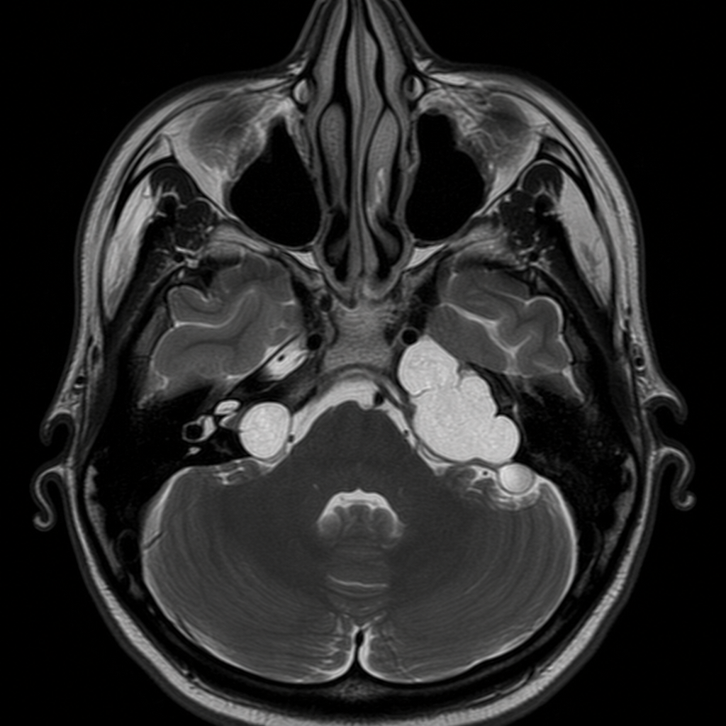
Surgeries used in management of Meniere's disease are all Except
Most definitive treatment for large symptomatic acoustic neuroma is
True about acoustic neuroma:
All of the following cranial nerves are involved in Acoustic neuroma, except?
The most common brain tumour in an adult is
A patient has multiple meningiomas, acoustic neuroma and hyperpigmented skin lesions; most likely diagnosis is –
Explanation: ***Acoustic neuroma*** - Acoustic neuromas, also known as **vestibular schwannomas**, are the most common tumors of the **cerebellopontine angle (CPA)**, accounting for **80-90%** of CPA tumors. [1] - They arise from the **Schwann cells** of the **vestibular portion of cranial nerve VIII** (vestibulocochlear nerve). [1] - Typical clinical presentation includes **progressive unilateral sensorineural hearing loss**, **tinnitus**, **vertigo**, and in advanced cases, involvement of adjacent cranial nerves (V and VII). *Meningioma* - Meningiomas are the **second most common** CPA tumor (10-15% of cases), originating from the **arachnoid cap cells**. [1] - They typically grow more slowly than acoustic neuromas and may present with different cranial nerve deficits depending on their exact location. - On imaging, they show characteristic **dural tail sign** and homogeneous enhancement. *Neurofibroma* - While neurofibromas can affect cranial nerves, they are **far less common** in the CPA than acoustic neuromas. [1] - They are often associated with **Neurofibromatosis type 1 (NF1)**, whereas bilateral acoustic neuromas are characteristic of **NF2**. [1] - Isolated CPA neurofibromas are rare. *None of the options* - This option is incorrect as **acoustic neuroma** is the well-established most common tumor of the cerebellopontine angle. [1] - This is a fundamental concept in neurology, neurosurgery, and pathology. **References:** [1] Cross SS. Underwood's Pathology: A Clinical Approach. 6th ed. Common Clinical Manifestations Of Central And Peripheral Nervous System Disease, pp. 727-728.
Explanation: ***Brain tumours or lesions 4 cm or larger in diameter*** - **Gamma Knife radiosurgery** is typically used for **small to medium-sized lesions** (generally less than 3-4 cm in diameter). - Larger lesions carry a **higher risk of cerebral edema** and radiation necrosis when treated with radiosurgery, making conventional surgery or fractionated radiotherapy more appropriate. *Acoustic neuroma* - **Gamma Knife radiosurgery** is a well-established treatment option for **acoustic neuromas** (vestibular schwannomas). - It aims to control tumor growth and preserve hearing and facial nerve function with a high success rate. *Arteriovenous malformation* - **Arteriovenous malformations (AVMs)** are commonly treated with **Gamma Knife radiosurgery** to induce thrombosis and obliteration of the abnormal vascular nidus. - This treatment helps in preventing future hemorrhage and reducing seizure risk. *Trigeminal neuralgia* - **Gamma Knife radiosurgery** is an effective treatment for **refractory trigeminal neuralgia**. - It delivers a highly focused dose of radiation to the trigeminal nerve root, creating a lesion that disrupts the pain signals.
Explanation: ***Acoustic neuroma*** - The combination of **progressive unilateral hearing loss**, **balance difficulties**, and **headaches**, along with an **enhancing mass in the cerebellopontine angle** on MRI, is highly characteristic of an acoustic neuroma (vestibular schwannoma). - These are benign tumors that arise from the **vestibulocochlear nerve (cranial nerve VIII)**, causing compression of adjacent structures. *Vestibular neuritis* - This condition presents with **sudden onset, severe vertigo** often triggered by head movement, but it is typically **acute and self-limiting**, without a progressive course or an intracranial mass. - **Hearing is usually spared** in vestibular neuritis, which is inconsistent with the patient's progressive hearing loss. *Otosclerosis* - Otosclerosis is a disease of abnormal bone remodeling in the middle ear, leading to **progressive conductive hearing loss**, often bilateral. - It does **not typically cause balance difficulties** or present as a **cerebellopontine angle mass** on MRI. *Meniere's disease* - Characterized by a triad of **fluctuating hearing loss**, **episodic vertigo**, and **tinnitus**, often with a sensation of aural fullness. - While it causes hearing loss and balance issues, it does **not involve an enhancing mass** in the cerebellopontine angle.
Explanation: Acoustic neuroma typically presents with unilateral progressive hearing loss and sometimes tinnitus [1]. While acoustic neuromas can cause unsteadiness and gait imbalance due to gradual growth, they typically do not present with acute, severe, isolated vertigo because slow growth allows compensatory brainstem mechanisms to develop [1]. Acute vertigo is more characteristic of peripheral vestibular disorders like benign paroxysmal positional vertigo (BPPV) or Meniere's disease. As an acoustic neuroma (vestibular schwannoma) grows, it can compress the adjacent cerebellum, leading to cerebellar signs [1]. These signs include ataxia, dysmetria, and nystagmus, reflecting cerebellar dysfunction. The facial nerve (CN VII) runs in close proximity to the vestibulocochlear nerve (CN VIII), so it can be affected by an acoustic neuroma, leading to facial paresis or paralysis, though facial weakness is unusual at presentation [1]. Unilateral deafness is a classic and early symptom of acoustic neuroma due to compression of the cochlear portion of CN VIII [1]. The trigeminal nerve (CN V) has sensory innervation to the cornea, and its fibers pass near the cerebellopontine angle where acoustic neuromas grow. Compression of the trigeminal nerve by the tumor can lead to a reduced or absent corneal reflex on the affected side.
Explanation: ***Stapedectomy*** - **Stapedectomy** is a surgical procedure primarily used to treat **otosclerosis**, a condition causing conductive hearing loss due to abnormal bone growth in the middle ear. - It involves removing the stapes bone and replacing it with a prosthesis to restore sound conduction, which is not a treatment for **Meniere's disease**. *Sacculotomy* - This procedure involves making a small incision in the **saccule** (part of the labyrinth) to decompress the inner ear in Meniere's disease. - Its goal is to reduce inner ear pressure and improve symptoms like **vertigo**, but it carries a risk of hearing loss. *Endolymphatic shunt operation* - This surgery aims to create a drainage path for excess **endolymph** from the endolymphatic sac, reducing pressure in the inner ear. - It is a common surgical option for intractable Meniere's disease to control vertigo attacks while preserving hearing. *Labyrinthectomy* - **Labyrinthectomy** is a destructive surgical procedure where the entire **labyrinth**, including the vestibular and cochlear structures, is removed. - It is typically reserved for severe, intractable Meniere's disease in patients with **non-serviceable hearing** in the affected ear, as it results in complete hearing loss.
Explanation: ***Surgery*** - **Surgical resection** is the most definitive treatment for **large, symptomatic acoustic neuromas** (typically >3 cm), especially those causing **mass effect** on the brainstem and cerebellum. - It aims for **complete tumor removal** to alleviate symptoms (hearing loss, facial nerve dysfunction, brainstem compression) and prevent further neurological compromise. - Surgical approaches include **translabyrinthine**, **retrosigmoid**, or **middle cranial fossa** approaches depending on tumor size and hearing status. *Steroids* - **Steroids** may be used to manage acute symptoms like **edema** or inflammation associated with the tumor, but they are not a definitive treatment. - They do not address the underlying tumor growth or remove the mass. - Used only as **temporary symptomatic relief** or perioperative adjunct. *Radiotherapy* - **Stereotactic radiosurgery** (Gamma Knife, CyberKnife) is effective for **small to medium-sized tumors** (<3 cm) with good tumor control rates. - For **large tumors**, radiotherapy is **insufficient** as it only aims to **control tumor growth** rather than remove the mass, and cannot provide immediate decompression. - Large tumors with mass effect require **surgical decompression** for definitive management. *Anti-neoplastic drugs* - **Anti-neoplastic drugs** (chemotherapy) are generally **ineffective** against acoustic neuromas, which are **benign vestibular schwannomas**. - They are typically reserved for malignant tumors or specific genetic syndromes (e.g., bevacizumab in NF2-related schwannomas), but not for standard sporadic acoustic neuromas. - Chemotherapy is **not a definitive treatment** for these benign tumors.
Explanation: ***Arises from vestibular nerve*** - An **acoustic neuroma**, also known as a **vestibular schwannoma**, is a benign tumor that originates from the **Schwann cells** of the **vestibular nerve (cranial nerve VIII)**. - This tumor typically grows in the **internal auditory canal** and cerebellopontine angle. *Malignant tumour* - Acoustic neuromas are almost always **benign tumors**, meaning they are non-cancerous and do not typically spread to other parts of the body. - While they are benign, their growth can compress adjacent nerves and brain structures, leading to significant neurological deficits. *Upper pole compresses IX,X,XI nerves* - The **glossopharyngeal (IX), vagus (X), and accessory (XI) nerves** originate lower in the brainstem and are more commonly compressed by tumors in the **jugular foramen** region, not typically by the upper pole of an acoustic neuroma. - An acoustic neuroma primarily affects the **vestibulocochlear nerve (VIII)** and, if large enough, the **facial nerve (VII)** and **trigeminal nerve (V)** in the **cerebellopontine angle**. *Lower pole compresses trigeminal cranial nerve* - The **trigeminal nerve (V)** is located more superiorly and medially in the **cerebellopontine angle** relative to the usual growth pattern of an acoustic neuroma. - Compression of the trigeminal nerve by an acoustic neuroma is more likely to occur with a **large tumor** expanding into the superior part of the cerebellopontine angle, rather than by its lower pole.
Explanation: ***Oculomotor*** - The **oculomotor nerve (CN III)** is located in the midbrain, far from the cerebellopontine angle where acoustic neuromas typically grow. - Its involvement is not characteristic of an acoustic neuroma and would suggest a different intracranial pathology. *Vagus* - The **vagus nerve (CN X)** exits the brainstem near the cerebellopontine angle, and can be affected by larger acoustic neuromas. - Compression can lead to symptoms like **dysphagia**, **hoarseness**, or **vocal cord paralysis**. *Glossopharyngeal* - The **glossopharyngeal nerve (CN IX)** is also in close proximity to the cerebellopontine angle. - Compression can result in symptoms such as **dysphagia**, **loss of taste** in the posterior tongue, or **loss of gag reflex**. *Facial* - The **facial nerve (CN VII)** is anatomically very close to the vestibulocochlear nerve (CN VIII) within the internal auditory canal. - **Facial nerve palsy**, characterized by **facial weakness** or **paralysis**, is a common symptom of acoustic neuromas due to direct compression or surgical manipulation.
Explanation: ***Cerebral metastasis*** - **Cerebral metastases** are the **most common brain tumours in adults**, originating from primary cancers elsewhere in the body (e.g., lung, breast, melanoma). - They often present as **multiple lesions** and can cause focal neurological deficits, seizures, and increased intracranial pressure. *Glioma* - While **gliomas** (including astrocytomas, glioblastoma multiforme) are the most common primary brain tumours, they are less common than metastatic lesions overall in adults. - They arise from **glial cells** within the brain and can be highly aggressive. *Pituitary tumour* - **Pituitary tumours** are benign adenomas originating from the pituitary gland. - They are common but constitute a smaller proportion of all adult brain tumours compared to metastases or gliomas, and often present with **endocrine disturbances** or **visual field defects**. *Vestibular schwannoma* - **Vestibular schwannomas** (acoustic neuromas) are benign tumours arising from the **vestibulocochlear nerve (cranial nerve VIII)**. - They are relatively rare and typically present with **hearing loss**, **tinnitus**, and **balance issues**.
Explanation: ***Neurofibromatosis*** - The combination of **multiple meningiomas**, **acoustic neuroma** (specifically bilateral in NF2) [1], and **hyperpigmented skin lesions** (cafe-au-lait spots in NF1, or multiple cutaneous neurofibromas) is highly characteristic of neurofibromatosis. - This presentation suggests either **Neurofibromatosis type 1 (NF1)** with meningiomas (less common but possible) or, more strongly, **Neurofibromatosis type 2 (NF2)** due to the bilateral acoustic neuromas and the presence of meningiomas, with hyperpigmentation being a variable feature [1]. *Von Hippel-Lindau disease* - This disorder is characterized by **hemangioblastomas** of the retina and central nervous system, **renal cell carcinoma**, and **pheochromocytomas**. - It does not typically involve meningiomas, acoustic neuromas, or hyperpigmented skin lesions. *Sturge-Weber syndrome* - This is a neurocutaneous disorder featuring a **port-wine stain (facial nevus flammeus)**, leptomeningeal angioma, and **glaucoma**. - The clinical picture of meningiomas, acoustic neuroma, and hyperpigmented skin lesions does not align with Sturge-Weber syndrome. *Tuberous sclerosis* - This condition is characterized by the growth of numerous **benign tumors in many different organs**, including the brain (tubers, subependymal giant cell astrocytomas), skin (facial angiofibromas, shagreen patches, ash-leaf spots), kidneys (angiomyolipomas), and heart (rhabdomyomas). - While it has **skin lesions** and **brain tumors**, these are distinct from meningiomas and acoustic neuromas, which are not typical features.
Get full access to all questions, explanations, and performance tracking.
Start For Free