
Neurotology — MCQs

On this page
Defective function of which of the following causes hyperacusis?
Battle's sign is defined as:
Unilateral sensorineural hearing loss may occur in which of the following conditions?
A patient presented with high-frequency sensorineural hearing loss and ipsilateral cerebellar ataxia. What is the likely site of the lesion?
Herpes zoster infection of the geniculate ganglion is known to cause which of the following conditions?
All of the following are true for Ramsay Hunt syndrome, EXCEPT:
Facial nerve palsy can be caused by which of the following conditions?
A patient presents with painful vesicles in the external auditory meatus and over the tympanic membrane. In addition to that, he also has facial nerve palsy. What is the most likely diagnosis and which is the site that is affected?
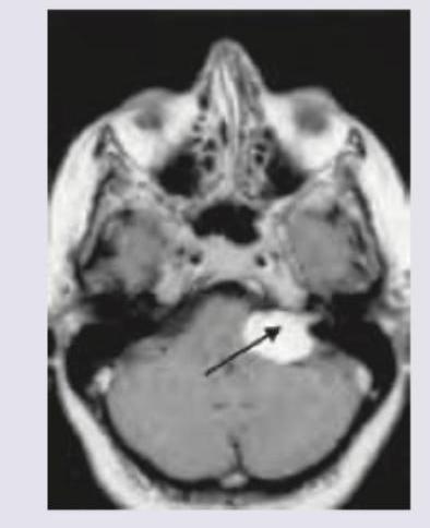
A 45-year-old male presents with progressive hearing loss in the right ear over 2 years, tinnitus, and occasional imbalance. Audiometry shows unilateral sensorineural hearing loss with speech discrimination score of 60%. MRI reveals a 2.5 cm enhancing mass at the cerebellopontine angle. What is the most likely diagnosis?
A 40-year-old female complains of progressive unilateral hearing loss and tinnitus. She also has developed numbness around posterior aspect of concha. MRI head was performed. What is the clinical diagnosis?
Practice by Chapter
Vestibular System Anatomy and Physiology
Practice Questions
Vestibular Testing
Practice Questions
Benign Paroxysmal Positional Vertigo
Practice Questions
Ménière's Disease
Practice Questions
Vestibular Neuritis
Practice Questions
Labyrinthitis
Practice Questions
Acoustic Neuroma
Practice Questions
Other Cerebellopontine Angle Tumors
Practice Questions
Facial Nerve Disorders
Practice Questions
Skull Base Surgery
Practice Questions
Cochlear Implantation
Practice Questions
Vestibular Schwannoma Management
Practice Questions
Want unlimited practice?
Get full access to all questions, explanations, and performance tracking.
Scan to download app