A 40-year-old female complains of progressive unilateral hearing loss and tinnitus. She also has developed numbness around posterior aspect of concha. MRI head was performed. What is the clinical diagnosis?

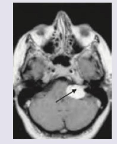
MRI Brain of a 40-year-old patient with progressive unilateral SNHL and tinnitus is shown below. Which is the most common extracanalicular nerve to be involved?

Which of the following hearing aid or implant is shown below?

True about acoustic neuroma:
Acoustic neuroma causes:
All of the following are true about glomus-jugulare tumor except:
The first clinical presentation of acoustic neuroma is characterized by ____________
Among the following non-auditory signs, which appears earliest in acoustic neuroma?
Which of the following would be the most appropriate treatment for rehabilitation of a patient, who has bilateral profound deafness following surgery for bilateral acoustic schwannoma?
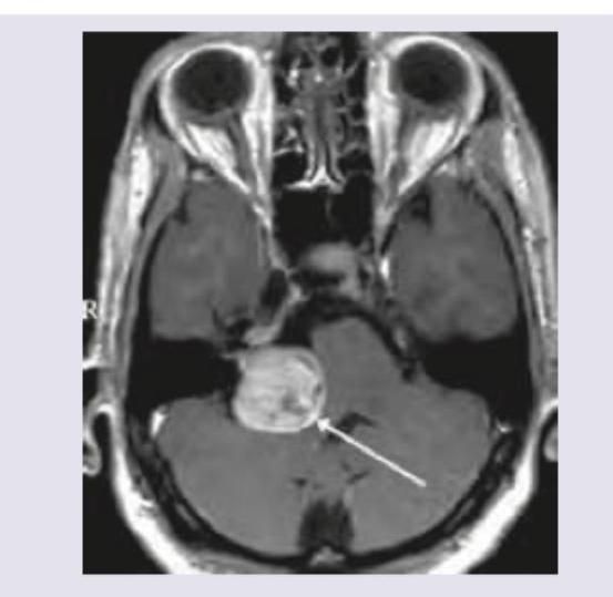
A 35-year-old man presents with progressive right-sided hearing loss, balance difficulties, and headaches. MRI reveals an enhancing mass in the cerebellopontine angle. Most likely diagnosis?
Explanation: ***Acoustic neuroma*** - Progressive **unilateral hearing loss** and **tinnitus** are classic symptoms due to compression of the **vestibulocochular nerve (CN VIII)**. - **Numbness around the posterior aspect of concha** indicates involvement of **Arnold's nerve (auricular branch of vagus nerve, CN X)**, which supplies sensory innervation to this area. This finding suggests tumor extension and is consistent with a larger acoustic neuroma. - The presented MRI shows a mass in the **cerebellopontine angle**, typical for an acoustic neuroma (a vestibular schwannoma arising from CN VIII). *Astrocytoma* - Astrocytomas are primary brain tumors that typically arise within the **brain parenchyma** and rarely present with isolated otologic symptoms. - They would not typically cause **concha numbness** through cranial nerve compression in this specific pattern. *Meningioma* - Meningiomas are typically **dural-based tumors** and can occur in the cerebellopontine angle, but they are less common than acoustic neuromas in this location. - While CPA meningiomas can cause cranial nerve symptoms, the specific combination of **CN VIII symptoms with Arnold's nerve involvement** is more characteristic of acoustic neuroma. *Straight sinus thrombosis* - Straight sinus thrombosis causes symptoms related to **increased intracranial pressure**, such as severe headache, seizures, and focal neurological deficits. - It would not cause **unilateral hearing loss**, **tinnitus**, or **localized concha numbness** through cranial nerve compression.
Explanation: ***V*** - The image shows a large **right cerebellopontine angle (CPA) mass**, consistent with an **acoustic neuroma** (vestibular schwannoma), which commonly arises from CN VIII in the internal auditory canal and extends into the CPA. - The **trigeminal nerve (CN V)** is the most common extracanalicular cranial nerve to be compressed and involved by larger CPA tumors due to its proximity and superior-anterior position relative to CN VIII. *VI* - The abducens nerve (CN VI) controls the lateral rectus muscle of the eye, and its involvement typically causes **diplopia** and **lateral gaze palsy**. While possible with very large and medially extending CPA tumors, it is less common than CN V involvement. - CN VI is located more medially and anteriorly within the brainstem and generally requires significant mass effect rather than direct compression from a typical CPA tumor. *VII* - The facial nerve (CN VII) travels with the vestibulocochlear nerve (CN VIII) through the **internal auditory canal**, and is frequently involved by acoustic neuromas within the canal, leading to **facial weakness or paralysis**. - However, the question specifically asks for the most common **extracanalicular** nerve involvement. While CN VII is intimately associated, its most common site of involvement is within the canal, or early in the CPA, and V is often implicated with larger CPA masses. *VIII* - The vestibulocochlear nerve (CN VIII) is the nerve from which **acoustic neuromas (vestibular schwannomas)** originate, causing the presenting symptoms of **unilateral SNHL and tinnitus**. Therefore, it is the primary affected nerve, not an extracanalicular one involved due to compression. - The question asks for the most common **extracanalicular nerve to be involved**, implying a nerve *other than* the one the tumor originates from or primarily affects locally within the canal.
Explanation: ***Cochlear implant*** - The image clearly shows an **electrode array** inserted into the **cochlea**, bypassing damaged hair cells and directly stimulating the auditory nerve. - This is characteristic of a **cochlear implant**, which is used for individuals with severe to profound sensorineural hearing loss. *Brainstem auditory implant* - A **brainstem auditory implant** (BAI) electrode is placed on the **cochlear nucleus** in the brainstem, not the cochlea itself. - BAIs are typically used for conditions where the auditory nerve is damaged or absent, such as in cases of **neurofibromatosis type 2** with bilateral acoustic neuromas. *Bone anchored hearing aid* - A **bone-anchored hearing aid** (BAHA) works by transmitting sound vibrations directly to the cochlea via bone conduction, typically through a surgically implanted post or magnet in the **skull bone**. - The image does not depict an external processor or bone conduction mechanism, but rather an internal electrode within the inner ear. *In the canal hearing aid* - An **in-the-canal (ITC) hearing aid** is an external device that sits within the ear canal and amplifies sound acoustically. - The image shows an **invasive internal device** implanted into the cochlea, which is distinct from a non-invasive, external ITC hearing aid.
Explanation: ***Arises from vestibular nerve*** - An **acoustic neuroma**, also known as a **vestibular schwannoma**, is a benign tumor that originates from the **Schwann cells** of the **vestibular nerve (cranial nerve VIII)**. - This tumor typically grows in the **internal auditory canal** and cerebellopontine angle. *Malignant tumour* - Acoustic neuromas are almost always **benign tumors**, meaning they are non-cancerous and do not typically spread to other parts of the body. - While they are benign, their growth can compress adjacent nerves and brain structures, leading to significant neurological deficits. *Upper pole compresses IX,X,XI nerves* - The **glossopharyngeal (IX), vagus (X), and accessory (XI) nerves** originate lower in the brainstem and are more commonly compressed by tumors in the **jugular foramen** region, not typically by the upper pole of an acoustic neuroma. - An acoustic neuroma primarily affects the **vestibulocochlear nerve (VIII)** and, if large enough, the **facial nerve (VII)** and **trigeminal nerve (V)** in the **cerebellopontine angle**. *Lower pole compresses trigeminal cranial nerve* - The **trigeminal nerve (V)** is located more superiorly and medially in the **cerebellopontine angle** relative to the usual growth pattern of an acoustic neuroma. - Compression of the trigeminal nerve by an acoustic neuroma is more likely to occur with a **large tumor** expanding into the superior part of the cerebellopontine angle, rather than by its lower pole.
Explanation: ***Retrocochlear deafness*** - An acoustic neuroma is a **benign tumor** that grows on the **vestibulocochlear nerve (cranial nerve VIII)**, which is responsible for hearing and balance. - As the tumor grows, it compresses the nerve, causing **sensorineural hearing loss** that originates distal to the cochlea and is therefore termed retrocochlear. *Any of the above* - This option is incorrect because acoustic neuromas specifically cause **retrocochlear deafness**, not all types of hearing loss. - The type of deafness is defined by the **location of the lesion**, and acoustic neuromas compress the nerve beyond the cochlea. *Conductive deafness* - **Conductive deafness** results from problems in the outer or middle ear that prevent sound waves from reaching the inner ear. - Acoustic neuromas affect the **nerve itself** (inner ear/brain pathway), not the sound conduction mechanisms of the outer or middle ear. *Cochlear deafness* - **Cochlear deafness** is a type of sensorineural hearing loss originating within the **cochlea** (inner ear). - While an acoustic neuroma causes sensorineural hearing loss, the lesion is **distal to the cochlea** (on the nerve), making it retrocochlear rather than cochlear.
Explanation: ***Most commonly affects male*** - **Glomus jugulare tumors** are more common in **females** (3:1 to 5:1 ratio), making this statement **incorrect**. - This tumor type is related to **paragangliomas** and typically affects middle-aged individuals, with a clear predilection for the female sex. *Are paragangliomas* - **Glomus jugulare tumors** are indeed **paragangliomas**, arising from neuroendocrine cells (chemoreceptor cells) of the jugular bulb. - These are **chemodectomas**, originating from neural crest cells associated with the parasympathetic nervous system. *Arises from the adventitia of jugular bulb* - **Glomus jugulare tumors** arise from the **paraganglia in the adventitia of the jugular bulb**. - These are **glomus bodies** (chemoreceptor tissue) located in the temporal bone, specifically in the jugular foramen region. *Pulsatile tinnitus and conductive deafness seen* - **Pulsatile tinnitus** is a classic symptom due to the tumor's highly vascular nature and proximity to the ear. - **Conductive hearing loss** can result from the tumor encroaching on the middle ear ossicles or filling the tympanic cavity.
Explanation: ***Cochleovestibular symptoms*** - Acoustic neuromas, arising from Schwann cells of the **vestibulocochlear nerve**, commonly cause symptoms related to this nerve first. - Patients typically present with **unilateral hearing loss**, tinnitus, and/or vestibular dysfunction (e.g., disequilibrium, vertigo). *Facial nerve involvement* - **Facial nerve** symptoms (e.g., weakness, numbness) are less common as an initial presentation because the facial nerve is typically compressed later as the tumor grows. - While the facial nerve runs in close proximity, it is usually more resilient to early compression than the vestibulocochlear nerve fibers. *Brainstem involvement* - **Brainstem compression** symptoms (e.g., cranial nerve palsies beyond VII and VIII, long tract signs) occur with larger tumors that extend into the posterior fossa. - These are typically **late manifestations**, indicating significant tumor growth beyond the internal auditory canal. *Cerebellar involvement* - **Cerebellar symptoms** (e.g., ataxia, dysmetria) are also late findings, occurring when the tumor is large enough to compress the cerebellum. - These are not usually the initial clinical presentation due to the tumor's origin from the eighth cranial nerve.
Explanation: ***Diminished corneal sensitivity*** - The **corneal reflex** is mediated by the **trigeminal nerve (CN V)**, which has a close anatomical relationship with the vestibulo-cochlear nerve (CN VIII) within the cerebellopontine angle. - Early compression of CN V by an enlarging **acoustic neuroma** can lead to diminished corneal sensitivity before more widespread neurological deficits appear. *Congestion of conjunctiva* - This symptom is non-specific and not directly associated with the early stages of an **acoustic neuroma**. - Conjunctival congestion can be caused by various factors, including irritation, allergies, or infections, and does not indicate cranial nerve involvement. *Diplopia* - **Diplopia (double vision)** typically occurs due to involvement of the extraocular motor nerves (CN III, IV, or VI) or brainstem compression. - This is generally a later sign and requires significant tumor growth to affect these structures, making it less likely to be an earliest non-auditory symptom. *Ptosis* - **Ptosis (drooping eyelid)** is caused by dysfunction of the oculomotor nerve (CN III) or the sympathetic nervous system (Horner's syndrome). - These nerves are usually affected later in the course of an **acoustic neuroma** once the tumor has grown significantly and compressed adjacent structures distant from its initial growth site.
Explanation: ***Brainstem implant*** - A **brainstem implant** is the most appropriate treatment when the auditory nerve has been damaged or destroyed, as can occur during bilateral acoustic schwannoma surgery. - This device bypasses the cochlea and auditory nerve by directly stimulating the **cochlear nucleus** in the brainstem, allowing sound perception. *Bilateral cochlear implant* - A **cochlear implant** requires an intact auditory nerve to transmit signals from the cochlea to the brain. - In this scenario, bilateral profound deafness post-surgery for acoustic schwannoma often implies damage to both **auditory nerves**, rendering cochlear implants ineffective. *Unilateral cochlear implant* - Similar to a bilateral cochlear implant, a **unilateral cochlear implant** relies on the functionality of at least one auditory nerve. - Since the patient has **bilateral profound deafness** following bilateral surgery, the auditory nerves are likely compromised on both sides, making even a unilateral implant unsuitable. *Bilateral high powered digital hearing aid* - Hearing aids amplify sound and rely on the presence of residual hair cell function in the **cochlea** and an intact auditory pathway. - Profound deafness indicates severe damage to the inner ear or auditory nerve, which hearing aids cannot overcome as they only provide *amplification*, not direct neural stimulation.
Explanation: ***Acoustic neuroma*** - The combination of **progressive unilateral hearing loss**, **balance difficulties**, and **headaches**, along with an **enhancing mass in the cerebellopontine angle** on MRI, is highly characteristic of an acoustic neuroma (vestibular schwannoma). - These are benign tumors that arise from the **vestibulocochlear nerve (cranial nerve VIII)**, causing compression of adjacent structures. *Vestibular neuritis* - This condition presents with **sudden onset, severe vertigo** often triggered by head movement, but it is typically **acute and self-limiting**, without a progressive course or an intracranial mass. - **Hearing is usually spared** in vestibular neuritis, which is inconsistent with the patient's progressive hearing loss. *Otosclerosis* - Otosclerosis is a disease of abnormal bone remodeling in the middle ear, leading to **progressive conductive hearing loss**, often bilateral. - It does **not typically cause balance difficulties** or present as a **cerebellopontine angle mass** on MRI. *Meniere's disease* - Characterized by a triad of **fluctuating hearing loss**, **episodic vertigo**, and **tinnitus**, often with a sensation of aural fullness. - While it causes hearing loss and balance issues, it does **not involve an enhancing mass** in the cerebellopontine angle.
Vestibular System Anatomy and Physiology
Practice Questions
Vestibular Testing
Practice Questions
Benign Paroxysmal Positional Vertigo
Practice Questions
Ménière's Disease
Practice Questions
Vestibular Neuritis
Practice Questions
Labyrinthitis
Practice Questions
Acoustic Neuroma
Practice Questions
Other Cerebellopontine Angle Tumors
Practice Questions
Facial Nerve Disorders
Practice Questions
Skull Base Surgery
Practice Questions
Cochlear Implantation
Practice Questions
Vestibular Schwannoma Management
Practice Questions
Get full access to all questions, explanations, and performance tracking.
Start For Free