Which of the following tests is recommended for neonatal screening of hearing?
Angular movement is sensed by which structure?
A 70-year-old male presents with loss of sensation in the external auditory meatus (Hitselberger sign positive). What is the likely diagnosis?
Auditory neurotherapy is an effective modality of treatment for which of the following abnormalities of hearing?
A laparoscopic intranasal approach is used for accessing all of the following EXCEPT?
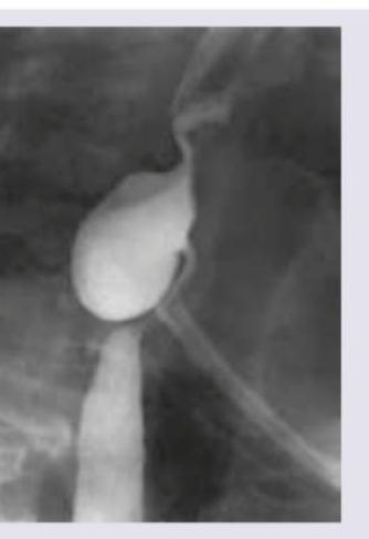
An 80-year-old female presents with complaints of difficulty swallowing and foul breath for 4 months. On auscultation, her lung fields have crepitations. A barium swallow finding is shown. What is your diagnosis?

Fluctuating deafness is a characteristic symptom of which condition?
Malignant otitis externa is caused by:
Scanty, foul-smelling, painless discharge from the ear is a characteristic feature of which of the following lesions?
The organ of Corti is located within which part of the inner ear?
Explanation: **Explanation:** The primary goal of neonatal hearing screening is to identify hearing loss early (the "1-3-6" rule). **Evoked Otoacoustic Emissions (EOAEs)** are the gold standard for initial screening because they are non-invasive, quick, and highly sensitive. 1. **Why Evoked OAE is correct:** EOAEs measure the sound generated by the outer hair cells (OHCs) of the cochlea in response to an acoustic stimulus. In a healthy neonate, the cochlea "echoes" this sound back. If these emissions are present, it indicates that the conductive mechanism and the cochlea are functioning normally (up to the level of OHCs). 2. **Why other options are incorrect:** * **Automated ABR (AABR):** While highly accurate and used as a second-tier test or for high-risk neonates (NICU graduates), it is more expensive and time-consuming than OAE for universal primary screening. * **Spontaneous OAE (SOAE):** These occur in only about 50-70% of the normal-hearing population without any external stimulus. Their absence does not necessarily indicate hearing loss, making them clinically useless for screening. * **Distortion Product OAE (DPOAE):** This is a *type* of evoked OAE. While used clinically, "Evoked OAE" is the broader, more appropriate categorical term for the screening protocol which includes both Transient Evoked (TEOAE) and DPOAE. **High-Yield Clinical Pearls for NEET-PG:** * **Universal Neonatal Hearing Screening (UNHS):** The recommended protocol is **OAE** first; if the baby fails twice, proceed to **AABR**. * **Auditory Neuropathy:** OAEs will be **normal** (as OHCs are intact), but ABR will be **abnormal**. This is why AABR is mandatory for NICU babies. * **1-3-6 Rule:** Screening by **1** month, Diagnosis by **3** months, Intervention by **6** months. * OAEs disappear if there is a conductive loss or sensorineural loss >30-35 dB.
Explanation: **Explanation:** The inner ear consists of the bony and membranous labyrinth, housing the organs for both hearing and equilibrium. The vestibular apparatus, responsible for balance, is divided into two functional units: the **Semicircular Canals (SCC)** and the **Otolith organs** (Saccule and Utricle). 1. **Why Semicircular Canals are correct:** There are three SCCs (Lateral, Superior, and Posterior) oriented at right angles to each other. They contain **endolymph** and a sensory receptor called the **Crista Ampullaris**. When the head undergoes **angular (rotational) acceleration**, the inertia of the endolymph causes it to lag behind, displacing the cupula and stimulating hair cells. This allows the brain to sense rotation in all three planes of space. 2. **Why other options are incorrect:** * **Cochlea:** This is the sensory organ for **hearing**, not balance. It converts sound waves into nerve impulses via the Organ of Corti. * **Utricle and Saccule:** These are the Otolith organs. They contain the **Macula** and sense **linear acceleration** and **static tilt** (gravity). Specifically, the Utricle senses horizontal acceleration (e.g., a car moving forward), while the Saccule senses vertical acceleration (e.g., riding an elevator). **Clinical Pearls for NEET-PG:** * **Receptor Cells:** Crista Ampullaris (SCC) vs. Macula (Otolith organs). * **BPPV (Benign Paroxysmal Positional Vertigo):** Most commonly involves the **Posterior SCC** due to displaced otoconia (canalithiasis). * **Caloric Testing:** Primarily tests the **Lateral (Horizontal) SCC**. * **Scarpa’s Ganglion:** The vestibular ganglion where first-order neurons of the vestibular nerve are located.
Explanation: ### Explanation **Correct Option: A. Vestibular Schwannoma** The **Hitselberger sign** is a classic clinical finding in Vestibular Schwannoma (Acoustic Neuroma). It refers to **hypesthesia (loss of sensation) of the posterior-superior wall of the external auditory canal**. **Pathophysiology:** This occurs due to compression of the **sensory fibers of the Facial Nerve (Nervus Intermedius)** as the tumor expands within the internal auditory canal or the cerebellopontine angle. Since the facial nerve and vestibulocochlear nerve travel together, large tumors often involve the facial nerve's sensory component before motor weakness (facial palsy) becomes clinically evident. **Why the other options are incorrect:** * **B. Mastoiditis:** This is an acute bacterial infection of the mastoid air cells. It presents with ear pain, fever, and post-auricular swelling/tenderness, not localized sensory loss in the canal. * **C. Bell's palsy:** This is an idiopathic lower motor neuron facial nerve paralysis. While it involves the facial nerve, it primarily presents with acute motor weakness (drooping of the face). Hitselberger sign is specifically associated with the slow compression seen in tumors. * **D. Cholesteatoma:** While a cholesteatoma can erode the canal wall or cause facial nerve palsy via bony destruction, it does not typically present with the isolated Hitselberger sign. **High-Yield Clinical Pearls for NEET-PG:** * **Earliest Symptom:** Progressive unilateral sensorineural hearing loss (SNHL) and tinnitus. * **Earliest Sign:** Loss of corneal reflex (due to Trigeminal nerve involvement). * **Hitselberger Sign:** Indicates involvement of the sensory branch of the VIIth cranial nerve. * **Gold Standard Investigation:** Gadolinium-enhanced MRI of the Brain/CP angle. * **Audiometry:** Characterized by "Retrocochlear" pathology (Poor speech discrimination score out of proportion to the hearing loss).
Explanation: ### Explanation **Correct Answer: B. Meniere’s Disease** **Concept:** Auditory neurotherapy (often referred to in the context of **Tinnitus Retraining Therapy (TRT)** or specialized auditory rehabilitation) is a modality used to manage the neuro-sensory symptoms associated with inner ear disorders. In **Meniere’s disease**, the pathology involves endolymphatic hydrops, leading to a triad of fluctuating sensorineural hearing loss (SNHL), vertigo, and tinnitus. While medical management (diuretics, low-salt diet) addresses the fluid imbalance, auditory neurotherapy/rehabilitation is employed to help the brain habituate to the abnormal signals (tinnitus) and compensate for the fluctuating auditory input. It focuses on the neuroplasticity of the auditory pathway to improve sound tolerance and speech perception. **Why the other options are incorrect:** * **A. CSOM (Chronic Suppurative Otitis Media):** This is a structural/infectious pathology of the middle ear causing conductive hearing loss. Treatment is surgical (Tympanomastoidectomy) and medical (antibiotics). * **C. Malignant Otitis Externa:** This is a life-threatening skull base osteomyelitis (usually caused by *Pseudomonas*). It requires urgent IV antibiotics and glycemic control, not neuro-rehabilitation. * **D. Otosclerosis:** This involves stapes fixation leading to conductive hearing loss. The definitive treatment is surgical (Stapedotomy) or the use of conventional hearing aids. **NEET-PG High-Yield Pearls:** * **Meniere’s Disease Triad:** Episodic Vertigo, Fluctuating SNHL (initially low frequency), and Tinnitus. * **Lermoyez Syndrome:** A variant of Meniere’s where hearing *improves* during a vertigo attack. * **Burnout Phenomenon:** In late-stage Meniere’s, vertigo spells may cease as the vestibular system is destroyed, leaving the patient with permanent SNHL and imbalance. * **Tinnitus Retraining Therapy (TRT):** Combines directive counseling and low-level sound therapy to achieve habituation.
Explanation: **Explanation:** The **Endoscopic Endonasal Approach (EEA)** is a minimally invasive surgical technique that utilizes the natural corridor of the nasal cavity and sinuses to access the skull base. **Why Cerebellum is the Correct Answer:** The cerebellum is located in the **posterior cranial fossa**, situated behind the brainstem and beneath the tentorium cerebelli. While endoscopic endonasal approaches can reach the clivus (anterior to the brainstem), they cannot safely bypass the brainstem and major vascular structures to access the cerebellum. The cerebellum is traditionally accessed via a **suboccipital craniotomy** or retrosigmoid approach. **Analysis of Incorrect Options:** * **Lacrimal Sac (A):** Accessed via **Endoscopic Dacryocystorhinostomy (DCR)**. The sac lies lateral to the lateral nasal wall (near the agger nasi cell), making it easily accessible intranasally. * **Pituitary Gland (C):** This is the classic indication for the **Transsphenoidal approach**. The sella turcica forms the posterior-superior limit of the sphenoid sinus. * **Optic Nerve (D):** The optic nerve can be accessed for **decompression** (e.g., in traumatic optic neuropathy or Graves' ophthalmopathy) via the medial wall of the orbit and the lateral wall of the sphenoid sinus (recess of the optic nerve). **High-Yield Clinical Pearls for NEET-PG:** * **Limits of EEA:** It can access the Anterior Fossa (Crista galli to Planum sphenoidale), Middle Fossa (Sella, Cavernous sinus), and the Upper Clivus. * **Critical Landmark:** The **Sphenoid Sinus** is the "gateway" to the skull base in endoscopic surgery. * **Optico-Carotid Recess (OCR):** A key landmark in the lateral wall of the sphenoid sinus; it is the space between the internal carotid artery and the optic nerve. * **CSF Leak:** The most common complication of expanded endonasal approaches; often repaired using a **Hadad-Bassagasteguy flap** (nasoseptal flap based on the posterior septal artery).
Explanation: ***Zenker's diverticulum*** - The classic triad of **dysphagia**, **halitosis** (foul breath from food stagnation), and **pulmonary crepitations** (from aspiration) in an elderly patient strongly suggests Zenker's diverticulum. - Barium swallow typically shows a **posterior pharyngeal pouch** at **Killian's triangle** (between the thyropharyngeus and cricopharyngeus muscles). *Plummer-Vinson syndrome* - Characterized by **iron deficiency anemia**, **esophageal webs**, and **dysphagia** in middle-aged women, not elderly patients. - Associated with **koilonychia** (spoon-shaped nails) and **glossitis**, which are not mentioned in this case. *Schatzki's ring* - Presents with **intermittent dysphagia** to solids only, typically triggered by eating bread or meat ("steakhouse syndrome"). - Barium swallow shows a **thin circumferential ring** at the gastroesophageal junction, not a posterior pouch. *Corkscrew esophagus* - Associated with **diffuse esophageal spasm** causing severe **chest pain** and dysphagia to both liquids and solids. - Barium swallow shows **tertiary contractions** creating a corkscrew appearance, not a diverticular pouch.
Explanation: **Explanation:** **Meniere’s Disease (Endolymphatic Hydrops)** is characterized by the classic triad of episodic vertigo, tinnitus, and **fluctuating sensorineural hearing loss (SNHL)**. The underlying pathology is the distension of the endolymphatic system due to increased pressure (hydrops). During an attack, the increased pressure affects the hair cells, leading to low-frequency hearing loss. As the pressure subsides between episodes, hearing often improves, creating the "fluctuating" pattern. Over time, however, the hearing loss may become permanent and involve all frequencies. **Why other options are incorrect:** * **Otosclerosis:** Characterized by **progressive**, painless, bilateral conductive hearing loss (CHL). It is not fluctuating. A key finding is Carhart’s notch at 2000 Hz. * **CSOM:** Presents with persistent or recurrent ear discharge and a permanent perforation of the tympanic membrane, leading to a **stable or slowly progressive** conductive hearing loss. * **ASOM:** Presents with acute ear pain and fever. While hearing is reduced due to fluid/pus in the middle ear, it is an **acute, temporary** impairment that resolves once the infection is treated, rather than a chronic fluctuating pattern. **High-Yield Clinical Pearls for NEET-PG:** * **Hearing Loss Pattern:** In early Meniere’s, the SNHL typically affects **low frequencies** (rising curve on audiometry). * **Lermoyez Syndrome:** A variant of Meniere’s where hearing *improves* during a vertigo attack. * **Glycerol Test:** Used for diagnosis; glycerol (an osmotic diuretic) temporarily reduces hydrops, leading to an improvement in speech discrimination and hearing thresholds. * **Tinnitus:** Classically described as "low-pitched roaring" or "seashell" tinnitus.
Explanation: **Explanation:** **Malignant Otitis Externa (MOE)**, also known as Necrotizing Otitis Externa, is a life-threatening infection of the external auditory canal that spreads to the skull base (osteomyelitis). 1. **Why Pseudomonas Aeruginosa is Correct:** * **Pseudomonas aeruginosa** is the causative organism in over **95% of cases**. It is an opportunistic, gram-negative aerobe that thrives in moist environments. * In patients with **uncontrolled diabetes mellitus** or **immunocompromised states**, the microangiopathy and high tissue glucose levels provide an ideal environment for Pseudomonas to invade the soft tissue and bone. * The organism produces exotoxins and enzymes (like collagenase and elastase) that facilitate rapid tissue destruction and neurovascular invasion. 2. **Why Other Options are Incorrect:** * **A & C (H. Influenzae & S. Pyogenes):** These are common causes of Acute Otitis Media (AOM) but are rarely implicated in the invasive, necrotizing pathology of the external ear. * **D (E. coli):** While it can cause various systemic infections, it is not a primary pathogen for MOE. Fungal causes (like Aspergillus) are the second most common after Pseudomonas, but E. coli remains irrelevant here. **High-Yield Clinical Pearls for NEET-PG:** * **Patient Profile:** Typically an elderly diabetic or an HIV-positive patient. * **Hallmark Sign:** Presence of **granulation tissue** at the bony-cartilaginous junction of the external auditory canal. * **Key Symptom:** Severe, deep-seated otalgia (ear pain) that is out of proportion to clinical findings and worsens at night. * **Cranial Nerve Involvement:** The **Facial Nerve (CN VII)** is the most commonly affected nerve as the infection spreads to the stylomastoid foramen. * **Investigation of Choice:** **CT Scan** to assess bone destruction; **Technetium-99m** scan for initial diagnosis (detects osteoblastic activity); **Gallium-67** scan for monitoring treatment response (shows resolution of infection). * **Treatment:** Long-term intravenous anti-pseudomonal antibiotics (e.g., Ciprofloxacin, Ceftazidime).
Explanation: **Explanation:** The clinical presentation of **scanty, foul-smelling, and painless ear discharge** is a classic hallmark of **Atticoantral type** of Chronic Suppurative Otitis Media (CSOM), which is typically associated with a **Cholesteatoma**. **1. Why Cholesteatoma is Correct:** Cholesteatoma is a keratinizing squamous epithelium collection in the middle ear. The discharge is **scanty** because it arises from the desquamation of keratin rather than active mucosal secretion. The **foul smell (putrid)** is due to the anaerobic infection and bone destruction (osteitis) caused by enzymes like collagenases. It is typically **painless** unless complicated by otitis externa or an intracranial complication. **2. Analysis of Incorrect Options:** * **ASOM (Acute Suppurative Otitis Media):** Characterized by **severe earache (otalgia)** and fever. The discharge, if present after perforation, is usually profuse and may be blood-stained, but the hallmark is the preceding acute pain. * **Central Perforation:** This is seen in **Tubotympanic CSOM** (Safe type). The discharge is typically **profuse, mucoid/mucopurulent, and odorless**. It increases during upper respiratory tract infections (URTI) or when water enters the ear. * **Otitis Externa:** While it can cause discharge, it is almost always associated with **significant pain (otalgia)**, tenderness on moving the pinna or tragus, and itching. **3. High-Yield Clinical Pearls for NEET-PG:** * **Safe vs. Unsafe Ear:** Tubotympanic is "Safe" (Central perforation, no bone erosion); Atticoantral is "Unsafe" (Marginal/Attic perforation, bone erosion/cholesteatoma). * **Bone Erosion:** Cholesteatoma causes bone destruction via **osteoclast activation** and cytokines (IL-1, TNF-α). * **Hearing Loss:** Usually conductive; however, if the cholesteatoma involves the labyrinth, it can lead to sensorineural hearing loss (SNHL). * **Management:** The definitive treatment for Cholesteatoma is surgical (e.g., Canal Wall Down Mastoidectomy).
Explanation: **Explanation:** The **organ of Corti** is the sensory organ of hearing, containing specialized hair cells that convert mechanical sound vibrations into electrical nerve impulses. **Why Option A is correct:** The inner ear consists of a "tube within a tube" structure. The **membranous labyrinth** is the inner system of ducts filled with **endolymph**. Specifically, the organ of Corti is situated on the **basilar membrane** within the **scala media** (cochlear duct), which is the part of the membranous labyrinth located inside the cochlea. **Why the other options are incorrect:** * **Option B (Bony labyrinth):** This is the rigid outer shell of the inner ear made of bone. It contains **perilymph** and houses the membranous labyrinth. While the cochlea itself is part of the bony labyrinth, the sensory organ (Corti) is specifically contained within the membranous portion. * **Option C (Middle ear):** This contains the ossicles (malleus, incus, stapes) and functions to conduct and amplify sound, but it does not house the sensory receptors. * **Option D (Medulla oblongata):** This is part of the brainstem. While it contains the cochlear nuclei where the auditory nerve fibers synapse, it is not the anatomical location of the organ of Corti. **High-Yield Clinical Pearls for NEET-PG:** * **Fluid Dynamics:** The organ of Corti is bathed in **endolymph** (high $K^+$, low $Na^+$), which is unique as it resembles intracellular fluid. * **Hair Cells:** Inner hair cells (single row) are primarily responsible for sensory transduction, while outer hair cells (three rows) act as "cochlear amplifiers." * **Otoacoustic Emissions (OAEs):** These are generated by the outer hair cells and are used for neonatal hearing screening. * **Innervation:** The organ of Corti is innervated by the **spiral ganglion** (bipolar neurons of the CN VIII).
Vestibular System Anatomy and Physiology
Practice Questions
Vestibular Testing
Practice Questions
Benign Paroxysmal Positional Vertigo
Practice Questions
Ménière's Disease
Practice Questions
Vestibular Neuritis
Practice Questions
Labyrinthitis
Practice Questions
Acoustic Neuroma
Practice Questions
Other Cerebellopontine Angle Tumors
Practice Questions
Facial Nerve Disorders
Practice Questions
Skull Base Surgery
Practice Questions
Cochlear Implantation
Practice Questions
Vestibular Schwannoma Management
Practice Questions
Get full access to all questions, explanations, and performance tracking.
Start For Free