What is the commonest cause of acute otitis media in children?
A child presents with ear infection with foul-smelling discharge. On further exploration, a small perforation is found in the pars flaccida of the tympanic membrane. What is the most appropriate next step in management?
The organ of Corti is situated in which of the following structures?
Which of the following is NOT caused by an acoustic neuroma?
What is the cause of myringosclerosis?
What is the average length of the adult external auditory canal?
What is the cause of unilateral secretory otitis media in an adult?
The Arnold nerve is a branch of which of the following nerves?
All of the following are of the size of an adult ear except?
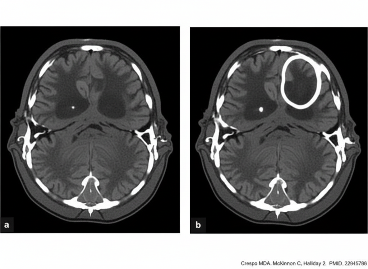
A patient presents with foul-smelling ear discharge and convulsions. What is the appropriate management for this patient, given the CECT findings?

Explanation: **Explanation:** **Streptococcus pneumoniae** is the most common bacterial pathogen responsible for Acute Otitis Media (AOM) across all age groups, particularly in children. It accounts for approximately 30–50% of bacterial isolates. The pathogenesis typically involves an antecedent viral upper respiratory tract infection that causes Eustachian tube dysfunction, allowing nasopharyngeal flora to migrate into the middle ear. **Analysis of Options:** * **A. Haemophilus influenzae:** This is the second most common cause (non-typeable strains). However, since the introduction of the pneumococcal conjugate vaccine (PCV), the relative prevalence of *H. influenzae* has increased, sometimes rivaling *S. pneumoniae* in specific populations, but *S. pneumoniae* remains the standard answer for exams. * **C. Staphylococcus aureus:** While it can cause middle ear infections, it is a much less common primary cause of acute AOM and is more frequently associated with chronic suppurative otitis media (CSOM) or postoperative infections. * **D. Pseudomonas:** This is the hallmark pathogen for **Otitis Externa** and **CSOM**, but it is rarely a cause of primary AOM in immunocompetent children. **High-Yield Clinical Pearls for NEET-PG:** * **Top 3 Pathogens in AOM:** 1. *S. pneumoniae*, 2. *H. influenzae*, 3. *Moraxella catarrhalis*. * **Viral Etiology:** Viruses (RSV, Rhinovirus) are often the initial triggers, but bacterial secondary infection is what defines clinical AOM. * **Drug of Choice:** Amoxicillin remains the first-line antibiotic. * **Complication:** The most common extracranial complication of AOM is **Acute Mastoiditis**.
Explanation: ### Explanation **1. Why the Correct Answer is Right:** The clinical presentation of **foul-smelling ear discharge** and a **pars flaccida perforation** is a classic indicator of **Attico-antral type** of Chronic Suppurative Otitis Media (CSOM). This type is frequently associated with **cholesteatoma**, a destructive keratinizing squamous epithelium collection. Unlike the tubotympanic type (safe), the attico-antral type is considered "unsafe" because cholesteatoma can erode bony structures, leading to intracranial and extracranial complications. The definitive management for cholesteatoma is surgical. **Tympanomastoid exploration** (which may include a Canal Wall Down or Canal Wall Up Mastoidectomy) is necessary to visualize the extent of the disease, remove the cholesteatoma, and exteriorize the attic/mastoid to prevent recurrence. **2. Why the Incorrect Options are Wrong:** * **Options A & B:** Medical management (topical or IV antibiotics) may temporarily control secondary infection and reduce discharge, but it **cannot cure** a cholesteatoma. Delaying surgery increases the risk of complications like facial nerve palsy, labyrinthitis, or brain abscess. * **Option C:** **Tympanoplasty** is the surgical repair of the tympanic membrane and/or ossicles. While it may be part of the procedure, it is insufficient on its own for attico-antral disease because it does not address the disease pathology hidden within the mastoid antrum and attic. **3. Clinical Pearls for NEET-PG:** * **Pars Flaccida Perforation:** Pathognomonic for Attico-antral CSOM (Unsafe ear). * **Foul smell:** Suggests bone erosion and anaerobic infection. * **Cholesteatoma Theory:** The most accepted theory for primary acquired cholesteatoma is the **Invagination Theory (Wittmaack’s)** due to negative middle ear pressure. * **Radiology:** HRCT Temporal Bone is the gold standard imaging to assess the extent of bone erosion before surgery.
Explanation: **Explanation:** The **organ of Corti** is the sensory organ of hearing, located within the cochlea of the inner ear. It is situated on the **basilar membrane**, which forms the floor of the scala media (cochlear duct). 1. **Why Option A is correct:** The organ of Corti consists of hair cells (sensory receptors) and supporting cells. These cells rest directly on the basilar membrane. When sound waves travel through the cochlear fluids, they cause the basilar membrane to vibrate. This mechanical displacement bends the stereocilia of the hair cells against the overlying tectorial membrane, converting mechanical energy into electrical nerve impulses (transduction). 2. **Why Options B and C are incorrect:** * **Utricle and Saccule:** These are parts of the **vestibular apparatus** (specifically the otolith organs) responsible for maintaining equilibrium and balance, not hearing. * The sensory epithelium in the utricle and saccule is called the **Macula**, which detects linear acceleration and head tilt. **High-Yield Clinical Pearls for NEET-PG:** * **Endolymph vs. Perilymph:** The organ of Corti is bathed in **endolymph** (high in $K^+$), which is found in the scala media. * **Outer vs. Inner Hair Cells:** There are three rows of outer hair cells (act as amplifiers) and one row of inner hair cells (primary transducers of sound). * **Tonotopic Organization:** The basilar membrane is narrow and stiff at the **base** (responds to high frequencies) and wide and compliant at the **apex** (responds to low frequencies). * **Modiolus:** The central bony pillar of the cochlea around which the basilar membrane winds.
Explanation: **Explanation:** Acoustic Neuroma (Vestibular Schwannoma) is a benign tumor arising from the Schwann cells of the **vestibular nerve** (CN VIII), typically within the internal auditory canal or the cerebellopontine (CP) angle. **Why Ptosis is the Correct Answer:** Ptosis (drooping of the upper eyelid) is caused by a lesion of the **Oculomotor nerve (CN III)** or sympathetic nerve fibers (Horner’s syndrome). The Oculomotor nerve is located in the midbrain and cavernous sinus, far superior to the CP angle. Acoustic neuromas do not typically involve CN III; therefore, ptosis is not a clinical feature of this tumor. **Analysis of Other Options:** * **Unilateral Deafness:** This is the most common presenting symptom. The tumor compresses the cochlear division of **CN VIII**, leading to progressive sensorineural hearing loss. * **Nystagmus:** As the tumor originates from the vestibular nerve, it disrupts vestibular input, leading to vertigo and vestibular nystagmus (typically horizontal-torsional). * **Loss of Corneal Reflex:** This is the **earliest sign** of trigeminal nerve (**CN V**) involvement. As the tumor expands in the CP angle, it compresses the trigeminal nerve, leading to diminished corneal sensitivity before overt facial numbness occurs. **High-Yield Clinical Pearls for NEET-PG:** * **Earliest Symptom:** Progressive unilateral sensorineural hearing loss and tinnitus. * **Earliest Sign:** Loss of corneal reflex (CN V involvement). * **Facial Nerve (CN VII):** Surprisingly resistant to pressure; facial weakness is usually a late feature despite the nerve's proximity. * **Gold Standard Investigation:** Gadolinium-enhanced MRI of the Internal Auditory Canal. * **Bilateral Acoustic Neuromas:** Pathognomonic for **Neurofibromatosis Type 2 (NF2)**.
Explanation: **Explanation:** **Myringosclerosis** refers to the deposition of hyaline and calcium in the lamina propria of the tympanic membrane. It is a localized form of tympanosclerosis, often appearing as chalky white patches on the eardrum. **Why Grommet Insertion is the Correct Answer:** The most common cause of myringosclerosis is trauma to the tympanic membrane, specifically **Grommet (Ventilation Tube) insertion**. The procedure triggers an inflammatory response and oxidative stress within the fibrous layer of the drum. Studies show that up to 40–50% of children undergoing grommet insertion develop these white patches. Other causes include chronic otitis media and recurrent episodes of otitis media with effusion. **Analysis of Incorrect Options:** * **Genetic predisposition:** While some individuals may be more prone to scarring, there is no established genetic inheritance pattern for myringosclerosis. * **Otosclerosis:** This is a metabolic bone disease involving the otic capsule (specifically the stapes footplate), leading to conductive hearing loss. It does not involve the tympanic membrane. * **Idiopathic:** While some cases occur without a clear history, the vast majority are secondary to identifiable clinical triggers like trauma or infection. **High-Yield Clinical Pearls for NEET-PG:** * **Tympanosclerosis vs. Myringosclerosis:** If the calcification is limited to the drum, it is *myringosclerosis*. If it involves the ossicles or middle ear mucosa (leading to "frozen" ossicles), it is *tympanosclerosis*. * **Clinical Significance:** Myringosclerosis is usually asymptomatic and rarely causes significant hearing loss unless it is extensive. * **Horseshoe Appearance:** Extensive myringosclerosis can sometimes present as a "horseshoe" shaped white patch on the tympanic membrane. * **Chalky White Patch:** This is the classic otoscopic description used in exam questions.
Explanation: The **external auditory canal (EAC)** is an S-shaped passage extending from the concha of the auricle to the tympanic membrane. In an adult, its average length is **24 mm**. ### **Detailed Breakdown:** The EAC is divided into two distinct parts, which is a high-yield concept for exams: 1. **Cartilaginous Part (Outer 1/3rd):** Approximately **8 mm** long. It is a continuation of the auricular cartilage and contains hair follicles, sebaceous glands, and ceruminous glands (the site of wax formation). 2. **Bony Part (Inner 2/3rd):** Approximately **16 mm** long. It is formed by the tympanic part of the temporal bone and lacks hair and glands. ### **Analysis of Options:** * **A (12 mm):** This represents only the bony portion (approximate) or is too short for the entire canal. * **B (24 mm):** **Correct.** This is the standard anatomical length (8 mm cartilaginous + 16 mm bony). * **C & D (36 mm / 48 mm):** These lengths are significantly longer than the human anatomy; a 3.6 cm or 4.8 cm canal would place the tympanic membrane much deeper into the skull than it naturally resides. ### **High-Yield Clinical Pearls for NEET-PG:** * **The Isthmus:** The narrowest part of the EAC, located at the junction of the cartilaginous and bony portions (or slightly into the bony part). Foreign bodies lodged beyond the isthmus are difficult to remove. * **Fissures of Santorini:** Deficiencies in the cartilaginous part of the EAC that allow infections (like Malignant Otitis Externa) or parotid tumors to spread between the canal and the parotid gland. * **Foramen of Huschke:** A deficiency in the anteroinferior aspect of the bony canal (common in children) that can persist in adults, allowing spread of infection to the infratemporal fossa. * **Nerve Supply:** The EAC is supplied by the **Auriculotemporal nerve** (V3) and **Arnold’s nerve** (Vagus branch). Stimulation of Arnold's nerve during ear cleaning can trigger a "cough reflex."
Explanation: **Explanation:** The presence of **unilateral secretory otitis media (SOM)** in an adult is a classic "red flag" clinical sign that must be considered **Nasopharyngeal Carcinoma (NPC)** until proven otherwise. **1. Why Nasopharyngeal Carcinoma is correct:** The nasopharynx contains the opening of the **Eustachian tube** in the Fossa of Rosenmüller. A tumor in this region (NPC) can mechanically obstruct the tube or infiltrate the *tensor veli palatini* muscle, which is responsible for opening the tube. This leads to persistent negative middle ear pressure, resulting in a sterile transudate (effusion). In adults, while SOM is often post-viral, a unilateral presentation without a preceding URTI strongly suggests a proximal obstructive lesion. **2. Why other options are incorrect:** * **CSOM:** This involves a chronic infection with a perforated tympanic membrane and otorrhea. SOM, by definition, occurs behind an intact tympanic membrane. * **Mastoiditis:** This is a complication of acute otitis media (AOM) characterized by retroauricular pain, fever, and erythema. It does not typically present as an isolated, painless serous effusion. * **Foreign body of the external ear:** This affects the external auditory canal and may cause conductive hearing loss or otitis externa, but it does not cause fluid accumulation in the middle ear space. **Clinical Pearls for NEET-PG:** * **Trotter’s Triad for NPC:** 1. Conductive hearing loss (due to SOM), 2. Ipsilateral facial/temporoparietal pain (Trigeminal nerve involvement), 3. Palatal paralysis (Vagus nerve involvement). * **Diagnostic Step:** Any adult with unilateral SOM must undergo **flexible fiberoptic nasopharyngoscopy** to visualize the Fossa of Rosenmüller. * **Most common site for NPC:** Fossa of Rosenmüller. * **Association:** Strongly linked with **Epstein-Barr Virus (EBV)**.
Explanation: **Explanation:** The **Arnold nerve**, also known as the **auricular branch of the Vagus nerve (CN X)**, provides sensory innervation to the floor of the external auditory canal and the posterior part of the tympanic membrane. It arises from the superior ganglion of the vagus nerve and enters the temporal bone through the mastoid canaliculus. **Why Option A is Correct:** The Arnold nerve is a direct branch of the Vagus nerve. Its clinical significance lies in the **"Ear-Cough Reflex"** (Arnold’s reflex), where stimulation of the external ear canal (e.g., during syringing or wax removal) triggers a cough due to the shared vagal innervation with the larynx and lungs. **Why Other Options are Incorrect:** * **B. Hypoglossal nerve (CN XII):** This is a purely motor nerve supplying the muscles of the tongue; it has no auricular branches. * **C. Glossopharyngeal nerve (CN IX):** This nerve gives off the **Jacobson’s nerve** (tympanic branch), which forms the tympanic plexus. While it also supplies the ear, it is distinct from Arnold’s nerve. * **D. Trigeminal nerve (CN V):** The mandibular division (V3) gives off the **auriculotemporal nerve**, which supplies the anterior part of the external ear and canal, not the Arnold nerve. **High-Yield Clinical Pearls for NEET-PG:** * **Jacobson’s Nerve:** Branch of CN IX (Glossopharyngeal). * **Arnold’s Nerve:** Branch of CN X (Vagus). * **Referred Otalgia:** Pain in the ear can be caused by pathologies in the throat (CN IX/X) or teeth/TMJ (CN V) due to these shared nerve pathways. * **Vagal Stimulation:** In some individuals, stimulating Arnold’s nerve can cause bradycardia or fainting (vasovagal syncope).
Explanation: **Explanation:** In the study of embryology and anatomy for NEET-PG, it is a high-yield fact that several structures of the ear are unique because they reach their **full adult size at birth**. **Why the Mastoid Process is the Correct Answer:** The **mastoid process** is absent or rudimentary at birth. It begins to develop during the first year of life as the sternocleidomastoid muscle pulls on the petrous bone when the infant starts to hold their head up. It continues to grow and undergo pneumatization (air cell development) until puberty. Therefore, it is the only structure in the list that is significantly smaller in a neonate compared to an adult. **Analysis of Incorrect Options:** * **Tympanic Membrane:** It reaches its full adult dimensions (approx. 9-10 mm vertically and 8-9 mm horizontally) by birth. However, its orientation changes from almost horizontal in infants to more vertical in adults. * **Ossicles:** The Malleus, Incus, and Stapes are the only bones in the human body that are fully ossified and at their adult size at the time of birth. * **Cochlea (and Bony Labyrinth):** The internal ear structures, including the cochlea and semicircular canals, reach adult size by the 20th week of intrauterine life. **Clinical Pearls for NEET-PG:** 1. **Facial Nerve Safety:** Because the mastoid process is not developed at birth, the **stylomastoid foramen** is superficial. This makes the facial nerve vulnerable to injury during birth (forceps delivery) or behind-the-ear incisions in infants. 2. **Antrum:** While the mastoid process is absent, the **mastoid antrum** is present at birth and is already adult-sized. 3. **Tympanic Ring:** In neonates, the external auditory canal is purely cartilaginous because the bony canal (tympanic ring) is not yet developed.
Explanation: ***Abscess drainage followed by Modified Radical Mastoidectomy (MRM)*** - **Brain abscess** with raised **intracranial pressure** requires immediate drainage to prevent **herniation** and neurological deterioration. - After stabilizing the patient with abscess drainage, **MRM** is performed to remove the **mastoid source** and prevent recurrence. *Modified Radical Mastoidectomy (MRM) followed by abscess drainage* - Performing **MRM first** delays treatment of the life-threatening **intracranial complication** and may worsen **raised ICP**. - The **brain abscess** poses immediate risk of **herniation** and requires priority management over the mastoid source. *Myringoplasty* - This procedure only repairs the **tympanic membrane** and does not address the underlying **chronic suppurative otitis media (CSOM)**. - Completely inadequate for managing **intracranial complications** like brain abscess requiring urgent neurosurgical intervention. *Modified Radical Mastoidectomy (MRM) only* - While **MRM** addresses the **mastoid source**, it ignores the life-threatening **brain abscess** causing convulsions. - **Intracranial complications** require **neurosurgical drainage** first to prevent immediate mortality from raised ICP.
Vestibular System Anatomy and Physiology
Practice Questions
Vestibular Testing
Practice Questions
Benign Paroxysmal Positional Vertigo
Practice Questions
Ménière's Disease
Practice Questions
Vestibular Neuritis
Practice Questions
Labyrinthitis
Practice Questions
Acoustic Neuroma
Practice Questions
Other Cerebellopontine Angle Tumors
Practice Questions
Facial Nerve Disorders
Practice Questions
Skull Base Surgery
Practice Questions
Cochlear Implantation
Practice Questions
Vestibular Schwannoma Management
Practice Questions
Get full access to all questions, explanations, and performance tracking.
Start For Free