To distinguish between cochlear and post-cochlear damage, which test is done?
Which of the following would be the most appropriate treatment for rehabilitation of a patient, who has bilateral profound deafness following surgery for bilateral acoustic schwannoma?
Arrange the following in the sequence of auditory pathway: 1. Cochlear nucleus 2. Spiral ganglion 3. Superior olivary nucleus 4. Inferior colliculus 5. Medial geniculate body
A child aged 3 yrs, presented with severe sensorineural deafness was prescribed hearing aids, but showed no improvement. What is the next line of management:
Site for placing an electrode in auditory brain stem implant is?
Electrode of cochlear implant is placed in:
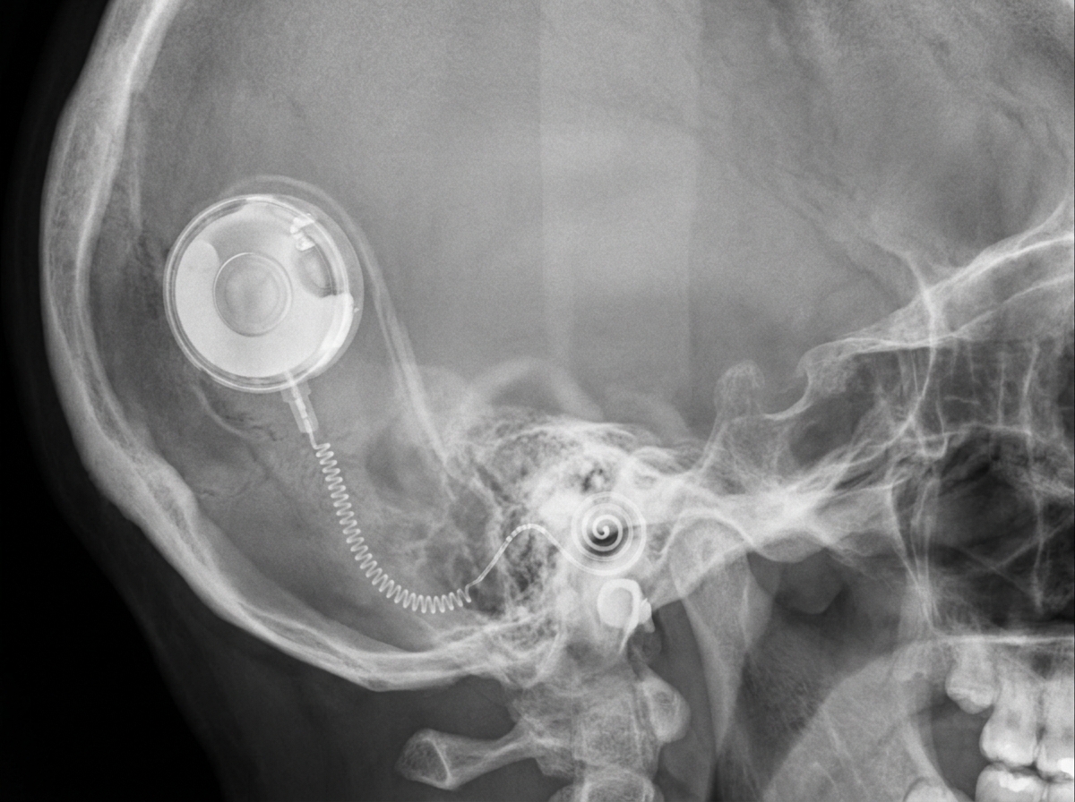
Which device is depicted below?

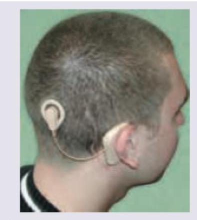
Identify the device shown in the image below:

Which of the following devices typically requires the most stringent safety protocols before MRI scanning?
Meniere's disease is characterized by which triad of symptoms?
Explanation: ***Auditory brainstem response (ABR)*** - This test evaluates the integrity of the **auditory pathway from the cochlea through the brainstem**, making it excellent for differentiating between cochlear (sensory) and post-cochlear (retrocochlear/neural) lesions. - Abnormalities in wave latencies or interpeak intervals suggest **retrocochlear pathology** (e.g., acoustic neuroma), while normal ABR responses despite hearing loss point towards cochlear damage. - ABR records **five characteristic waves (I-V)** representing neural transmission from the auditory nerve through the brainstem. *Impedance audiometry* - Primarily assesses the **middle ear function**, including the eardrum and ossicles, by measuring **tympanic membrane compliance** and **acoustic reflexes**. - It does not directly evaluate the function of the **cochlea or the retrocochlear pathways**, making it unsuitable for this differentiation. *Pure tone audiometry* - Measures a person's **hearing sensitivity** at different frequencies and provides information on the **degree and type of hearing loss (conductive, sensorineural, or mixed)**. - While it identifies sensorineural hearing loss, it cannot pinpoint whether the damage is **cochlear or retrocochlear** within the sensorineural category. *Electrocochleography (ECochG)* - Records **electrical potentials generated by the cochlea and auditory nerve** in response to sound, including **cochlear microphonics, summating potentials, and compound action potentials**. - While it evaluates cochlear function and is useful in diagnosing **Meniere's disease** and **auditory neuropathy**, it does not adequately assess the **integrity of the brainstem auditory pathways** needed to differentiate retrocochlear lesions.
Explanation: ***Brainstem implant*** - A **brainstem implant** is the most appropriate treatment when the auditory nerve has been damaged or destroyed, as can occur during bilateral acoustic schwannoma surgery. - This device bypasses the cochlea and auditory nerve by directly stimulating the **cochlear nucleus** in the brainstem, allowing sound perception. *Bilateral cochlear implant* - A **cochlear implant** requires an intact auditory nerve to transmit signals from the cochlea to the brain. - In this scenario, bilateral profound deafness post-surgery for acoustic schwannoma often implies damage to both **auditory nerves**, rendering cochlear implants ineffective. *Unilateral cochlear implant* - Similar to a bilateral cochlear implant, a **unilateral cochlear implant** relies on the functionality of at least one auditory nerve. - Since the patient has **bilateral profound deafness** following bilateral surgery, the auditory nerves are likely compromised on both sides, making even a unilateral implant unsuitable. *Bilateral high powered digital hearing aid* - Hearing aids amplify sound and rely on the presence of residual hair cell function in the **cochlea** and an intact auditory pathway. - Profound deafness indicates severe damage to the inner ear or auditory nerve, which hearing aids cannot overcome as they only provide *amplification*, not direct neural stimulation.
Explanation: ***2-1-3-4-5*** - The auditory pathway begins with the **spiral ganglion**, which contains the cell bodies of the first-order neurons that innervate the hair cells of the cochlea. - Signals then proceed to the **cochlear nucleus** in the brainstem, followed by the **superior olivary nucleus**, the **inferior colliculus**, and finally the **medial geniculate body** in the thalamus before reaching the auditory cortex [1]. *5-4-3-2-1* - This sequence represents a nearly reverse order of the ascending auditory pathway, starting from a higher processing center (medial geniculate body) and moving backward, which is incorrect for sensory input. - The **medial geniculate body** is the thalamic relay for auditory information, receiving input from lower centers and projecting to the auditory cortex [1]. *3-4-5-1-2* - This sequence incorrectly places the **superior olivary nucleus** as the initial processing stage, preceding the lower-level **spiral ganglion** and **cochlear nucleus**. - Auditory information must first be transduced by hair cells and then relayed by the spiral ganglion neurons to the cochlear nucleus before further processing in the olivary complex. *1-2-3-4-5* - This sequence incorrectly places the **cochlear nucleus** before the **spiral ganglion**. - The **spiral ganglion** contains the primary afferent neurons that receive input from the hair cells and project their axons to the cochlear nucleus.
Explanation: ***Cochlear implant*** - For **severe sensorineural deafness** where conventional hearing aids provide no benefit, a cochlear implant is the most effective next step for restoring hearing. - A cochlear implant directly stimulates the **auditory nerve**, bypassing damaged hair cells in the cochlea, which is crucial for severe sensorineural hearing loss. - In children aged **12 months to 5 years**, early cochlear implantation is critical for optimal **speech and language development**. *Conservative* - This typically refers to observation or non-invasive treatments like hearing aids, which have already failed in this case. - Continuing a conservative approach would delay effective intervention for severe deafness, potentially impacting the child's **speech and language development**. *Fenestration surgery* - This is a surgical procedure primarily used for some types of **conductive hearing loss**, especially **otosclerosis**, by creating an opening in the bony labyrinth. - It is not indicated for **sensorineural deafness**, as the problem lies with the inner ear or auditory nerve, not the sound conduction pathway. *Stapes mobilisation* - This procedure aims to restore mobility to the **stapes bone** in cases of **otosclerosis**, a form of conductive hearing loss where the stapes becomes fixed. - It is not appropriate for **sensorineural hearing loss**, where the primary issue is damage to the inner ear's sensory cells or the auditory nerve.
Explanation: ***Recess of fourth ventricle*** - The auditory brainstem implant (ABI) electrode arrays are typically placed on the surface of the **cochlear nucleus**, which lies in the **lateral recess of the fourth ventricle and cerebellopontine angle**. - This placement allows direct stimulation of the central auditory pathways, bypassing a damaged or absent auditory nerve. *Round window* - The round window is the site for electrode placement in a **cochlear implant**, not an auditory brainstem implant. - A cochlear implant stimulates the **auditory nerve terminals** within the cochlea. *Sinus tympani* - The sinus tympani is an **anatomical space** within the middle ear. - It is not a site for implant electrode placement for either cochlear or brainstem implants. *Lateral ventricle* - The lateral ventricles are spaces within the brain that contain **cerebrospinal fluid** and are not directly involved in the auditory pathway for implant stimulation. - Implants for hearing are generally directed towards the auditory neural structures.
Explanation: ***Scala tympani*** - The electrode array of a **cochlear implant** is carefully inserted into the **scala tympani** of the cochlea. - This placement allows the electrodes to directly stimulate the **spiral ganglion neurons**, bypassing damaged hair cells and transmitting electrical signals to the auditory nerve. *Horizontal semicircular canal* - The **horizontal semicircular canal** is part of the **vestibular system**, responsible for sensing angular head movements, not hearing. - Placing an electrode here would cause **vestibular dysfunction** and would not restore hearing. *Scala media* - The **scala media** (cochlear duct) contains the **organ of Corti** and **endolymph**, which has a high potassium concentration. - Inserting an electrode here would damage the delicate structures essential for natural sound transduction and could lead to electric potential imbalances. *Scala vestibuli* - The **scala vestibuli** is filled with **perilymph** and receives sound vibrations from the stapes. - While it's adjacent to the scala tympani, the **scala tympani** offers a safer and more direct path for optimal electrode insertion with less trauma to the sensory structures.
Explanation: ***Cochlear implant*** - A cochlear implant is an **electronic medical device that replaces the function of a damaged inner ear (cochlea)** and provides sound signals directly to the brain - On imaging (X-ray, CT, or skull radiograph), it appears as a **characteristic circular receiver-stimulator device** under the skin behind the ear with an **electrode array extending into the cochlea** - The **internal receiver has a distinctive appearance** with visible magnets and electrode contacts, making it easily identifiable on radiographic images - Used for patients with **severe to profound sensorineural hearing loss** who do not benefit from conventional hearing aids *Auditory brainstem implant (ABI)* - An ABI **bypasses both the cochlea and auditory nerve**, directly stimulating the **cochlear nucleus in the brainstem** - On imaging, the electrode array would be located at the **cerebellopontine angle** near the brainstem, not in the cochlea - Reserved for patients with **absent or non-functional auditory nerves** (e.g., bilateral vestibular schwannomas, neurofibromatosis type 2) *Bone anchored hearing aid (BAHA)* - A BAHA consists of a **titanium implant osseointegrated into the skull bone** behind the ear with an external sound processor - On X-ray, only the **small titanium fixture/abutment** would be visible in the mastoid bone, without any cochlear or intracranial components - Works by **bone conduction**, transmitting sound vibrations directly to the inner ear, bypassing the outer and middle ear - Used for **conductive hearing loss, mixed hearing loss, or single-sided deafness** *Hearing aid* - A conventional hearing aid is a **completely external electronic device** that amplifies sound - It would **not be visible on X-ray or CT imaging** as it contains no implanted components - Simply amplifies sound for individuals with mild to moderate hearing loss
Explanation: ***Cochlear implant*** - The image displays the external components of a **cochlear implant**: a **speech processor** worn behind the ear connected to an external transmitter that sends signals to an implanted receiver. - This device is designed to provide a sense of sound to individuals with **severe-to-profound hearing loss** by directly stimulating the auditory nerve. *Transcranial magnetic stimulation* - This therapy involves a **coil placed on the scalp** that delivers magnetic pulses to stimulate nerve cells in the brain, typically for depression or migraines. - It does not involve ear-worn components or internal surgical implants of the type seen in the image. *Vagus nerve stimulation* - This involves a device surgically implanted under the skin in the chest, with wires connected to the **vagus nerve** in the neck. - It is used to treat epilepsy and depression and does not have external components positioned around the ear or on the head as depicted. *Deep brain stimulation* - This neurosurgical procedure involves implanting electrodes into specific areas of the brain, connected to a pulse generator (similar to a pacemaker) implanted in the chest. - It is primarily used for movement disorders like Parkinson's disease and does not feature external ear-worn components visible in the image.
Explanation: ***Automatic Cardioverter-defibrillators*** - **Implantable cardioverter-defibrillators (ICDs)**, while increasingly MRI-conditional, require stringent protocols due to potential for **heating, lead damage, and inappropriate pacing/shocks**. - Detailed device interrogation, programming to asynchronous modes or MRI mode, and continuous monitoring are often required to mitigate risks and ensure patient safety. *Prosthetic cardiac valves* - Most modern **prosthetic cardiac valves** (both mechanical and bioprosthetic) are **MRI-conditional** or **MRI-safe** and generally do not pose significant risks. - They are typically made of non-ferromagnetic materials, reducing concerns about displacement or heating. *Insulin pump* - External insulin pumps are generally considered **MR-unsafe** and must be **removed from the patient** before entering the MRI scan room. - While removal is a safety protocol, they don't involve complex internal electronic interactions in the same way an implanted ICD does, making their protocol simpler (remove and resume). *Cochlear implants* - Many **cochlear implants** are now **MRI-conditional**, but they often require specific protocols such as removing the external processor and sometimes applying a head bandage to secure the implant. - Older models or certain configurations may still be considered MR-unsafe due to potential for magnet displacement or device damage.
Explanation: **Explanation** Meniere’s disease (Endolymphatic Hydrops) is a disorder of the inner ear characterized by an abnormal accumulation of endolymph within the membranous labyrinth. The classic clinical triad consists of: 1. **Episodic Vertigo:** Sudden, rotatory vertigo lasting 20 minutes to several hours, often accompanied by nausea and vomiting. 2. **Sensorineural Hearing Loss (SNHL):** Characteristically fluctuating and low-frequency in the early stages. 3. **Tinnitus:** Often described as a low-pitched "roaring" or "seashell" sound. *(Note: Many authorities include a fourth symptom—**Aural Fullness**—making it a tetrad).* **Analysis of Options:** * **Option A:** Incorrect. Meniere’s causes **Sensorineural** hearing loss, not conductive. Conductive loss suggests pathology in the external or middle ear (e.g., ASOM, Otosclerosis). * **Option B:** Incorrect. **Ear discharge (Otorrhea)** is a hallmark of middle ear infections (CSOM) and is never seen in Meniere’s, which is an inner ear pathology with an intact tympanic membrane. * **Option C:** Incorrect. While headache can occur, it is not a defining component of the diagnostic triad. Its presence might instead suggest Vestibular Migraine. * **Option D:** **Correct.** This captures the classic diagnostic triad essential for NEET-PG. **Clinical Pearls for NEET-PG:** * **Pathology:** Distension of the endolymphatic system (Reissner’s membrane bulges into the scala vestibuli). * **Lermoyez Syndrome:** A variant where hearing improves during a vertigo attack ("the phenomenon of reverse symptoms"). * **Tuning Fork Tests:** Rinne positive (SNHL) and Weber lateralized to the better ear. * **Audiometry:** Shows a "rising curve" in early stages (low-frequency loss). * **Glycerol Test:** Used for diagnosis; oral glycerol (osmotic diuretic) temporarily improves hearing by reducing endolymphatic pressure. * **Management:** Low salt diet, Betahistine (drug of choice), and diuretics. Intratympanic Gentamicin is used for refractory cases.
Get full access to all questions, explanations, and performance tracking.
Start For Free