One of the most important complication of tracheostomy is:
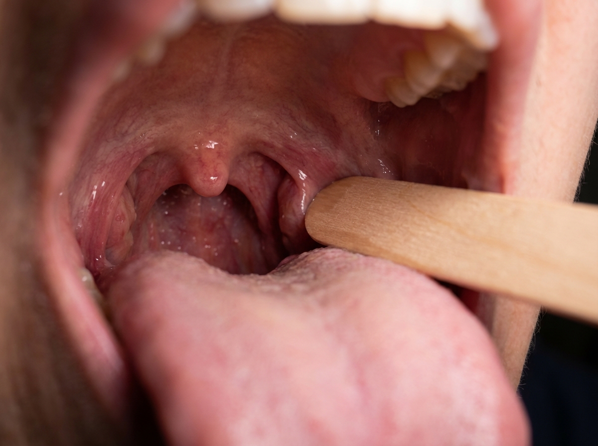
A patient was admitted with skull base trauma. The doctor was testing the marked structure in the pharyngeal region. Which of the following nerves was being tested?

A patient has a lacerated, untidy wound of the leg and attended the casualty department after 2 hours. His wound should be:
Frey's syndrome is associated with-
Which of the following statements is false regarding postpartum hemorrhage and pelvic hematomas?
Reactionary Hemorrhage occurs due to?
Carcinoma of pyriform fossa usually presents with :
In patient of head injuries with rapidly increasing intracranial tension without hematoma, the drug of choice for initial management would be :
Which of the following is not directly implicated as a cause of squamous cell carcinoma of the head and neck?
Burns involving the head and neck region are particularly dangerous because :
Explanation: ***Displacement of tube*** - **Accidental decannulation** or displacement of the tracheostomy tube is considered one of the most serious and common complications, particularly in the immediate post-operative period. - This can lead to **loss of airway**, requiring immediate intervention to prevent severe hypoxia and potential brain injury or death. *Hemorrhage* - While hemorrhage can occur during or after tracheostomy, it is often a concern during the procedure or in the immediate postoperative period and is usually managed effectively. - Significant, life-threatening hemorrhage such as **tracheo-innominate fistula** is a rare but severe complication. *Surgical emphysema* - Surgical emphysema (subcutaneous emphysema) is a relatively common but usually benign complication that occurs when air leaks from the trachea into the subcutaneous tissues. - It typically resolves spontaneously and rarely poses a direct threat to the airway unless severe and rapidly progressive. *Recurrent laryngeal nerve palsy* - **Recurrent laryngeal nerve injury** is a rare complication of tracheostomy, as the nerve is usually well clear of the incision site in the neck. - While it can cause hoarseness or vocal cord paralysis, it typically does not present an immediate life-threatening situation or emergency comparable to airway compromise.
Explanation: ***Glossopharyngeal nerve*** - The image shows a probe stimulating the posterior part of the **pharynx**, which elicits the **gag reflex**. - The afferent limb of the **gag reflex** is mediated primarily by the **glossopharyngeal nerve (CN IX)**, which detects sensation from the posterior tongue and pharynx. *Trigeminal nerve* - The **trigeminal nerve (CN V)** primarily mediates sensation from the face, teeth, and anterior two-thirds of the tongue, and motor control of the **muscles of mastication**. - It does not have a primary role in the sensation or reflex of the posterior pharyngeal wall. *Facial nerve* - The **facial nerve (CN VII)** is responsible for the **muscles of facial expression**, taste from the anterior two-thirds of the tongue, and parasympathetic innervation to several glands. - While it contributes to some aspects of swallowing, it is not the main sensory nerve for the gag reflex from the posterior pharynx. *Vagus* - The **vagus nerve (CN X)** provides the efferent limb of the gag reflex, causing pharyngeal muscle contraction. - However, the sensory input from the posterior pharynx (the afferent limb being tested by the probe) is primarily carried by the **glossopharyngeal nerve**. *Hypoglossal nerve* - The **hypoglossal nerve (CN XII)** provides motor innervation to the intrinsic and extrinsic muscles of the tongue. - While it is relevant in skull base trauma, it does not mediate sensation from the pharynx or the gag reflex being tested in the image.
Explanation: ***Debrided and sutured secondarily*** - An **untidy wound** indicates contamination, irregular edges, and devitalized tissue, which significantly increases the **risk of wound infection**. - The standard management involves **thorough debridement** to remove all contaminated and non-viable tissue, followed by **delayed primary closure** (suturing after 3-5 days once the wound shows healthy granulation) or **healing by secondary intention**. - This approach is especially important for **lower extremity wounds**, which have a higher infection risk due to relatively poorer blood supply compared to facial wounds. - Even though the patient presented within 2 hours (well within the "golden period"), the **untidy nature** of the wound makes **immediate primary closure risky** and secondary closure the safer, preferred option. *Debrided and sutured immediately* - While **debridement is essential** for untidy wounds, **immediate primary closure** after debridement is generally reserved for **tidy wounds** with minimal contamination. - For untidy wounds, immediate closure increases the risk of **trapping bacteria and devitalized tissue**, leading to **wound infection**, abscess formation, or dehiscence despite being within the golden period. - Primary closure may be considered in select cases with minimal contamination and excellent debridement, but this is not the standard teaching for untidy wounds. *Sutured immediately* - **Immediate suturing without debridement** of an untidy wound would be dangerous, as it would trap contaminants, foreign material, and devitalized tissue. - This approach would significantly increase the risk of **serious wound infection**, including **gas gangrene** or necrotizing fasciitis in contaminated wounds. - Proper wound preparation is mandatory before any closure is considered. *Cleaned and dressed* - Simple **cleaning and dressing** is insufficient for an untidy wound as it does not address the devitalized tissue that requires **surgical debridement**. - While this avoids the risk of premature closure, it fails to provide adequate treatment for a wound that needs formal surgical debridement to remove non-viable tissue and reduce bacterial load. - This approach might be acceptable only as a temporary measure if surgical debridement cannot be performed immediately.
Explanation: ***Parasympathetic fibres of auriculo temporal nerve*** - **Frey's syndrome**, or **gustatory sweating**, occurs due to aberrant regeneration of damaged **auriculotemporal nerve fibers** after **parotid gland surgery** or trauma. - **Parasympathetic secretomotor fibers** that originally innervated the **parotid gland** mistakenly reinnervate overlying **sweat glands** and **blood vessels** of the skin. *Motor fibres of facial nerve* - **Motor fibers of the facial nerve** control **facial expression muscles** and are not directly involved in the pathogenesis of Frey's syndrome. - Damage to these fibers would result in **facial paralysis**, not gustatory sweating. *Sympathetic fibres of auriculo temporal nerve* - The **auriculotemporal nerve** contains **sensory fibers** to the temporal region and **parasympathetic secretomotor fibers** to the parotid gland, but its sympathetic fibers are primarily **vasomotor**. - **Sympathetic fibers** control vasoconstriction and eccrine sweating generally, but their aberrant regeneration is not the cause of Frey's syndrome. *Parasympathetic fibres of trigeminal nerve* - The **trigeminal nerve** is primarily **sensory** to the face and **motor** to the muscles of mastication; it does not directly innervate the parotid gland. - **Parasympathetic innervation** to the parotid gland is carried by the glossopharyngeal nerve via the otic ganglion, not the trigeminal nerve.
Explanation: ***The vulva is the most common site for pelvic hematoma.*** - While vulvar hematomas are common, the **vagina is actually the most common site** for puerperal hematomas. - **Retroperitoneal hematomas** are the least common but most dangerous type, often associated with a higher mortality rate due to delayed diagnosis. *Hematomas less than 5 cm can often be managed conservatively.* - **Small, stable hematomas** (typically less than 2-5 cm) that are not expanding can often be managed with observation, pain control, and ice packs. - Close monitoring for continued bleeding, signs of infection, or hemodynamic instability is crucial even with conservative management. *Uterine atony is the most common cause of postpartum hemorrhage.* - **Uterine atony** (failure of the uterus to contract after birth) accounts for approximately 70-80% of all cases of postpartum hemorrhage. - This condition leads to excessive bleeding from the placental site due to the inability of uterine muscle fibers to compress blood vessels effectively. *The most common artery to form a vulvar hematoma is the pudendal artery.* - Vulvar hematomas primarily arise from injury to branches of the **pudendal artery**, particularly during lacerations or episiotomies. - Trauma to the **perineum** during childbirth can cause these arteries or their venous counterparts to bleed into the surrounding loose connective tissue.
Explanation: ***Dislodgement of clot*** - **Reactionary hemorrhage** occurs within the first 24 hours post-surgery as the initial **vasoconstriction** and **blood pressure drop** from anesthesia resolve. - As blood pressure normalizes and peripheral vessels dilate, a **clot** that formed in a previously bleeding vessel becomes dislodged, leading to bleeding. *Infection* - **Infection** can cause secondary hemorrhage, but this typically occurs later, usually several days to weeks after surgery, due to tissue necrosis and erosion of blood vessels. - It is not the primary mechanism for hemorrhage occurring within the first 24 hours. *Damage to a blood vessel* - **Damage to a blood vessel** during surgery is a cause of primary hemorrhage, which occurs during or immediately after the procedure. - While it initiates the potential for bleeding, reactionary hemorrhage specifically refers to bleeding that resumes due to changes in patient physiology post-operatively, rather than ongoing vessel damage. *Pressure necrosis* - **Pressure necrosis** refers to tissue death due to sustained external pressure, often leading to skin breakdown or deep tissue injury. - It does not directly cause reactionary hemorrhage, although necrotic tissue could potentially contribute to later secondary hemorrhage if a vessel erodes.
Explanation: ***Dysphagia*** - Carcinoma of the **pyriform fossa** is a type of hypopharyngeal cancer, and given its anatomical location, it commonly interferes with swallowing [1]. - The pyriform fossa lies immediately lateral to the laryngeal inlet, and involvement here directly impacts the ability to form a **food bolus** and propel it into the esophagus. *Lump in the neck* - A neck lump can occur, especially if there is **lymph node metastasis**, but it's often a later symptom [1]. - **Dysphagia** usually precedes the development of a palpable neck mass as the primary tumor expands within the pyriform fossa [1]. *Cough* - While aspiration might lead to coughing, it's not the primary presenting symptom. - Cough is more commonly associated with laryngeal involvement or **tracheal invasion**, which can occur with advanced disease. *Hoarseness* - **Hoarseness** is a prominent symptom if the **vocal cords** or recurrent laryngeal nerve are directly involved [2]. - The pyriform fossa is adjacent but distinct from the vocal cords, so hoarseness is not typically the initial or most common symptom unless the tumor extends medially.
Explanation: ***20% Mannitol*** - **Mannitol** is an osmotic diuretic that reduces **intracranial pressure (ICP)** by creating an osmotic gradient, drawing water from the brain parenchyma into the intravascular space [1]. - Its rapid onset of action and significant ICP-reducing effects make it the drug of choice for acute management of elevated ICP in head injuries without hematoma. *Lasix* - **Furosemide (Lasix)** is a loop diuretic that can reduce ICP by decreasing cerebrospinal fluid production and promoting diuresis. - However, its effects are generally slower and less potent than mannitol for acute, rapidly increasing ICP. *Glycine* - **Glycine** is an amino acid and neurotransmitter; it has no direct role in the acute management of increased ICP. - It is sometimes used as an irrigating solution in urological procedures but is not indicated for brain injury. *Steroids* - **Steroids**, particularly **dexamethasone**, are effective in reducing vasogenic edema associated with brain tumors or abscesses. - They are generally **not recommended** for acute traumatic brain injury due to lack of benefit and potential for increased mortality or complications.
Explanation: ***Vitamin A*** - Vitamin A deficiency is associated with increased risk of squamous metaplasia but not a direct cause of squamous cell carcinoma in the head and neck. - Adequate levels of Vitamin A are actually protective against various epithelial cancers. *EBV* - Epstein-Barr Virus (EBV) is linked to certain types of cancers, including nasopharyngeal carcinoma, but is not a major causative factor for squamous cell carcinoma [1]. - It can contribute to **lymphoproliferative disorders** but not primarily to squamous cell carcinoma of the head and neck [1]. *HPV* - Human Papillomavirus (HPV), particularly types 16 and 18, are recognized as significant contributors to oropharyngeal squamous cell carcinoma [1]. - HPV infection can lead to **malignant transformation** of epithelial cells [1]. *Betel Nut* - Betel nut chewing is a well-established risk factor for oral squamous cell carcinoma, associated with its carcinogenic properties [2]. - It can cause **oral lesions** and dysplasia, contributing significantly to the etiology of head and neck cancers [2].
Explanation: ***There may be thermal damage to the respiratory passage*** - Burns to the **head and neck** often indicate exposure to heat or flame around the face, increasing the risk of inhaling hot air, smoke, or toxic fumes. - This can lead to **thermal damage** to the upper and lower **respiratory passages**, causing edema, airway obstruction, and acute respiratory distress. *Face is a very vascular area* - While the face is indeed **vascular**, this property primarily impacts **healing time** (often faster due to good blood supply) and the potential for swelling, but does not inherently make burns in this region "particularly dangerous" in the immediate, life-threatening sense compared to airway compromise. - The vascularity itself doesn't directly cause a unique danger that surpasses the risk of **airway obstruction** or systemic complications. *Renal failure is more frequent* - **Acute renal failure** can be a complication of severe burns due to hypovolemia, rhabdomyolysis, or sepsis, but it is not specific to burns of the head and neck region. - It is a systemic complication related to the overall burn severity and total body surface area (TBSA) involved, rather than the specific anatomical location of the burn. *Blood loss may be more severe* - Significant **blood loss** is not typically a direct primary concern in burn injuries unless there are associated trauma or very deep burns to highly vascular areas. - While fluid shifts in burns can be massive, initial blood loss is not the defining factor that makes head and neck burns particularly dangerous from a life-threatening perspective.
Get full access to all questions, explanations, and performance tracking.
Start For Free