A 25-year-old woman presents with episodes of dizziness, tinnitus, and hearing loss in the right ear. What is the most likely diagnosis?
In a patient with acoustic neuroma all are seen except:
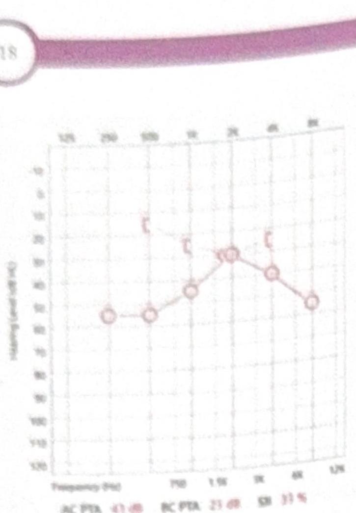
A female patient presents with mild conductive hearing loss (CHL) and tinnitus. Based on the pure tone audiometry (PTA) shown in the image, what is the most likely diagnosis?

A child aged 3 yrs, presented with severe sensorineural deafness was prescribed hearing aids, but showed no improvement. What is the next line of management:
35 years old female presents with tinnitus, vertigo and aural fullness. Likely diagnosis:
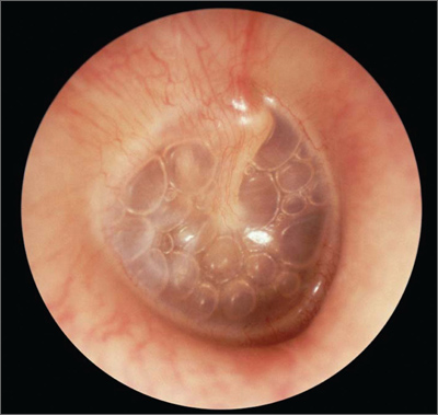
A 5-year-old child presents with reduced hearing for the past 2-3 months. Based on the otoscopy findings shown, what is the most likely diagnosis?

Most common cause of sensorineural hearing loss (SNHL)
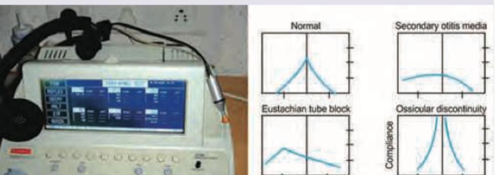
A patient presents with fullness in the ear and hearing loss. Tympanometry shows a type B curve. What does this indicate?
The following test is useful for diagnosis of all except:

A 72-year-old man presents to his primary care physician with progressively worsening hearing loss. He states that his trouble with hearing began approximately 7-8 years ago. He is able to hear when someone is speaking to him; however, he has difficulty with understanding what is being said, especially when there is background noise. In addition to his current symptoms, he reports a steady ringing in both ears, and at times experiences dizziness. Medical history is significant for three prior episodes of acute otitis media. Family history is notable for his father being diagnosed with cholesteatoma. His temperature is 98.6°F (37°C), blood pressure is 138/88 mmHg, pulse is 74/min, and respirations are 13/min. On physical exam, when a tuning fork is placed in the middle of the patient's forehead, sound is appreciated equally on both ears. When a tuning fork is placed by the external auditory canal and subsequently on the mastoid process, air conduction is greater than bone conduction. Which of the following is most likely the cause of this patient's symptoms?
Explanation: ***Ménière's disease*** - This condition is characterized by a classic triad of **episodic vertigo (dizziness)**, fluctuating **sensorineural hearing loss**, and **tinnitus**, often accompanied by aural fullness, typically affecting one ear. - The symptoms arise from an accumulation of **endolymph** in the inner ear, leading to increased pressure and dysfunction. *Labyrinthitis* - **Labyrinthitis** is an inflammation of the inner ear, usually viral, causing sudden, severe **vertigo** potentially with hearing loss and tinnitus. - Unlike Meniere's disease, **hearing loss** and **tinnitus** in labyrinthitis are usually constant rather than episodic or fluctuating. *Acoustic neuroma* - An **acoustic neuroma** (vestibular schwannoma) is a benign tumor on the eighth cranial nerve, often causing **gradual, progressive unilateral hearing loss**, tinnitus, and **balance issues**, but typically not episodic severe dizziness. - While it can cause hearing loss and tinnitus, the **episodic nature of vertigo** is less common than in Ménière's disease. *Benign paroxysmal positional vertigo* - **BPPV** is characterized by sudden, **brief episodes of vertigo** triggered by specific head movements, caused by dislodged **otoconia** in the semicircular canals. - Critically, BPPV does **not** typically cause associated **hearing loss or tinnitus**, which are prominent symptoms in the presented case.
Explanation: Acoustic neuroma typically presents with unilateral progressive hearing loss and sometimes tinnitus [1]. While acoustic neuromas can cause unsteadiness and gait imbalance due to gradual growth, they typically do not present with acute, severe, isolated vertigo because slow growth allows compensatory brainstem mechanisms to develop [1]. Acute vertigo is more characteristic of peripheral vestibular disorders like benign paroxysmal positional vertigo (BPPV) or Meniere's disease. As an acoustic neuroma (vestibular schwannoma) grows, it can compress the adjacent cerebellum, leading to cerebellar signs [1]. These signs include ataxia, dysmetria, and nystagmus, reflecting cerebellar dysfunction. The facial nerve (CN VII) runs in close proximity to the vestibulocochlear nerve (CN VIII), so it can be affected by an acoustic neuroma, leading to facial paresis or paralysis, though facial weakness is unusual at presentation [1]. Unilateral deafness is a classic and early symptom of acoustic neuroma due to compression of the cochlear portion of CN VIII [1]. The trigeminal nerve (CN V) has sensory innervation to the cornea, and its fibers pass near the cerebellopontine angle where acoustic neuromas grow. Compression of the trigeminal nerve by the tumor can lead to a reduced or absent corneal reflex on the affected side.
Explanation: ***Otosclerosis*** - The audiogram shows a **conductive hearing loss** with a notable **Carhart notch** (bone conduction dip at 2000 Hz), which is characteristic of otosclerosis. - The patient's symptoms of **mild CHL** and **tinnitus** are consistent with the presentation of otosclerosis, a condition involving abnormal bone growth in the middle ear. *Ménière's disease* - This condition primarily causes **sensorineural hearing loss**, often fluctuating and affecting low frequencies initially, along with **vertigo, tinnitus, and aural fullness**. - The audiogram indicates **conductive hearing loss**, not sensorineural, and **vertigo** is not mentioned as a primary symptom. *Ototoxicity* - Ototoxicity typically results in **sensorineural hearing loss**, often bilateral and affecting high frequencies first. - The audiogram demonstrates **conductive hearing loss**, and there is no information about exposure to ototoxic medications. *Noise-Induced Hearing Loss (NIHL)* - NIHL is characterized by **sensorineural hearing loss**, typically with a **notch at 3000-6000 Hz** (most commonly 4000 Hz notch) on the audiogram. - The audiogram reflects **conductive hearing loss**, and the specific pattern does not match that of NIHL.
Explanation: ***Cochlear implant*** - For **severe sensorineural deafness** where conventional hearing aids provide no benefit, a cochlear implant is the most effective next step for restoring hearing. - A cochlear implant directly stimulates the **auditory nerve**, bypassing damaged hair cells in the cochlea, which is crucial for severe sensorineural hearing loss. - In children aged **12 months to 5 years**, early cochlear implantation is critical for optimal **speech and language development**. *Conservative* - This typically refers to observation or non-invasive treatments like hearing aids, which have already failed in this case. - Continuing a conservative approach would delay effective intervention for severe deafness, potentially impacting the child's **speech and language development**. *Fenestration surgery* - This is a surgical procedure primarily used for some types of **conductive hearing loss**, especially **otosclerosis**, by creating an opening in the bony labyrinth. - It is not indicated for **sensorineural deafness**, as the problem lies with the inner ear or auditory nerve, not the sound conduction pathway. *Stapes mobilisation* - This procedure aims to restore mobility to the **stapes bone** in cases of **otosclerosis**, a form of conductive hearing loss where the stapes becomes fixed. - It is not appropriate for **sensorineural hearing loss**, where the primary issue is damage to the inner ear's sensory cells or the auditory nerve.
Explanation: ***Meniere's Disease*** - The classic triad of symptoms for Meniere's disease includes **tinnitus**, **vertigo**, and **aural fullness**, along with fluctuating sensorineural hearing loss. - This condition is thought to be caused by an excess of **endolymphatic fluid** within the inner ear. *Ototoxicity* - This condition typically presents with **bilateral, symmetrical hearing loss** and tinnitus, often induced by certain medications (e.g., aminoglycosides, aspirin in high doses). - It usually does not involve episodic vertigo or aural fullness, which are characteristic of Meniere's. *Noise Induced Hearing Loss* - Characterized primarily by **permanent sensorineural hearing loss**, often at specific frequencies (e.g., 4000 Hz notch), and **tinnitus** after prolonged exposure to loud noise. - It does not typically cause the episodic vertigo or sense of aural fullness seen in Meniere's disease. *Otosclerosis* - This condition causes **progressive conductive hearing loss** due to abnormal bone growth around the stapes bone, impairing its movement. - While it can cause tinnitus, it typically does not present with vertigo or aural fullness, and the primary hearing loss is conductive, not sensorineural.
Explanation: ***Otitis media with effusion*** - The image clearly shows the presence of **bubbles behind the tympanic membrane**, indicating fluid accumulation in the middle ear. - This fluid leads to **reduced hearing**, as reported in the 5-year-old child, consistent with otitis media with effusion (OME), also known as "glue ear." *Myringitis* - Myringitis typically presents with **inflammation of the tympanic membrane**, often with vesicles or bullae on the drum, which are not visible here. - While it can cause pain, it does not typically show the characteristic bubbles signifying middle ear effusion. *Acute ear infection* - An **acute otitis media** would show a **bulging, erythematous (red), and opaque tympanic membrane** due to inflammation and pus, which is not consistent with the image. - Systemic symptoms like fever and severe ear pain would also be expected with an acute infection. *Air in the middle ear* - The presence of **air in the middle ear is normal** and indicates a healthy, functioning Eustachian tube. - The visible bubbles in the image are consistent with **fluid and air-fluid levels**, not just air, and indicate a pathological condition affecting hearing.
Explanation: ***Presbycusis*** - **Presbycusis**, or age-related hearing loss, is the most common cause of **sensorineural hearing loss (SNHL)**, affecting a significant portion of the elderly population. - It typically results from **degenerative changes** in the inner ear, specifically the hair cells and nerve fibers, leading to a gradual, symmetrical, high-frequency SNHL. *Labyrinthitis* - **Labyrinthitis** is an inflammation of the inner ear that can cause sudden SNHL, often accompanied by **vertigo** and **tinnitus**. - While it causes SNHL, it is an acute condition and not the most common overall cause, especially when considering the prevalence of age-related hearing loss. *Meniere's disease* - **Meniere's disease** is characterized by recurrent episodes of **vertigo, fluctuating low-frequency SNHL, tinnitus**, and aural fullness. - It is a specific condition causing SNHL, but its prevalence is much lower than presbycusis, making it a less common overall cause. *Vestibular Schwannoma* - A **vestibular schwannoma** (acoustic neuroma) is a benign tumor that arises from the Schwann cells of the **vestibulocochlear nerve**. - It can cause **progressive unilateral SNHL**, tinnitus, and balance issues, but it is a relatively rare condition compared to presbycusis.
Explanation: ***Middle ear effusion*** - A **Type B tympanogram** indicates a stiff, non-compliant middle ear system, typically due to fluid (effusion) filling the middle ear space. - This fluid prevents the eardrum from moving freely, leading to the **flat curve** on the tympanogram and resulting in hearing loss and a sense of fullness. *Normal middle ear function* - **Normal middle ear function** would be indicated by a **Type A tympanogram**, which shows a characteristic peak pressure near 0 daPa and normal compliance. - This type of curve signifies an air-filled middle ear with a mobile tympanic membrane. *Eustachian tube dysfunction* - **Eustachian tube dysfunction** can lead to negative middle ear pressure, often shown as a **Type C tympanogram** (a peak shifted to negative pressure). - While it can precede effusion, a Type B curve specifically suggests the presence of fluid, not just pressure imbalance. *Otosclerosis* - **Otosclerosis** often results in increased stiffness of the ossicular chain, leading to a **Type As (shallow A) tympanogram**. - This curve shows normal middle ear pressure but reduced compliance with a shallow peak because of the fixation of the stapes.
Explanation: ***Postmeningitis deafness*** - The image illustrates a **tympanometer**, which primarily assesses the function of the **middle ear** and **eardrum mobility** within the context of air pressure changes. - **Postmeningitis deafness** typically results from **sensorineural hearing loss** due to damage to the **cochlea** or **auditory nerve**, which is a condition of the inner ear and cannot be directly diagnosed by tympanometry. *Ossicular discontinuity* - This condition involves a break or separation in the **ossicular chain**, leading to excessive mobility of the tympanic membrane. - Tympanometry in **ossicular discontinuity** typically shows a **Type Ad tympanogram**, characterized by abnormally high compliance due to the lack of resistance from the damaged ossicles. *Otosclerosis* - **Otosclerosis** involves abnormal bone growth around the **stapes** footplate, leading to its fixation and reduced mobility. - Tympanometry in otosclerosis typically yields a **Type As tympanogram**, indicating abnormally low compliance or a shallow peak. *Serous otitis media* - Also known as **otitis media with effusion**, this condition involves the accumulation of fluid in the middle ear without signs of acute infection. - Tympanometry in **serous otitis media** typically presents with a **Type B tympanogram**, characterized by a flat curve due to reduced or absent eardrum mobility caused by the fluid.
Explanation: ***Cochlear hair cell degeneration*** - The patient's **progressive, bilateral hearing loss** over several years, difficulty understanding speech in noise, and **tinnitus** are classic symptoms of **presbycusis**, which results from age-related **degeneration of cochlear hair cells**. - The **normal Weber test** (no lateralization) and **Rinne test** (air conduction > bone conduction) indicate a **sensorineural hearing loss**, consistent with cochlear pathology rather than conductive issues. *Stapedial abnormal bone growth* - This condition (**otosclerosis**) causes **conductive hearing loss** due to fixation of the stapes, which would present with an **abnormal Rinne test** (bone conduction > air conduction) in the affected ear. - While it can cause progressive hearing loss and tinnitus, the normal Rinne test contradicts this diagnosis. *Endolymphatic hydrops* - This is the underlying pathology of **Ménière's disease**, which typically presents with episodic vertigo, fluctuating sensorineural hearing loss, tinnitus, and aural fullness. - The patient's dizziness is non-episodic, and the absence of fluctuating hearing loss and aural fullness makes Ménière's less likely. *Accumulation of desquamated keratin debris* - This describes a **cholesteatoma**, which typically causes **conductive hearing loss** and often presents with otorrhea, earache, and possibly vestibular symptoms. - The normal Rinne test (indicating sensorineural loss) and lack of otorrhea or earache make cholesteatoma unlikely, despite a family history.
Get full access to all questions, explanations, and performance tracking.
Start For Free