What is the preferred treatment for Serous Otitis Media?
A 70-year-old patient with long-standing type 2 diabetes mellitus presents with complaints of pain in the left ear with purulent drainage. On physical exam, the patient is afebrile. The pinna of the left ear is tender, and the external auditory canal is swollen and edematous. The peripheral white blood cell count is normal. What is the organism most likely to grow from the purulent drainage?
Myringitis bullosa is caused by which of the following?
Which of the following is not a common site for paraganglioma?
Herpes zoster oticus is caused by which of the following viruses?
A 11-year-old boy presented with increasing left-sided pain below his ear for 4 days along with high-grade fever. He also had nasal congestion for the past 3 weeks, coinciding with high pollen counts. The pain worsened despite decongestants and acetaminophen. The patient has a history of allergic rhinitis and multiple episodes of otitis media. On examination, there is tenderness and swelling of the left mastoid process and fluid behind the left tympanic membrane. CT scan of the concerned region was performed. Which of the following is the most likely diagnosis?
One of the following abscesses occurs in the sternocleidomastoid sheath in relation to mastoid infections?
In chronic secretory otitis media, what is the treatment of choice?
Which of the following is a feature of tympanic membrane perforation?
A 30-year-old woman presents with progressive bilateral conductive deafness. What is the most common cause?
Explanation: **Explanation:** **Serous Otitis Media (SOM)**, also known as Otitis Media with Effusion (OME) or "Glue Ear," is characterized by the accumulation of non-purulent fluid in the middle ear cleft, typically due to Eustachian tube dysfunction. **Why Grommet Surgery is the Correct Answer:** The primary goal of treatment is to ventilate the middle ear and drain the fluid. **Myringotomy with Grommet (ventilation tube) insertion** is the definitive surgical treatment. The grommet bypasses the dysfunctional Eustachian tube, providing a continuous source of aeration to the middle ear, which allows the mucosa to return to normal and prevents the recurrence of fluid. **Analysis of Incorrect Options:** * **B. Oral Amoxicillin:** This is the first-line treatment for *Acute Suppurative Otitis Media (ASOM)*, where an active bacterial infection is present. SOM is typically a sterile effusion, making antibiotics ineffective as a primary treatment. * **C. Modified Radical Mastoidectomy:** This is indicated for *Chronic Suppurative Otitis Media (CSOM) - Atticoantral type* (cholesteatoma) to remove disease and exteriorize the mastoid. It is far too invasive for the simple fluid accumulation seen in SOM. * **D. Bed rest and antipyretics:** This is supportive management for viral upper respiratory infections or the early stages of ASOM. It does not address the mechanical obstruction/effusion in SOM. **Clinical Pearls for NEET-PG:** * **Most common cause of hearing loss in children:** Serous Otitis Media (Conductive Hearing Loss). * **Tympanometry finding:** Characterized by a **Type B (flat) tympanogram**. * **Otoscopy:** Appearance of a dull, retracted TM with air-fluid levels or bubbles; "Amber-colored" fluid. * **Red Flag:** Unilateral SOM in an adult is **Nasopharyngeal Carcinoma** until proven otherwise (due to obstruction of the Eustachian tube opening in the Fossa of Rosenmüller).
Explanation: ### Explanation The clinical presentation of an elderly diabetic patient with severe ear pain (otalgia), purulent discharge, and edema of the external auditory canal is classic for **Malignant Otitis Externa (MOE)**, also known as Necrotizing Otitis Externa. **1. Why Pseudomonas aeruginosa is correct:** *Pseudomonas aeruginosa* is the causative organism in more than **95% of cases** of Malignant Otitis Externa. It is an opportunistic gram-negative aerobe that thrives in the moist environment of the ear canal. In diabetic or immunocompromised patients, it invades the soft tissues and bony structures (skull base osteomyelitis), often starting at the junction of the cartilaginous and bony external auditory canal (fissures of Santorini). The "afebrile" status and "normal WBC count" are common in MOE, making the diagnosis rely heavily on clinical suspicion and imaging. **2. Why the other options are incorrect:** * **Staphylococcus aureus:** While a common cause of localized otitis externa (furunculosis), it is rarely the primary pathogen in the invasive, necrotizing form seen in diabetics. * **Candida albicans:** Fungal infections (Otomycosis) typically present with intense itching and "wet newspaper" debris rather than deep-seated pain and bony invasion. * **M. Tuberculosis:** Tuberculous otitis media usually presents with painless otorrhoea, multiple tympanic membrane perforations, and early facial nerve palsy, which does not fit this acute inflammatory picture. ### NEET-PG High-Yield Pearls * **Hallmark Sign:** Presence of **granulation tissue** at the floor of the external auditory canal (at the bony-cartilaginous junction). * **Diagnosis:** Technetium-99m bone scan is used for initial diagnosis (detects osteoblastic activity); Gallium-67 scan is used to monitor response to treatment. * **Complication:** Cranial nerve involvement (CN VII is most common). * **Treatment:** Long-term intravenous or oral antipseudomonal antibiotics (e.g., Ciprofloxacin) and strict glycemic control.
Explanation: **Explanation:** **Myringitis Bullosa** is a specific inflammatory condition of the tympanic membrane characterized by the sudden onset of severe ear pain (otalgia) and the formation of fluid-filled vesicles or bullae on the drum. **Why the correct answer (D) is right:** The etiology of Myringitis Bullosa is primarily **viral**. It is most commonly associated with the **Influenza virus**, though other respiratory viruses like Parainfluenza and Adenovirus can also be responsible. It often occurs as a sequela to an upper respiratory tract infection. While historically *Mycoplasma pneumoniae* was thought to be a major cause, modern studies have confirmed that viruses are the predominant pathogens. **Why the incorrect options are wrong:** * **A. Common causes of otitis externa:** Otitis externa is typically caused by *Pseudomonas aeruginosa* or *Staphylococcus aureus*. While these cause inflammation of the canal skin, they do not typically present with the isolated hemorrhagic bullae seen on the tympanic membrane. * **B. Fungus:** Otomycosis (fungal ear infection) usually presents with itching and debris (wet newspaper appearance for *Aspergillus niger*), not acute bullae formation. * **C. Bacteria:** While secondary bacterial infection (like *S. pneumoniae*) can lead to Acute Otitis Media (AOM), the primary pathology of "bullous" formation is classically viral. **High-Yield Clinical Pearls for NEET-PG:** * **Clinical Presentation:** Sudden, excruciating ear pain followed by serosanguinous discharge if a bulla ruptures. * **Key Finding:** Hemorrhagic or serous bullae on the outer surface of the tympanic membrane. * **Treatment:** Primarily symptomatic. Analgesics (NSAIDs) are the mainstay. Antibiotics are only indicated if there is a secondary AOM. * **Prognosis:** Usually self-limiting; hearing loss is typically conductive (if present) and temporary.
Explanation: **Explanation:** Paragangliomas (also known as glomus tumors) are neuroendocrine tumors arising from **extra-adrenal paraganglia**—clusters of neural crest cells associated with the autonomic nervous system. These cells are typically found near major blood vessels and specific cranial nerves. **Why Option D is Correct:** The **Geniculate ganglion** is a sensory ganglion of the Facial Nerve (CN VII) located at the first transition (first genu) of the nerve. It does not contain paraganglionic tissue. Therefore, it is not a site for paraganglioma. Tumors at this site are typically facial nerve schwannomas or hemangiomas. **Why the other options are incorrect:** * **A. Carotid bifurcation:** This is the most common site for head and neck paragangliomas, known as a **Carotid Body Tumor**. It typically presents as a painless, pulsatile neck mass (Fontaine’s sign positive). * **B. Jugular foramen:** This is the site for **Glomus Jugulare**, arising from the dome of the jugular bulb. It often involves cranial nerves IX, X, and XI. * **C. Promontory in the middle ear:** This is the site for **Glomus Tympanicum**, arising from the paraganglia along the tympanic branch of the glossopharyngeal nerve (Jacobson’s nerve). **Clinical Pearls for NEET-PG:** 1. **Rule of 10s:** 10% are familial, 10% are bilateral/multiple, and 10% are malignant (though head and neck paragangliomas are mostly benign). 2. **Phelps Sign:** Loss of bony outline between the carotid canal and jugular foramen (seen in Glomus Jugulare). 3. **Brown’s Sign:** Pulsation of the tympanic membrane that blanches with positive pressure (Pneumatic otoscopy). 4. **Aquino’s Sign:** Blanching of the tympanic mass on carotid artery compression. 5. **Investigation of Choice:** Contrast-enhanced MRI and MRA/MRV. On imaging, they show a characteristic **"Salt and Pepper" appearance**.
Explanation: **Explanation:** **Herpes zoster oticus**, also known as **Ramsay Hunt Syndrome**, is caused by the reactivation of the **Varicella-zoster virus (VZV)** within the geniculate ganglion of the facial nerve (CN VII). VZV is the same virus responsible for chickenpox (primary infection) and shingles. When the virus reactivates in the ear, it typically presents with a classic triad: facial nerve palsy, otalgia (ear pain), and vesicular eruptions on the auricle, external auditory canal, or oropharynx. **Analysis of Options:** * **Option A & B (HSV-1 & HSV-2):** While Herpes simplex viruses can cause oral or genital lesions and are implicated in Bell’s palsy, they do not cause the specific clinical entity of Herpes zoster oticus. * **Option D (Epstein-Barr virus):** EBV is primarily associated with Infectious Mononucleosis, Nasopharyngeal Carcinoma, and Burkitt’s lymphoma, not vesicular ear eruptions. **High-Yield Clinical Pearls for NEET-PG:** * **Ramsay Hunt Syndrome:** Defined as Herpes zoster oticus associated with lower motor neuron facial palsy. It is the second most common cause of non-traumatic facial paralysis. * **Nerves Involved:** Primarily the **Facial nerve (CN VII)**, but CN VIII is frequently involved, leading to vertigo and sensorineural hearing loss. * **Treatment:** Combination therapy with **Acyclovir/Valacyclovir** and **Corticosteroids** is the gold standard. * **Prognosis:** The recovery rate for facial nerve function in Ramsay Hunt Syndrome is significantly lower (approx. 21-27%) compared to Bell’s palsy.
Explanation: **Explanation:** **1. Why Acute Mastoiditis is Correct:** Acute mastoiditis is a common complication of **Acute Otitis Media (AOM)**, especially in children with pre-existing risk factors like allergic rhinitis and recurrent ear infections. The clinical triad of **fever, ear pain, and mastoid tenderness/swelling** (often with protrusion of the pinna) is classic. The pathophysiology involves the spread of infection from the middle ear to the mastoid air cells, leading to pus accumulation and bony destruction (coalescent mastoiditis). The CT scan (implied in the prompt) would typically show opacification of mastoid air cells and loss of bony septa. **2. Why Other Options are Incorrect:** * **Otitis Externa:** Presents with tragal tenderness and ear canal edema. While it causes pain, it does not typically cause mastoid swelling or fluid behind the tympanic membrane. * **Mucormycosis:** An invasive fungal infection usually seen in immunocompromised or diabetic patients. It typically presents with necrotic eschars in the nasal cavity or palate, not isolated mastoiditis in a child. * **Meningioma:** A slow-growing benign tumor. While it can occur in the posterior fossa, it would not present with acute fever, nasal congestion, and inflammatory mastoid swelling. **Clinical Pearls for NEET-PG:** * **Most common organism:** *Streptococcus pneumoniae* (followed by *H. influenzae* and *S. pyogenes*). * **Imaging of choice:** HRCT of the Temporal Bone (shows "clouding" of air cells). * **Reservoir Sign:** Sinking of the posterosuperior meatal wall is a characteristic finding. * **Management:** IV antibiotics (e.g., Ceftriaxone) + Myringotomy. If medical management fails or a subperiosteal abscess forms, a **Cortical Mastoidectomy (Schwartze operation)** is indicated.
Explanation: **Explanation:** **Bezold’s abscess** is a known extracranial complication of acute coalescent mastoiditis. It occurs when pus perforates the thin inner cortex of the **mastoid tip** (medial to the attachment of the sternocleidomastoid muscle). The pus tracks down along the **sternocleidomastoid (SCM) sheath**, presenting as a painful, fluctuant swelling in the upper part of the neck. **Analysis of Incorrect Options:** * **Meatal abscess:** This typically refers to a localized furuncle (infection of the hair follicle) in the cartilaginous part of the external auditory canal, unrelated to mastoid tip perforation. * **Luc’s abscess:** This is a subperiosteal abscess where pus tracks from the mastoid antrum through the **meatal wall** into the subperiosteal space of the external auditory canal. It occurs without involving the mastoid air cells or the SCM. * **Citelli’s abscess:** This occurs when pus tracks from the mastoid cells into the **digastric fossa**, often tracking posteriorly towards the occipital bone or the posterior belly of the digastric muscle. **High-Yield Clinical Pearls for NEET-PG:** * **Bezold’s Abscess:** Associated with a **well-pneumatized mastoid**. Because the SCM muscle is superficial to the abscess, the swelling is often deep and may not show early fluctuance. * **Post-auricular abscess:** The most common complication of mastoiditis; pus collects under the periosteum over the mastoid bone, displacing the pinna forward and downward. * **Zygomatic abscess:** Pus tracks into the zygomatic air cells, causing swelling over the zygomatic arch and potentially causing trismus.
Explanation: **Explanation:** **Chronic Secretory Otitis Media (SOM)**, also known as "Glue Ear," is characterized by the accumulation of non-purulent mucoid fluid in the middle ear due to prolonged Eustachian tube dysfunction. **Why Grommet Tube Insertion is the Correct Choice:** The primary goal of treatment is to ventilate the middle ear and equalize pressure, bypassing the dysfunctional Eustachian tube. A **Grommet (Myringotomy with ventilation tube)** acts as an artificial Eustachian tube. It allows continuous aeration of the middle ear, which helps the mucosa return to normal and prevents the recurrence of fluid. It is the gold standard for chronic cases that do not resolve with medical management. **Analysis of Incorrect Options:** * **A & B (Mastoidectomy):** These are surgical procedures for Chronic Suppurative Otitis Media (CSOM) or cholesteatoma. Secretory otitis media is a "sterile" condition involving fluid, not a bone-eroding infection or chronic suppuration, making mastoidectomy unnecessary. * **D (Antibiotics and Antihistamines):** While a short course (1–2 weeks) of medical therapy (decongestants/antibiotics) is tried initially, it is rarely effective for *chronic* (lasting >3 months) secretory otitis media. Antihistamines have no proven benefit unless there is documented underlying allergy. **High-Yield Clinical Pearls for NEET-PG:** * **Most common cause of hearing loss in children:** Secretory Otitis Media. * **Characteristic Audiometry:** Conductive hearing loss with a **Type B (Flat) Tympanogram**. * **Otoscopy finding:** Dull, retracted TM with restricted mobility; "Air-fluid levels" or "Hairline" may be seen. * **Unilateral SOM in an adult:** Always rule out **Nasopharyngeal Carcinoma** (fossa of Rosenmüller) obstructing the Eustachian tube. * **Grommet placement:** Usually done in the **Antero-inferior quadrant** of the tympanic membrane to avoid injury to ossicles.
Explanation: ### Explanation **1. Why Conductive Deafness is Correct:** The tympanic membrane (TM) plays a vital role in the **transformer mechanism** of the middle ear, converting sound waves into mechanical vibrations. A perforation reduces the surface area available for sound collection and disrupts the pressure gradient between the external and middle ear. This interference with sound conduction to the oval window results in **Conductive Hearing Loss (CHL)**. The degree of hearing loss typically correlates with the size and location of the perforation (posterior-inferior perforations often cause more significant loss than anterior ones). **2. Analysis of Incorrect Options:** * **A. Tinnitus:** While tinnitus can occasionally accompany ear pathologies, it is not a cardinal or diagnostic feature of a simple TM perforation. It is more commonly associated with sensory neural hearing loss or Meniere’s disease. * **B. Vertigo:** Vertigo indicates vestibular involvement. A simple perforation does not affect the inner ear. If vertigo is present with a perforation, one must suspect complications like a perilymph fistula or labyrinthitis. * **C. Fullness in ear:** This is a classic symptom of **Eustachian tube dysfunction** or serous otitis media (intact but retracted TM). Once the membrane perforates, the pressure equalizes, and the sensation of "fullness" usually disappears. **3. NEET-PG High-Yield Pearls:** * **Hearing Loss Range:** A TM perforation usually causes a conductive loss of **10–40 dB**. * **Audiometry:** On a Pure Tone Audiogram (PTA), look for an **Air-Bone Gap (ABG)**. * **Patch Test:** If hearing improves significantly when the perforation is temporarily covered with a paper patch, it confirms the hearing loss is due to the perforation alone. * **Management:** Small traumatic perforations often heal spontaneously (within 3–6 weeks). Persistent perforations require **Myringoplasty** (Type 1 Tympanoplasty).
Explanation: ### Explanation **Correct Answer: B. Otosclerosis** Otosclerosis is a primary metabolic bone disease of the otic capsule characterized by abnormal bone remodeling. It is the **most common cause of progressive conductive hearing loss (CHL)** in young adults (20–40 years), with a higher prevalence in females (2:1 ratio). * **Pathophysiology:** The stapes footplate becomes fixed in the oval window due to new spongy bone formation, preventing sound transmission to the inner ear. * **Clinical Presentation:** Patients typically present with bilateral, painless, progressive CHL and a normal-looking tympanic membrane. A classic finding is **Schwartz sign** (flamingo flush) on otoscopy and **Carhart’s notch** (dip at 2000 Hz in bone conduction) on pure tone audiometry. **Why other options are incorrect:** * **A. Tympanosclerosis:** This is a sequel of chronic otitis media characterized by chalky white patches of hyaline deposits on the tympanic membrane. While it causes CHL, it is usually unilateral and follows a history of ear infections. * **C. Meniere's disease:** This is an inner ear disorder characterized by a triad of vertigo, tinnitus, and **sensorineural hearing loss (SNHL)**, not conductive deafness. * **D. Bilateral wax:** While common, it causes sudden or fluctuating hearing loss rather than the chronic, progressive pattern typical of otosclerosis in this age group. **High-Yield NEET-PG Pearls:** * **Gelle’s Test:** Negative in Otosclerosis (indicates stapes fixation). * **Tympanometry:** Shows an **As type** curve (stiffened system). * **Treatment of Choice:** Stapedotomy or Stapedectomy. * **Paracusis Willisii:** A paradoxical phenomenon where the patient hears better in noisy surroundings; highly characteristic of Otosclerosis.
Otitis Externa
Practice Questions
Acute Otitis Media
Practice Questions
Chronic Otitis Media
Practice Questions
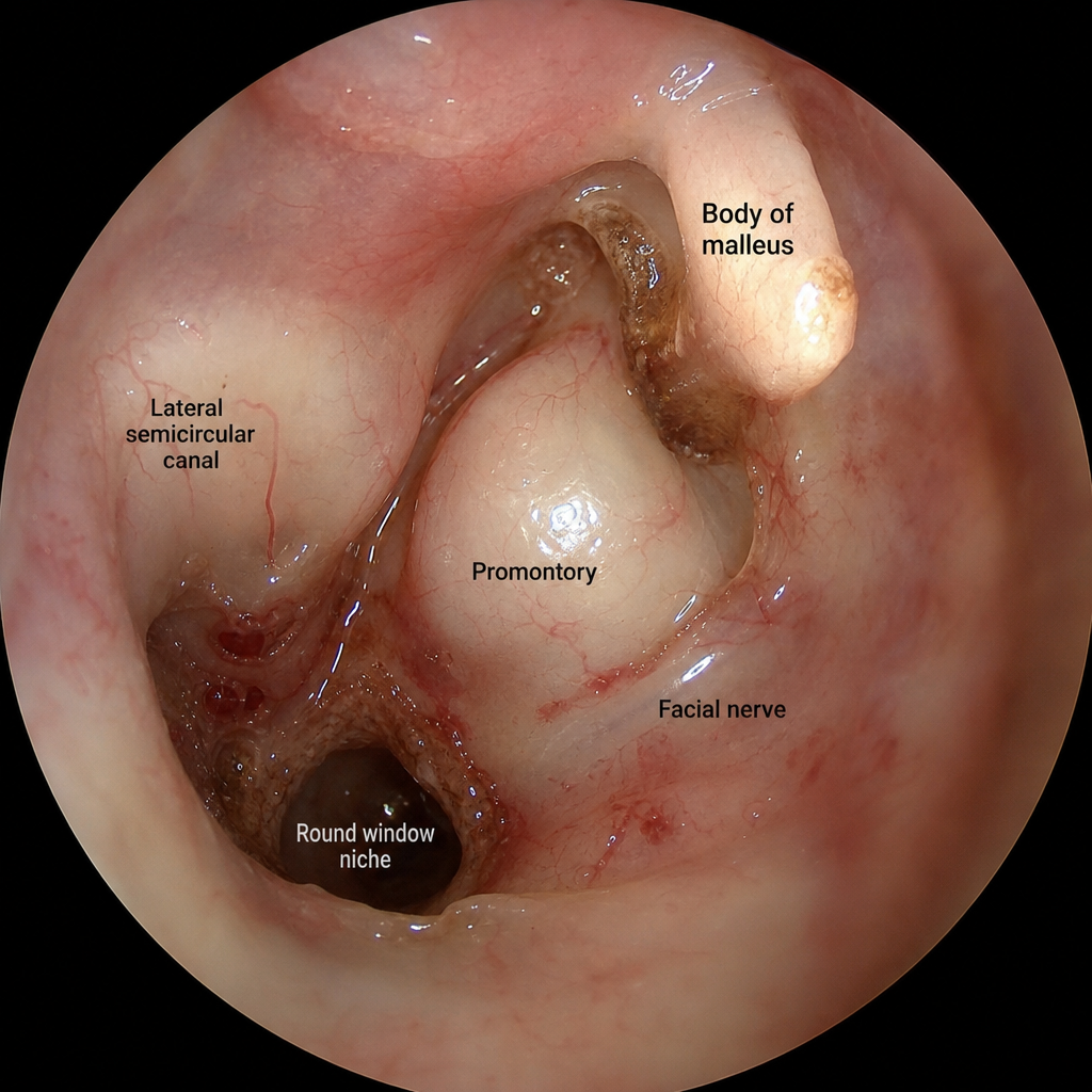
Complications of Otitis Media
Practice Questions
Otosclerosis
Practice Questions
Presbycusis
Practice Questions
Sudden Sensorineural Hearing Loss
Practice Questions
Noise-Induced Hearing Loss
Practice Questions
Ménière's Disease
Practice Questions
Benign Paroxysmal Positional Vertigo
Practice Questions
Vestibular Neuritis
Practice Questions
Tumors of the Ear and Temporal Bone
Practice Questions
Get full access to all questions, explanations, and performance tracking.
Start For Free