
Diseases of the Ear — MCQs

On this page
External auditory canal atresia has been associated with all of the following except?
The pressure difference that can cause atmosphere and middle ear barotrauma is:
Lateral sinus thrombosis is associated with all of the following except:
What is the most common cause of bilateral conductive deafness in a child?
A 35-year-old patient presents with 6 weeks of ear discharge that is not foul-smelling. Which of the following management options is NOT indicated?
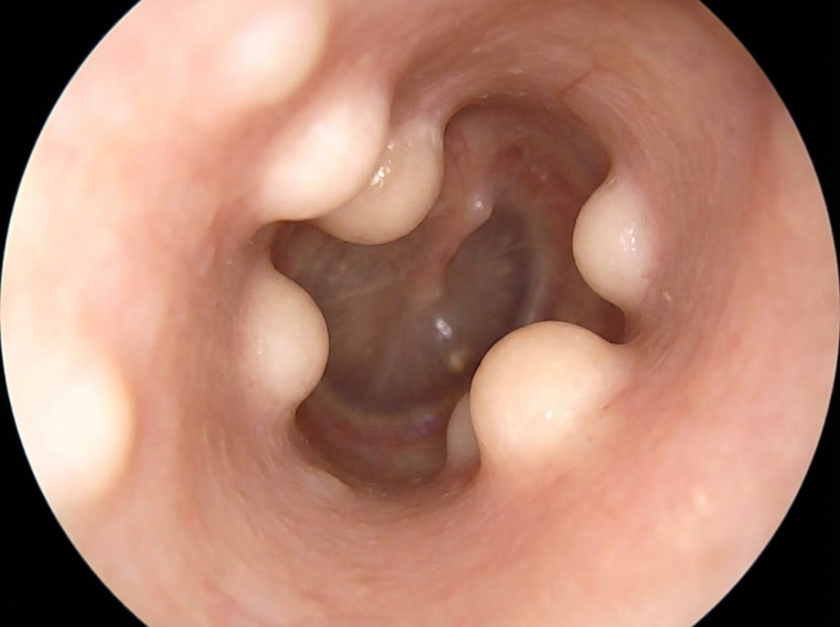
What is the diagnosis?
Ototoxic drugs generally affect the hearing of what frequencies of sound?
A 32-year-old teacher presents complaining of hearing loss in her right ear. Physical examination reveals cerumen completely obstructing the ear canal. Which of the following methods is recommended for ear wax removal?
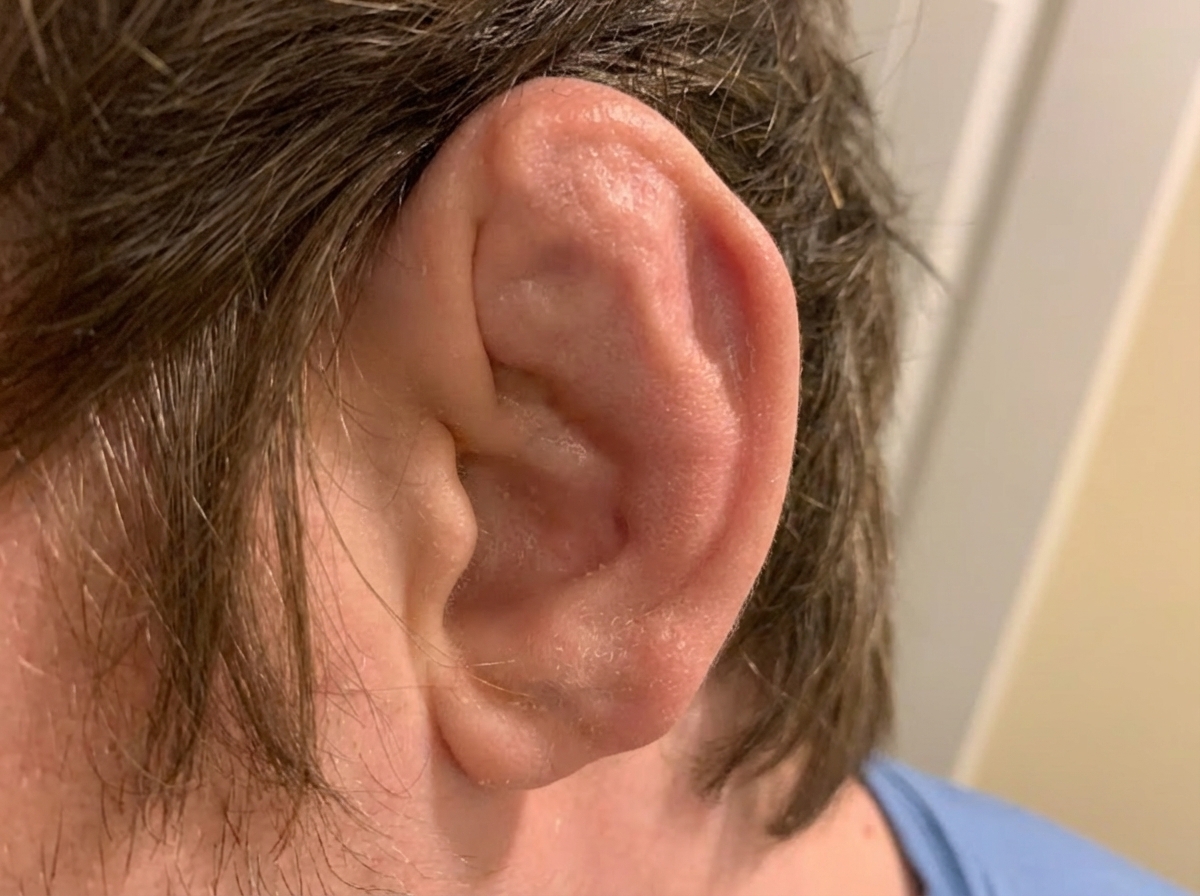
The given appearance of the pinna is suggestive of which of the following conditions?
Conductive hearing loss is seen in all of the following conditions EXCEPT:
Practice by Chapter
Otitis Externa
Practice Questions
Acute Otitis Media
Practice Questions
Chronic Otitis Media
Practice Questions
Complications of Otitis Media
Practice Questions
Otosclerosis
Practice Questions
Presbycusis
Practice Questions
Sudden Sensorineural Hearing Loss
Practice Questions
Noise-Induced Hearing Loss
Practice Questions
Ménière's Disease
Practice Questions
Benign Paroxysmal Positional Vertigo
Practice Questions
Vestibular Neuritis
Practice Questions
Tumors of the Ear and Temporal Bone
Practice Questions
Want unlimited practice?
Get full access to all questions, explanations, and performance tracking.
Scan to download app