
Diseases of the Ear — MCQs

On this page
A patient with CSOM presented to the OPD with seizures. On examination, homonymous hemianopia was present. Identify the condition based on the CT scan given below.
A 5-year-old child presents with sudden severe ear pain and hearing loss. On otoscopy, you observe hemorrhagic bullae on an inflamed tympanic membrane. What is the most likely diagnosis?
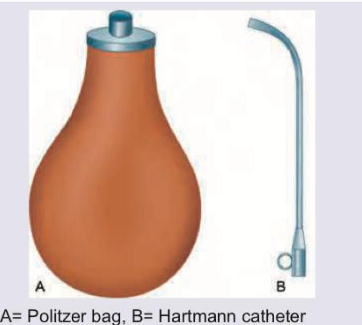
Identify the instruments shown in the image:
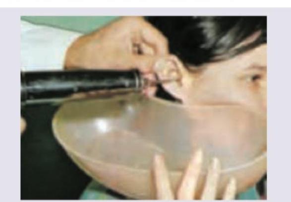
Name the procedure being done:
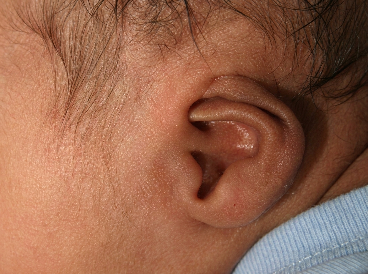
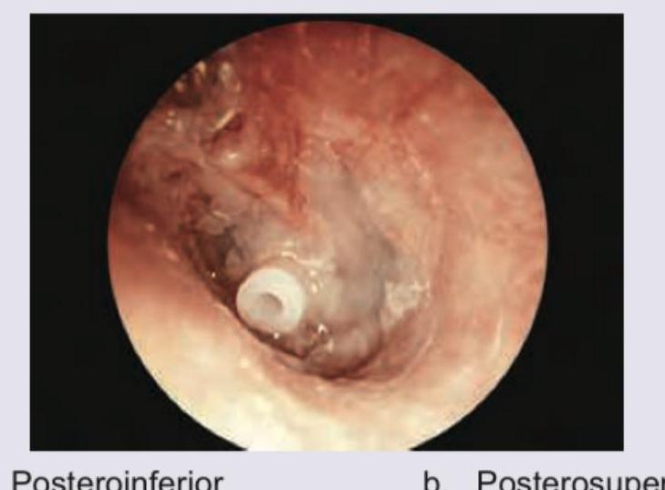
The image shows presence of the following:
Which is correct about the incisions in tympanic membrane?

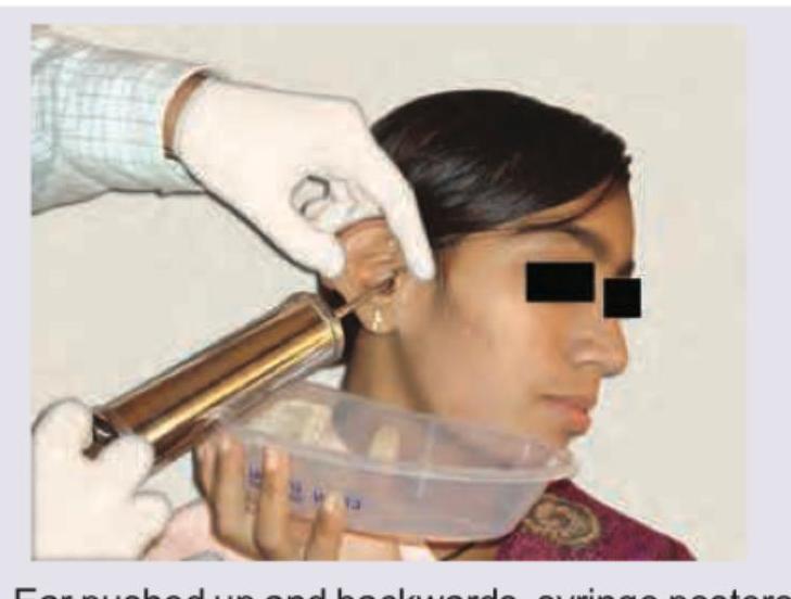
The image shows ear syringing/irrigation being performed in an adult. Which is the correct technique for this procedure?
What is the preferred site of insertion of device shown below?
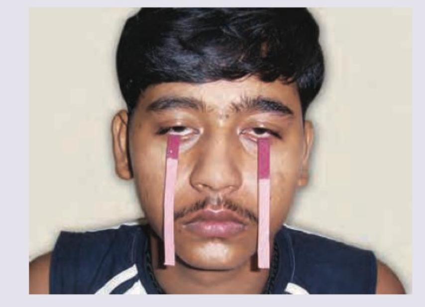
The test shown in the image is used for identifying a lesion in which cranial nerve?
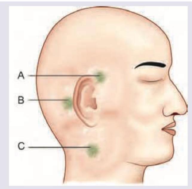
Which is correct about the location of abscess formation secondary to mastoiditis?
Practice by Chapter
Otitis Externa
Practice Questions
Acute Otitis Media
Practice Questions
Chronic Otitis Media
Practice Questions
Complications of Otitis Media
Practice Questions
Otosclerosis
Practice Questions
Presbycusis
Practice Questions
Sudden Sensorineural Hearing Loss
Practice Questions
Noise-Induced Hearing Loss
Practice Questions
Ménière's Disease
Practice Questions
Benign Paroxysmal Positional Vertigo
Practice Questions
Vestibular Neuritis
Practice Questions
Tumors of the Ear and Temporal Bone
Practice Questions
Want unlimited practice?
Get full access to all questions, explanations, and performance tracking.
Scan to download app