Impedance in audiology denotes:
In conductive deafness, to which ear does the Weber test lateralize?
A 63-year-old woman presents with a 5-year history of gradual hearing loss and recent intermittent tinnitus. Physical and neurologic examinations are normal except for sensorineural hearing loss in the left ear and no cranial nerve deficits. An MRI of the brain with gadolinium reveals an extra-axial tumor in the left cerebellopontine angle. What is the most likely diagnosis?
High-frequency audiometry, which uses frequencies higher than those required for speech comprehension, is used to monitor for which of the following conditions?
Which of the following is a subjective test in the assessment of hearing?
Deafness is seen with which of the following conditions?
Type Ad curve is seen in?
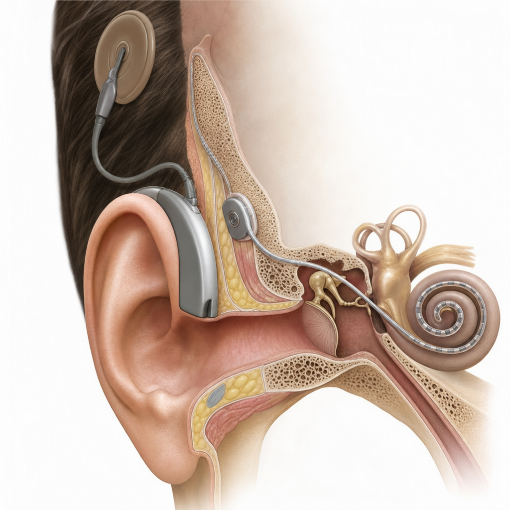
Which of the following methods is used to assess hearing loss in infants?
Carhart's notch in an audiogram represents the deepest frequency loss at which of the following frequencies?
What is the threshold for pain in the ear in decibels?
Explanation: **Explanation:** **Impedance Audiometry** is an objective test used to evaluate the functional status of the middle ear. In physics, impedance refers to the total opposition offered by a system to the flow of energy. In audiology, it specifically measures the resistance of the middle ear to the passage of sound. **Why Option C is correct:** The middle ear is an impedance-matching mechanism. Any pathology affecting the **ossicular chain** (e.g., Otosclerosis causing fixation or Trauma causing ossicular discontinuity) significantly alters the stiffness and mass of the system. Impedance audiometry (specifically tympanometry and acoustic reflex testing) detects these changes in the mobility of the tympanic membrane and ossicles, making it the gold standard for diagnosing middle ear and ossicular disorders. **Why other options are incorrect:** * **Option A (Site of perforation):** While a perforation affects the middle ear, it is diagnosed via otoscopy. In the presence of a perforation, impedance audiometry cannot be performed as a hermetic seal cannot be maintained. * **Option B (Disease of cochlea):** Cochlear diseases represent sensory hearing loss. While the acoustic reflex can indirectly suggest cochlear pathology (e.g., recruitment), impedance primarily measures the mechanical conductive apparatus. * **Option D (Higher function disorder):** These are evaluated using Central Auditory Processing tests or cortical evoked potentials, not by measuring middle ear resistance. **High-Yield Clinical Pearls for NEET-PG:** * **Compliance** is the inverse of impedance (ease of sound flow). * **Tympanogram Types:** * **Type As:** Ossicular fixation (Otosclerosis). * **Type Ad:** Ossicular discontinuity (Monomeric TM). * **Type B:** Fluid in middle ear (Serous Otitis Media). * **Type C:** Eustachian tube dysfunction (Negative pressure). * The **Stapedial Reflex** is absent in Otosclerosis (earliest sign) and Facial nerve palsy proximal to the nerve to stapedius.
Explanation: ### Explanation **Underlying Concept** The Weber test is a tuning fork test (using 512 Hz) that assesses bone conduction. In **conductive hearing loss (CHL)**, the sound lateralizes to the **affected ear (the ear with hearing loss)**. This occurs due to two primary mechanisms: 1. **The Masking Effect:** In a diseased ear with conductive pathology (e.g., wax, otosclerosis, or fluid), ambient room noise cannot reach the cochlea via air conduction. This lack of "background noise" makes the cochlea more sensitive to the bone-conducted vibrations of the tuning fork. 2. **The Occlusion Effect:** The conductive block prevents the escape of sound energy out of the external auditory canal, reflecting it back to the inner ear and increasing the perceived intensity. **Analysis of Options** * **Option A & B:** These are incorrect for CHL. Lateralization to the normal or better-hearing ear occurs in **Sensorineural Hearing Loss (SNHL)**. In SNHL, the cochlea or nerve of the affected ear is damaged; therefore, the sound is perceived only by the functioning cochlea of the better ear. * **Option D:** No lateralization (sound heard in the midline) occurs in individuals with normal hearing or those with symmetrical hearing loss of the same type and degree. **NEET-PG High-Yield Pearls** * **Rinne Test:** In CHL, Rinne is **Negative** (BC > AC). In SNHL, Rinne is **Positive** (AC > BC). * **False Negative Rinne:** Seen in severe unilateral SNHL; the patient perceives sound in the "dead ear" because vibrations travel across the skull to the opposite normal cochlea. * **ABC (Absolute Bone Conduction) Test:** This is the best test to differentiate between SNHL and normal hearing (decreased in SNHL, normal in CHL). * **Tuning Fork Choice:** 512 Hz is preferred because lower frequencies (256 Hz) produce a sense of vibration, and higher frequencies (1024 Hz) decay too quickly.
Explanation: **Explanation:** The clinical presentation of a middle-aged patient with **unilateral progressive sensorineural hearing loss (SNHL)** and tinnitus is a classic "red flag" for an **Acoustic Neuroma** (Vestibular Schwannoma). **1. Why Acoustic Neuroma is correct:** Acoustic neuroma is a benign, slow-growing tumor arising from the Schwann cells of the vestibular nerve (CN VIII). It is the **most common tumor of the cerebellopontine (CP) angle**, accounting for approximately 80-90% of cases. The tumor compresses the cochlear nerve, leading to gradual SNHL and tinnitus. MRI with gadolinium is the gold standard for diagnosis, typically showing an enhancing mass within the internal auditory canal (IAC) or CP angle. **2. Why other options are incorrect:** * **Meningioma:** The second most common CP angle tumor. While it can present similarly, it usually has a broader base along the petrous bone and often shows a "dural tail" on MRI, which is not mentioned here. * **Epidermoid Tumor:** These are congenital lesions that appear "pearly white" and are non-enhancing on MRI (unlike this case, which shows gadolinium enhancement). They typically present with cranial nerve palsies earlier than hearing loss. * **Glioblastoma Multiforme:** This is a highly malignant primary brain tumor (intra-axial) usually found in the cerebral hemispheres, not an extra-axial tumor in the CP angle. **High-Yield Clinical Pearls for NEET-PG:** * **Earliest symptom:** Progressive unilateral SNHL (high frequencies affected first). * **Earliest sign:** Loss of corneal reflex (due to CN V involvement). * **Gold Standard Investigation:** MRI with Gadolinium (shows "ice-cream cone" appearance). * **Bilateral Acoustic Neuromas:** Pathognomonic for **Neurofibromatosis Type 2 (NF2)**. * **Audiometry:** Shows "Retrocochlear" pathology (Poor speech discrimination scores out of proportion to pure tone loss and significant "Roll-over" phenomenon).
Explanation: **Explanation:** **1. Why Ototoxicity is Correct:** High-frequency audiometry (HFA) tests frequencies between **8,000 Hz and 20,000 Hz**, whereas standard pure-tone audiometry only goes up to 8,000 Hz. Ototoxic drugs (such as **Aminoglycosides** like Amikacin/Gentamicin and **Cisplatin**) typically damage the outer hair cells at the **basal turn of the cochlea** first. Since the base of the cochlea is responsible for high-frequency sounds, hearing loss begins in the ultra-high frequencies before progressing to the speech frequencies (500–3,000 Hz). Therefore, HFA is the gold standard for the **early detection** of ototoxicity, allowing clinicians to modify drug therapy before functional hearing loss occurs. **2. Why Other Options are Incorrect:** * **Otosclerosis/Otospongiosis:** These are characterized by stapes fixation, leading to **conductive hearing loss**. The hallmark finding on audiometry is **Carhart’s Notch** (a dip in bone conduction at 2,000 Hz), not high-frequency loss. * **Meniere’s Disease:** This condition typically presents with **fluctuating, low-frequency sensorineural hearing loss** (rising curve on an audiogram) due to endolymphatic hydrops. **3. Clinical Pearls for NEET-PG:** * **Cisplatin** is the most common drug requiring HFA monitoring due to its high incidence of permanent vestibulotoxicity and cochleotoxicity. * **Otoacoustic Emissions (OAEs)** are also used for early screening of ototoxicity as they specifically assess outer hair cell function. * **Presbycusis** (age-related hearing loss) also starts at high frequencies, but HFA’s primary *clinical monitoring* application is for drug toxicity.
Explanation: ### Explanation Hearing tests are broadly classified into two categories: **Subjective** and **Objective**. **1. Why Pure Tone Audiometry (PTA) is the Correct Answer:** Pure Tone Audiometry is a **subjective (behavioral)** test because it relies entirely on the patient's active participation and cooperation. The clinician presents sounds of varying frequencies and intensities, and the patient must signal (e.g., by pressing a button or raising a hand) when they perceive the sound. Because the results depend on the patient's honesty, alertness, and cognitive function, it is prone to human error or malingering. **2. Why the Other Options are Incorrect:** * **Brainstem Evoked Response Audiometry (BERA):** This is an **objective** electrophysiological test. It records electrical activity along the auditory nerve and brainstem in response to sound. It requires no response from the patient (often done under sedation in children). * **Impedance Audiometry (Tympanometry):** This is an **objective** test that measures the compliance of the tympanic membrane and the status of the middle ear. It is performed by changing air pressure in the ear canal; the patient remains passive. * **Otoacoustic Emissions (OAE):** This is an **objective** screening tool that measures the "echo" produced by the outer hair cells of the cochlea. It is used extensively in neonatal hearing screening. **Clinical Pearls for NEET-PG:** * **Gold Standard:** PTA remains the gold standard for describing the degree, type, and configuration of hearing loss in cooperative adults. * **Malingering:** If a patient is suspected of faking hearing loss (Non-organic hearing loss), **objective tests** like BERA or OAE are used to find the true threshold. * **Newborn Screening:** The most common sequence is **OAE** (initial screening) followed by **BERA** (confirmatory test). * **Tuning Fork Tests:** These (Rinne, Weber) are also considered **subjective** tests.
Explanation: ### Explanation **Correct Answer: B. Congenital QT syndrome** The association between deafness and Congenital Long QT Syndrome (LQTS) is a classic high-yield medical correlation. Specifically, **Jervell and Lange-Nielsen syndrome** is an autosomal recessive condition characterized by the combination of: 1. **Congenital sensorineural hearing loss (SNHL):** Caused by mutations in potassium channel genes (*KCNQ1* or *KCNE1*), which are essential for maintaining the endolymph potential in the inner ear. 2. **Prolonged QT interval:** Leading to ventricular arrhythmias (Torsades de Pointes), syncope, and sudden cardiac death. **Analysis of Incorrect Options:** * **A. Essential Hypertension:** While chronic hypertension can lead to microvascular changes in the cochlea over decades, it is not a primary "cause" of deafness in the context of syndromic associations. * **C. Marfan Syndrome:** This is a connective tissue disorder (Fibrillin-1 mutation) primarily affecting the skeletal, ocular (ectopia lentis), and cardiovascular (aortic aneurysm) systems. It is not typically associated with hearing loss. * **D. Turner Syndrome:** While Turner syndrome (45, XO) is associated with an increased risk of middle ear infections (otitis media) and progressive SNHL, it is not the "classic" association for deafness in this specific MCQ set compared to the definitive Jervell and Lange-Nielsen syndrome. **High-Yield Clinical Pearls for NEET-PG:** * **Romano-Ward Syndrome:** The most common Long QT syndrome; it is autosomal dominant and features a normal hearing profile. * **Usher Syndrome:** The most common cause of combined deafness and blindness (Retinitis Pigmentosa). * **Waardenburg Syndrome:** Characterized by SNHL, white forelock, and heterochromia iridis. * **Alport Syndrome:** SNHL associated with progressive renal failure and ocular defects (lenticonus).
Explanation: ### Explanation **Tympanometry** measures the compliance of the tympanic membrane and the ossicular chain. The **Type $A_d$ curve** is a variation of the normal Type A curve, characterized by a normal peak pressure (near 0 daPa) but an **abnormally high compliance** (peak off the chart or very high). The "d" stands for **Discontinuity** or **Deep**. #### Why "After Stapedectomy" is Correct: After a stapedectomy, the natural ossicular chain is interrupted and replaced with a prosthesis. This often results in increased mobility or a "floppy" system compared to the original rigid chain. Similarly, **ossicular discontinuity** (e.g., incudostapedial joint dissociation due to trauma) is the classic cause of a Type $A_d$ curve because the tympanic membrane meets very little resistance from the detached ossicles. #### Why Other Options are Incorrect: * **Eustachian Tube Obstruction:** Leads to negative pressure in the middle ear, resulting in a **Type C curve** (peak shifted to the left/negative side). * **Secretory Otitis Media:** The presence of fluid behind the drum restricts all movement, resulting in a **Type B curve** (flat curve with no peak). * **Middle Ear Tumours:** Masses like Glomus jugulare increase the stiffness and mass of the middle ear, typically leading to a **Type B curve** (if the space is filled) or a reduced-amplitude curve. #### High-Yield Clinical Pearls for NEET-PG: * **Type A:** Normal middle ear function. * **Type $A_s$ (S = Stiffness/Shallow):** Normal pressure but low compliance. Seen in **Otosclerosis** and Tympanosclerosis. * **Type B (Flat):** Seen in **Serous Otitis Media** (normal ear canal volume) or **TM perforation** (large ear canal volume). * **Type C:** Indicates negative middle ear pressure; a precursor to ASOM or seen in Eustachian tube dysfunction.
Explanation: **Explanation:** **Auditory Brainstem Response (ABR/BERA)** is the gold standard for objective hearing assessment in infants. Since infants cannot provide subjective feedback (like raising a hand), clinicians rely on electrophysiological tests. ABR measures the electrical activity of the auditory nerve and brainstem pathways in response to sound stimuli via surface electrodes. It is non-invasive, reliable, and does not require the patient's active participation, making it ideal for neonatal screening and diagnosing permanent childhood hearing loss. **Analysis of Incorrect Options:** * **Rinne’s Test:** This is a subjective tuning fork test used to compare air conduction and bone conduction. It requires the patient to verbally communicate when they stop hearing a sound, which is impossible for infants. * **Short Increment Sensitivity Index (SISI):** This is a subjective psychoacoustic test used to identify cochlear pathology (recruitment). It requires the patient to detect small 1dB increases in intensity, a task far too complex for an infant. * **Caloric Test:** This is a vestibular function test (part of the Fitzgerald-Hallpike maneuver) used to evaluate the lateral semicircular canal. It assesses balance, not hearing. **High-Yield Clinical Pearls for NEET-PG:** * **Universal Neonatal Hearing Screening (UNHS):** Usually follows a two-step protocol: **OAE** (Otoacoustic Emissions) for initial screening, followed by **ABR** for confirmation. * **ABR Waves:** Wave V is the most robust wave and is used to determine the hearing threshold. * **ASSR (Auditory Steady-State Response):** Another objective test used to provide frequency-specific audiograms in infants, often used alongside ABR. * **Behavioral Observation Audiometry (BOA):** Can be used for infants (0-6 months) but is highly subjective and less reliable than ABR.
Explanation: **Explanation:** **Carhart’s Notch** is a classic audiometric finding pathognomonic for **Otosclerosis**. It is characterized by a mechanical (pseudo) sensorineural hearing loss observed in Bone Conduction (BC) thresholds, despite the inner ear being functionally intact. 1. **Why 2 KHz is Correct:** The notch is most prominent at **2 KHz** because this frequency corresponds to the natural resonant frequency of the ossicular chain. In Otosclerosis, stapes fixation disrupts this normal resonance and reduces the efficiency of the inertial component of bone conduction. This results in an apparent dip in the BC curve specifically at 2 KHz. Importantly, this notch disappears after a successful stapedotomy, proving it is a mechanical artifact rather than true nerve damage. 2. **Why Incorrect Options are Wrong:** * **0.5 KHz (A):** Low frequencies are typically affected by the "Air-Bone Gap" in early Otosclerosis (conductive loss), but the specific BC dip does not occur here. * **4 KHz (C):** A dip at 4 KHz is characteristic of **Noise-Induced Hearing Loss (NIHL)**, known as the "Acoustic Notch" or Boiler-maker's notch. * **8 KHz (D):** High-frequency loss at 8 KHz is usually associated with presbycusis or ototoxicity, not mechanical ossicular fixation. **High-Yield Clinical Pearls for NEET-PG:** * **Schwartz Sign:** A flamingo-pink flush on the promontory seen through the TM (indicates active otosclerosis). * **Gelle’s Test:** Negative in Otosclerosis (no change in hearing with increased ear canal pressure). * **Tympanometry:** Typically shows an **As type** curve (stiffened system). * **Stapedial Reflex:** Usually absent or shows an "on-off" effect in early stages.
Explanation: **Explanation:** The human ear is capable of perceiving a vast range of sound intensities. The **Threshold of Pain** is defined as the intensity level at which sound ceases to be just a perception of loudness and begins to cause physical pain or discomfort in the ear. In clinical audiology, this is consistently established at **140 dB**. At this level, the mechanical energy of the sound waves is sufficient to cause significant stress on the tympanic membrane and the ossicular chain, potentially leading to acoustic trauma. **Analysis of Options:** * **140 dB (Correct):** This is the standard physiological limit for human hearing endurance. Sounds at or above this level can cause immediate, permanent damage to the cochlear hair cells. * **120 dB (Incorrect):** This is known as the **Threshold of Discomfort** (or the "tickle" sensation). While very loud (e.g., a thunderclap or a nearby chainsaw), it is generally the upper limit of the "uncomfortable" range rather than the "pain" range. * **80 dB (Incorrect):** This represents the level of loud noise (e.g., heavy city traffic). Prolonged exposure (8 hours+) to sounds above **85 dB** is considered the starting point for noise-induced hearing loss (NIHL), but it is not painful. * **160 dB (Incorrect):** This level is well beyond the pain threshold and usually results in immediate physical rupture of the tympanic membrane (e.g., a gunshot or explosion in close proximity). **Clinical Pearls for NEET-PG:** * **Reference Level:** 0 dB is the threshold of hearing (not the absence of sound). * **Normal Conversation:** Typically occurs at **60 dB**. * **Safety Standard:** OSHA recommends hearing protection for occupational exposure exceeding **85-90 dB**. * **Logarithmic Scale:** Remember that the decibel scale is logarithmic; an increase of 10 dB represents a 10-fold increase in sound intensity.
Hearing Assessment Techniques
Practice Questions
Tympanometry and Acoustic Reflexes
Practice Questions
Otoacoustic Emissions
Practice Questions
Auditory Brainstem Response
Practice Questions
Hearing Aids
Practice Questions
Cochlear Implants
Practice Questions
Bone-Anchored Hearing Devices
Practice Questions
Speech and Language Development
Practice Questions
Articulation Disorders
Practice Questions
Stuttering
Practice Questions
Dysphonia
Practice Questions
Rehabilitation of Hearing-Impaired Children
Practice Questions
Get full access to all questions, explanations, and performance tracking.
Start For Free