A 65-year-old patient presents with gradual progressive hearing loss over several years. Rinne test shows bone conduction > air conduction bilaterally. Weber test lateralizes to the worse ear. Tympanic membranes are intact and normal. Which is the most likely diagnosis?
Most common bone affected by otosclerosis?
A 45-year-old gentleman reports decreased hearing in the right ear for the last two years. On testing with a 512 Hz tuning fork, the Rinne's test without masking is negative on the right ear and positive on the left ear. With the Weber's test, the tone is perceived as louder in the left ear. Patient most likely has -
Which anatomical structure serves as the passageway connecting the middle ear cavity to the mastoid air cells, allowing the spread of infection?
Which of the following statements about Bone Anchored Hearing Aids (BAHA) is true?
What type of prosthesis is commonly used during stapes surgery for otosclerosis?
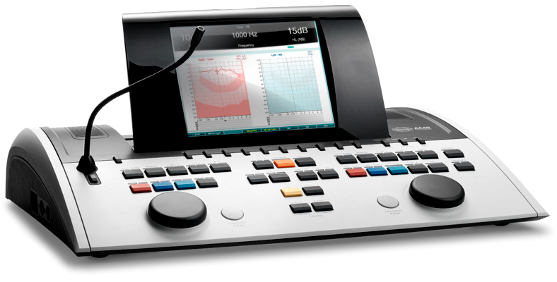
Which of the following is not a function of the instrument given below?

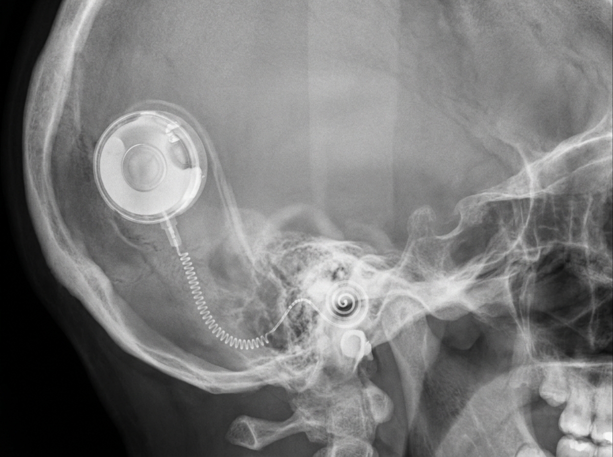
Which device is depicted below?

What should be the minimum hearing loss for Weber's test to lateralize?
A pure tone audiogram showing a bone conduction dip (Carhart notch) at 2000 Hz is characteristic of-
Explanation: ***Otosclerosis*** - **Otosclerosis** is characterized by abnormal bone remodeling in the otic capsule, leading to **stapes fixation** at the oval window - Classic presentation: **progressive bilateral conductive hearing loss** in adults (typically 20-40 years) - **Rinne negative** (bone conduction > air conduction) and **Weber lateralizes to worse ear** confirm conductive hearing loss - **Carhart notch** (bone conduction dip at 2000 Hz) is pathognomonic on audiometry - Tympanic membrane appears **normal** - key differentiating feature *Cerumen impaction* - Would cause conductive hearing loss but presents with **visible earwax obstruction** on otoscopy - Typically **acute onset**, not gradual progressive loss over years - Easily identified and removed during examination - Does not match the clinical picture of intact, normal tympanic membranes with chronic progressive bilateral loss *Acoustic neuroma* - Causes **sensorineural hearing loss**, not conductive - **Rinne would be positive** (air conduction > bone conduction) in both ears - Typically **unilateral** hearing loss with associated tinnitus and possible vestibular symptoms - Weber lateralizes to the **better ear** in sensorineural loss *Cholesteatoma* - Causes conductive hearing loss but presents with **abnormal tympanic membrane** findings - Typically shows **whitish mass visible behind or within the tympanic membrane** - Often associated with **chronic ear discharge** and history of chronic otitis media - Usually **unilateral** presentation - Does not match the description of intact, normal tympanic membranes bilaterally
Explanation: ***Stapes*** - **Otosclerosis** is characterized by abnormal bone remodeling in the otic capsule, primarily affecting the **stapes footplate**. - This leads to its fixation in the oval window, causing **conductive hearing loss**. - The **stapediovestibular joint** is the most common site, occurring in over 90% of cases. *Bony labyrinth* - While otosclerosis originates in the **otic capsule** (which forms the bony labyrinth), the term refers to a broader anatomical structure. - The specific site of clinical significance is the **stapes footplate**, not the labyrinth as a whole. *Mastoid process* - The **mastoid process** is part of the temporal bone but is structurally distinct from the middle ear and otic capsule. - It is not involved in otosclerosis pathology. *Incus* - While the **incus** is a middle ear ossicle, it is rarely affected by otosclerosis. - The disease process specifically targets the **stapes footplate** at the oval window, not other ossicles.
Explanation: ***Right conductive hearing loss*** - A **negative Rinne's test** (bone conduction louder than air conduction) in the right ear indicates **conductive hearing loss** on that side - However, in true conductive hearing loss, **Weber should lateralize to the affected (right) ear** due to the occlusion effect, not to the left ear - The Weber lateralizing to the left ear with a negative Rinne on the right suggests this may be a **false negative Rinne** due to lack of masking, where sound crosses over to the better left ear - This combination is atypical for pure conductive loss and requires repeat testing with proper masking *Right sensorineural hearing loss* - In **sensorineural hearing loss**, Rinne's test should be **positive** (air conduction > bone conduction) on both sides, though both may be reduced on the affected side - **Weber lateralizes to the unaffected (left) ear**, which matches the given finding - The **negative Rinne on the right ear without masking** is likely a **false negative** due to sound crossing over to the better left ear during bone conduction testing - This is the **most consistent interpretation** when Rinne testing is done without masking, but traditionally the question frame suggests conductive loss *Left sensorineural hearing loss* - Would show **positive Rinne bilaterally** with reduced hearing on the left - **Weber would lateralize to the right ear** (the better ear), contradicting the given findings - This option is clearly inconsistent with the clinical findings *Left conductive hearing loss* - Would show **negative Rinne on the left** and positive on the right - Weber would lateralize to the left ear (affected side in conductive loss) - The **Rinne findings contradict this**, as the right ear shows negative Rinne, not the left
Explanation: ***Aditus ad antrum*** - The **aditus ad antrum** is the direct anatomical connection between the **epitympanic recess** (attic) of the middle ear and the **mastoid antrum**. - This passageway allows the spread of infection from the middle ear cavity into the **mastoid air cells**, leading to conditions like **mastoiditis**. *Cochlea (hearing organ)* - The **cochlea** is an inner ear structure primarily involved in **hearing** and is not a direct route for bacterial entry from the middle ear to the mastoid. - Infections would need to breach the **oval** or **round window** to reach the cochlea, which is a rare pathway for mastoid involvement. *Internal acoustic meatus (nerve passage)* - The **internal acoustic meatus** is a bony canal that transmits the **facial nerve** (CN VII) and **vestibulocochlear nerve** (CN VIII) to the inner ear and brainstem. - It does not directly communicate with the middle ear cavity or the mastoid air cells, making it an unlikely route for typical middle ear infections to spread to the mastoid. *Eustachian tube* - The **Eustachian tube** connects the **nasopharynx** to the **middle ear**, primarily equalizing pressure and draining secretions from the middle ear [1]. - While it can be a route for bacteria to enter the middle ear, it does not directly connect the middle ear to the mastoid air cells.
Explanation: ***It is useful in patients with canal atresia and microtia.*** - BAHA operates by **bone conduction**, bypassing the external auditory canal and middle ear structures. - This makes it an ideal solution for conductive hearing loss caused by malformations like **canal atresia** and **microtia**, where air conduction is impaired. - BAHA is particularly effective when the cochlea is functional but sound cannot reach it via normal air conduction pathways. *It transmits sound through air conduction, making it ideal for sensorineural hearing loss.* - This statement is incorrect on both counts. - BAHA uses **bone conduction**, not air conduction. - BAHA is primarily indicated for **conductive or mixed hearing loss**, not sensorineural hearing loss, as it requires a functional cochlea. *It is used after surgery for acoustic neuroma in neurofibromatosis type 2.* - After acoustic neuroma surgery, especially in Neurofibromatosis type 2, the primary concern is often **unilateral sensorineural hearing loss** or **single-sided deafness**. - While BAHA can be used for single-sided deafness, the first-line option for single-sided deafness is **Cochlear Implants (CIs)** or contralateral routing of signal (CROS) hearing aids. *It is beneficial for patients with bilateral severe sensorineural hearing loss.* - For **bilateral severe sensorineural hearing loss**, the primary intervention is typically **cochlear implantation**, not BAHA. - BAHA is less effective for severe sensorineural loss because it still relies on a functional cochlea to convert bone-conducted vibrations into neural signals.
Explanation: ***Teflon piston*** - A **Teflon piston** is a common and effective prosthesis for replacing the **stapes** in **otosclerosis** surgery. - It connects the **incus** to the **oval window**, restoring sound conduction. - Other materials like **titanium** are also commonly used for stapes prostheses. *Ventilation tube* - A **ventilation tube** (grommet) is typically used for **middle ear effusions** or **eustachian tube dysfunction**, not for ossicular chain reconstruction in otosclerosis. - It creates an opening in the **tympanic membrane** to equalize pressure, which is unrelated to the stapes' function. *Total ossicular replacement prosthesis (TORP)* - A **TORP** is used when the entire **ossicular chain** (malleus, incus, and stapes) is significantly damaged or missing, not specifically for isolated stapes fixation from otosclerosis. - It bypasses the intact portions of the ossicular chain and connects directly to the **tympanic membrane** and **oval window**. *Partial ossicular replacement prosthesis (PORP)* - A **PORP** is used when the **stapes superstructure is intact** but the incus is damaged or absent. - In otosclerosis, the stapes **footplate is fixed**, requiring removal and replacement with a piston prosthesis, not a PORP. - PORP connects the tympanic membrane (or malleus) to the stapes head, which is different from stapes surgery.
Explanation: ***To measure the threshold of hearing by bone conduction only*** - This instrument, an **audiometer**, measures hearing thresholds for both **air conduction and bone conduction**, not exclusively bone conduction. - Measurement of both pathways is crucial to differentiate between **conductive, sensorineural, and mixed hearing loss**. *To predict speech reception threshold* - An audiometer can generate pure tones and speech stimuli, which are essential for determining the **Speech Reception Threshold (SRT)**. - SRT is a key measure in audiology to predict a person's ability to **understand speech**. *To find degree of handicap* - While not directly measuring a "handicap," the **audiogram** produced by this instrument, along with other audiometric tests, helps in assessing the **functional impact of hearing loss**. - This information contributes to understanding the overall **degree of impairment** and guiding rehabilitation. *Records can be kept for future reference* - Modern audiometers like the one pictured typically have internal memory or can be connected to computers to **store and retrieve patient data**. - This feature is vital for **monitoring hearing changes over time**, evaluating treatment efficacy, and legal or insurance purposes.
Explanation: ***Cochlear implant*** - A cochlear implant is an **electronic medical device that replaces the function of a damaged inner ear (cochlea)** and provides sound signals directly to the brain - On imaging (X-ray, CT, or skull radiograph), it appears as a **characteristic circular receiver-stimulator device** under the skin behind the ear with an **electrode array extending into the cochlea** - The **internal receiver has a distinctive appearance** with visible magnets and electrode contacts, making it easily identifiable on radiographic images - Used for patients with **severe to profound sensorineural hearing loss** who do not benefit from conventional hearing aids *Auditory brainstem implant (ABI)* - An ABI **bypasses both the cochlea and auditory nerve**, directly stimulating the **cochlear nucleus in the brainstem** - On imaging, the electrode array would be located at the **cerebellopontine angle** near the brainstem, not in the cochlea - Reserved for patients with **absent or non-functional auditory nerves** (e.g., bilateral vestibular schwannomas, neurofibromatosis type 2) *Bone anchored hearing aid (BAHA)* - A BAHA consists of a **titanium implant osseointegrated into the skull bone** behind the ear with an external sound processor - On X-ray, only the **small titanium fixture/abutment** would be visible in the mastoid bone, without any cochlear or intracranial components - Works by **bone conduction**, transmitting sound vibrations directly to the inner ear, bypassing the outer and middle ear - Used for **conductive hearing loss, mixed hearing loss, or single-sided deafness** *Hearing aid* - A conventional hearing aid is a **completely external electronic device** that amplifies sound - It would **not be visible on X-ray or CT imaging** as it contains no implanted components - Simply amplifies sound for individuals with mild to moderate hearing loss
Explanation: ***Correct Option: 5 dB*** - The **minimum hearing loss for Weber's test to lateralize is typically 5 dB**, though some sources cite 5-10 dB - Weber test involves placing a vibrating tuning fork (typically 512 Hz) on the midline of the skull (forehead or vertex) - In a patient with **unilateral conductive hearing loss**, the sound lateralizes to the affected (poorer) ear - In a patient with **unilateral sensorineural hearing loss**, the sound lateralizes to the better ear - A difference of **5 dB between the two ears** is generally sufficient for the patient to perceive lateralization *Incorrect Option: 10 dB* - While 10 dB is sometimes cited in clinical practice as a threshold for **consistent and reliable** lateralization - The actual **minimum detectable difference** for lateralization is lower, around 5 dB - 10 dB represents a more robust clinical difference but not the minimum *Incorrect Option: 15 dB* - 15 dB represents a **moderate hearing asymmetry** that would definitely cause clear lateralization - However, this exceeds the minimum threshold required for lateralization to occur - Lateralization can be detected at much smaller differences (5 dB) *Incorrect Option: 20 dB* - 20 dB represents a **significant hearing difference** between ears - While this would certainly cause obvious lateralization, it is **well above the minimum threshold** - The minimum for lateralization detection is much lower (5 dB)
Explanation: ***Otosclerosis*** - A **Carhart notch** is a characteristic feature on a pure tone audiogram in otosclerosis, specifically a **bone conduction dip at 2000 Hz**. - This notch is believed to be an **artifact of cochlear mechanics** caused by the fixation of the stapes in the oval window. *Presbyacusis* - Characterized by a **bilateral, symmetrical, high-frequency sensorineural hearing loss** that gradually worsens with age. - It does not present with a specific bone conduction dip like the Carhart notch. *Ototoxicity* - Hearing loss induced by ototoxic drugs (e.g., aminoglycosides, loop diuretics) typically causes **bilateral, progressive, high-frequency sensorineural hearing loss**. - A Carhart notch is not a typical finding in ototoxicity. *Noise induced hearing loss* - Often presents with a **sensorineural hearing loss notch at 4000 Hz** (or sometimes 3000 Hz or 6000 Hz) on the audiogram. - This pattern is distinct from the 2000 Hz bone conduction dip seen in a Carhart notch.
Get full access to all questions, explanations, and performance tracking.
Start For Free