AIDS, secondary infection will be all except
A 65-year-old patient presents with symptoms of bone pain, anemia, hypercalcemia, and renal impairment. A bone marrow biopsy confirms the diagnosis of multiple myeloma. The patient is started on a treatment regimen. Which of the following treatments is most likely associated with the reactivation of herpes zoster?
What could be the most appropriate provisional diagnosis for multiple nodular exophytic reddish lesions of oral mucosa in an AIDS patient?
Which of the following statements about molluscum contagiosum is FALSE?
A patient on steroids develops sudden onset painful vesicles in T4 dermatome. Best initial treatment is:
A 19-year-old woman presents with painful genital ulcers and vesicles for 4 days, accompanied by fever, malaise, and tender inguinal lymphadenopathy. What is the most appropriate initial management?
In which condition are Bull's eye lesions typically found?
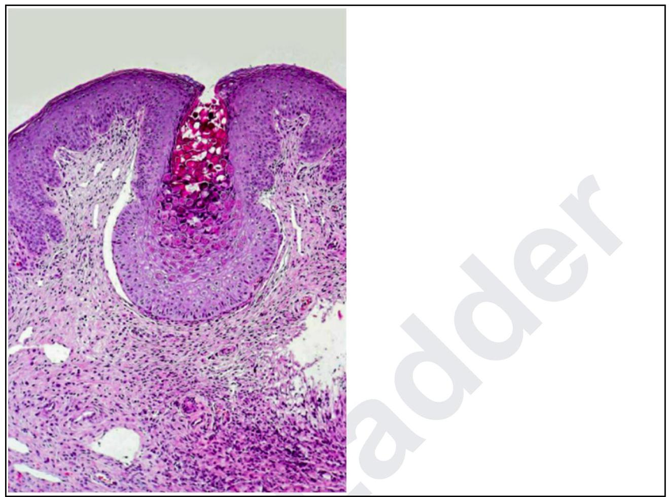
A woman presents with lesions on the inner thighs and peri-anal region. They are nodular, 4-6 mm in size and appear pale. The histopathological image shows multiple intracytoplasmic inclusion bodies consistent with Henderson-Patterson bodies. The diagnosis is:

In HIV patients, Kaposi's sarcoma is most likely caused by which of the following?
Neonatal conjunctivitis is caused by all of the following except:
Explanation: ***Kaposi's sarcoma*** - Kaposi's sarcoma is a **cancer** caused by human herpesvirus 8 (HHV-8) [2] that is common in patients with AIDS, but it is a **malignancy**, not a secondary infection [2],[3]. - While it arises due to immune suppression, it represents abnormal cell proliferation rather than direct microbial invasion. *Candida* - **Candidiasis** (e.g., oral thrush, esophageal candidiasis) is a common opportunistic fungal infection in AIDS patients due to their **impaired cellular immunity** [1]. - It often presents as **white plaques** on mucous membranes and is a clear example of a secondary infection. *HSV* - **Herpes Simplex Virus (HSV)** infections, including oral and genital herpes, are common and often severe in AIDS patients. - Due to immunocompromise, these infections can be **more widespread**, chronic, or recur frequently, qualifying as secondary infections. *Rubella* - **Rubella (German measles)** is a viral infection that is generally mild and self-limiting in immunocompetent individuals. - It is **not considered an opportunistic infection** or a common secondary infection specifically associated with AIDS; rather, it is listed as a differential diagnosis for the primary HIV infection rash [1].
Explanation: ***Bortezomib*** - **Bortezomib**, a **proteasome inhibitor**, is known to increase the risk of herpes zoster reactivation in patients with multiple myeloma due to its immunosuppressive effects. - Prophylaxis with antiviral agents (e.g., acyclovir) is often recommended during bortezomib treatment to prevent this complication. - Studies show herpes zoster incidence of 10-15% in bortezomib-treated patients without prophylaxis. *Lenalidomide* - While lenalidomide is an **immunomodulatory drug** used in multiple myeloma, it is generally associated with a lower risk of herpes zoster reactivation compared to proteasome inhibitors. - It primarily acts by inhibiting angiogenesis and stimulating T-cell and natural killer cell activity. *Daratumumab* - **Daratumumab** is a **monoclonal antibody** targeting CD38 on myeloma cells, leading to their destruction. - Although it has immunosuppressive effects, it is less commonly associated with herpes zoster reactivation than bortezomib. *Melphalan* - **Melphalan** is an **alkylating agent** used in chemotherapy for multiple myeloma, particularly in conditioning regimens for stem cell transplantation. - While it causes myelosuppression and general immunosuppression, the direct association with herpes zoster reactivation is not as prominent or specific as with bortezomib. *Dexamethasone* - **Dexamethasone** is a **corticosteroid** commonly used in combination regimens for multiple myeloma (e.g., RVD, VCD). - While corticosteroids cause immunosuppression and can increase infection risk, the specific association with herpes zoster reactivation is less pronounced than with bortezomib.
Explanation: ***Kaposi's sarcoma*** - **Kaposi's sarcoma (KS)** is a common malignancy in AIDS patients, often presenting with **reddish-purple to brown nodular lesions** on the skin and **mucous membranes**, including the oral cavity. - The description of **multiple nodular exophytic reddish lesions** of the oral mucosa in an AIDS patient is highly suggestive of KS. *Acute pseudo-membranous candidiasis* - This typically presents as **white, removable pseudomembranes** that, when scraped off, reveal an erythematous or bleeding surface. - It does not present as **reddish nodular exophytic lesions**. *Hemangioma* - While hemangiomas are benign vascular lesions that can be reddish, they are typically **solitary or develop earlier in life** and are not specifically associated with HIV/AIDS in this widespread, nodular form. - The presence of **multiple, exophytic nodular lesions** in an immunocompromised patient points to a more aggressive or opportunistic pathology. *Focal epithelial hyperplasia* - Also known as **Heck's disease**, this presents as **multiple, soft, flattened or rounded papules** that are usually the color of the normal mucosa or slightly paler. - It is caused by certain strains of **human papillomavirus (HPV)** and is not typically reddish or exophytic in the manner described.
Explanation: ***Laboratory confirmation is required for diagnosis*** - The diagnosis of **molluscum contagiosum** is primarily **clinical**, based on the characteristic appearance of the lesions (small, flesh-colored, dome-shaped papules with central umbilication). - While histology can confirm the diagnosis by revealing **molluscum bodies**, it is **not routinely required** for typical cases. *Lesions contain characteristic inclusion bodies* - This statement is **true**. Histological examination of molluscum contagiosum lesions reveals large, eosinophilic cytoplasmic inclusions, known as **molluscum bodies** or **Henderson-Paterson bodies**, within infected epidermal cells. - These inclusion bodies contain viral particles and are a **hallmark of the infection**. *Autoinoculation can spread the infection to new sites* - This statement is **true**. Molluscum contagiosum is highly contagious, and scratching or touching existing lesions can lead to the spread of the virus to previously unaffected skin areas on the same individual. - This process of **autoinoculation** explains why lesions often appear in clusters or linear arrays (Koebner phenomenon). *It is caused by a poxvirus* - This statement is **true**. Molluscum contagiosum is caused by the **molluscum contagiosum virus (MCV)**, which belongs to the **Poxviridae family**. - Poxviruses are known for their relatively large size and the ability to replicate entirely in the cytoplasm of host cells.
Explanation: ***IV Acyclovir*** - Patients on **steroids** are considered **immunocompromised**, and a sudden onset of painful vesicles in a dermatomal distribution strongly suggests **herpes zoster (shingles)** [1]. - In immunocompromised patients, **intravenous acyclovir** is the preferred initial treatment due to better bioavailability and more rapid systemic drug levels, helping to prevent complications like **postherpetic neuralgia** or disseminated disease [1]. *Oral Acyclovir* - While oral acyclovir is used for herpes zoster, it is generally less effective in **immunocompromised patients** due to lower bioavailability compared to IV administration. - The slower onset of action and lower peak plasma concentrations may not be sufficient to control the viral infection rapidly in this high-risk group. *Oral Valacyclovir* - **Valacyclovir** is a prodrug of acyclovir with improved oral bioavailability, making it a good option for immunocompetent patients with herpes zoster. - However, for **immunocompromised patients**, particularly those on steroids, **IV acyclovir** is still superior due to the need for rapid and high systemic drug levels to prevent severe complications [1]. *Topical Acyclovir* - **Topical acyclovir** is primarily used for **herpes simplex labialis (cold sores)** and has very limited efficacy for systemic viral infections like **herpes zoster**. - It does not achieve adequate systemic concentrations to treat the underlying viral replication or prevent complications in dermatomal zoster, especially in an immunocompromised individual.
Explanation: Acyclovir 400mg orally TID for 7-10 days - The patient's presentation with painful genital ulcers and vesicles, fever, malaise, and tender inguinal lymphadenopathy is highly suggestive of primary herpes simplex virus (HSV) infection [1]. - Acyclovir is an antiviral medication that effectively reduces the duration and severity of symptoms in primary HSV outbreaks [1]. Azithromycin 1g orally as single dose - Azithromycin is primarily used to treat bacterial infections, particularly chlamydia and gonorrhea, which typically present with urethritis or cervicitis, not painful vesicles. - It is ineffective against viral infections such as HSV. Benzathine penicillin G 2.4 million units IM - Benzathine penicillin G is the treatment of choice for syphilis, which causes a painless chancre in its primary stage, not painful vesicles. - This antibiotic has no efficacy against HSV. Doxycycline 100mg orally BID for 14 days - Doxycycline is an antibiotic used for various bacterial infections, including chlamydia, lymphogranuloma venereum, and granuloma inguinale [1]. - These conditions typically present with different clinical features (e.g., painless ulcers, buboes) and not the vesicular rash seen in HSV.
Explanation: ***Erythema multiforme*** - **Bull's eye lesions**, also known as **target lesions**, are the hallmark clinical finding in erythema multiforme. - These lesions feature a central dusky or blistered area, surrounded by a pale edematous ring, and an outermost erythematous halo. *Erythema nodosum* - Characterized by tender, red, subcutaneous nodules, typically located on the **shins**. - It represents a form of **panniculitis** (inflammation of subcutaneous fat) and does not exhibit targetoid morphology. *Erythema gangrenosum* - A specific skin lesion associated with **Pseudomonas aeruginosa** septicemia, characterized by hemorrhagic bullae that progress to necrotic ulcers with a black eschar. - It represents **necrotizing vasculitis** and does not present with bull's eye or target lesions. *Erythroderma* - Defined as generalized redness and scaling affecting more than **90% of the body surface area**. - This condition involves widespread inflammation of the skin and does not present with discrete target lesions.
Explanation: ***Molluscum contagiosum*** - The clinical presentation of **nodular, pale lesions** in the inner thighs and peri-anal region, combined with histopathology showing **Henderson-Patterson bodies** (large eosinophilic intracytoplasmic inclusions), is pathognomonic for molluscum contagiosum. - Henderson-Patterson bodies represent viral factories within infected keratinocytes, a key diagnostic feature of this **poxvirus** infection. *Trichodysplasia spinulosa* - This condition is characterized by **follicular papules** with prominent keratin spines, typically occurring on the face, and is associated with the **Trichodysplasia spinulosa-associated polyomavirus**. - It does not present with Henderson-Patterson bodies or the typical nodular lesions seen in molluscum contagiosum. *Condyloma acuminata* - These are **genital warts** caused by the **human papillomavirus (HPV)**, presenting as verrucous or cauliflower-like growths. - Histologically, they show **koilocytic atypia** (vacuolated cells with nuclear abnormalities), not Henderson-Patterson bodies. *Donovanosis* - Also known as **granuloma inguinale**, this is a chronic, progressive, ulcerative granulomatous disease caused by **Klebsiella granulomatis**. - Histopathology reveals **Donovan bodies** (intracellular bacteria within macrophages), which are distinct from Henderson-Patterson bodies and the clinical presentation of nodular lesions.
Explanation: ***Virus*** - The image likely depicts **Kaposi's sarcoma**, a common lesion in HIV patients, which is caused by **Human Herpesvirus 8 (HHV-8)**. - Other viral infections like **Herpes Simplex Virus (HSV)** can also cause mucocutaneous lesions in immunocompromised individuals. *Bacteria* - While HIV patients are susceptible to bacterial infections (e.g., **Staphylococcus aureus** causing skin abscesses), the described lesion type is not characteristic of common bacterial skin infections. - Bacterial lesions often present as pustules, cellulitis, or ulcers with purulent discharge, which differ from typical Kaposi's sarcoma. *Parasite* - Parasitic infections can occur in HIV patients (e.g., **scabies** or **leishmaniasis**), but these typically present with different dermatological features like intensely itchy papules or nodular ulcerative lesions. - Lesions caused by parasites do not usually manifest as the violaceous, nodular, or plaque-like appearances seen in Kaposi's sarcoma. *Fungus* - Fungal infections in HIV patients can cause skin lesions (e.g., **candidiasis** with oral thrush or esophagitis, or **cryptococcosis** with molluscum-like lesions). - However, the morphology of these fungal lesions generally differs from the classic appearance of Kaposi's sarcoma or other common viral lesions in HIV.
Explanation: ***Aspergillus*** - **Fungal infections** of the eye, particularly by *Aspergillus*, are extremely rare in neonates and typically present as **keratitis** rather than conjunctivitis. - While *Aspergillus* can cause severe infections in immunocompromised individuals, it is not a common cause of neonatal conjunctivitis. *Gonococcus* - **_Neisseria gonorrhoeae_** is a well-known cause of **ophthalmia neonatorum** (gonococcal conjunctivitis), presenting as severe, purulent discharge usually within the first 2-5 days of life. - This infection can lead to **corneal ulceration** and blindness if untreated. *Chlamydia* - **_Chlamydia trachomatis_** is the most common bacterial cause of **neonatal conjunctivitis**, typically appearing 5-14 days after birth. - It causes a **mucopurulent discharge** and can be associated with **chlamydial pneumonia** in infants. *Pseudomonas* - **_Pseudomonas aeruginosa_** can cause severe and rapidly progressive **neonatal conjunctivitis** and **keratitis**, especially in premature infants or those exposed to contaminated solutions. - It is a highly aggressive pathogen that can lead to significant ocular morbidity.
Get full access to all questions, explanations, and performance tracking.
Start For Free