
Viral Skin Infections — MCQs

On this page
A Tzanck smear prepared from a vesicle shows multinucleated giant cells. What is the diagnosis?
A female patient presents to the OPD with complaints of recurrent lesions on lips, which is associated with fever. Which of the following is the characteristic feature seen in Tzanck smear?
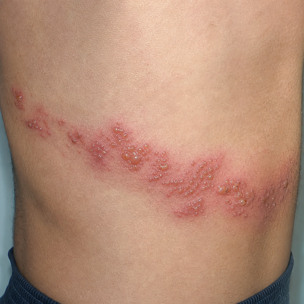
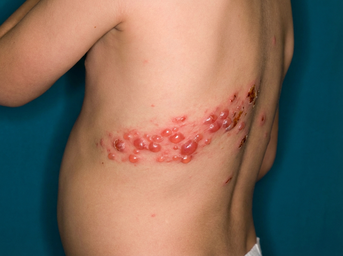
A patient presented with multiple painful blisters on an erythematous base along a dermatome on the trunk as shown in the image. What is the diagnosis?
A patient presented with multiple painful blisters on an erythematous base along a dermatome on the trunk, as shown in the image. What is the diagnosis? 
A patient presents with painful ulcer in the mouth and a past history of recurrent vesicular lesions in the genitalia. Bedside test findings are shown. What is the most appropriate drug for management?
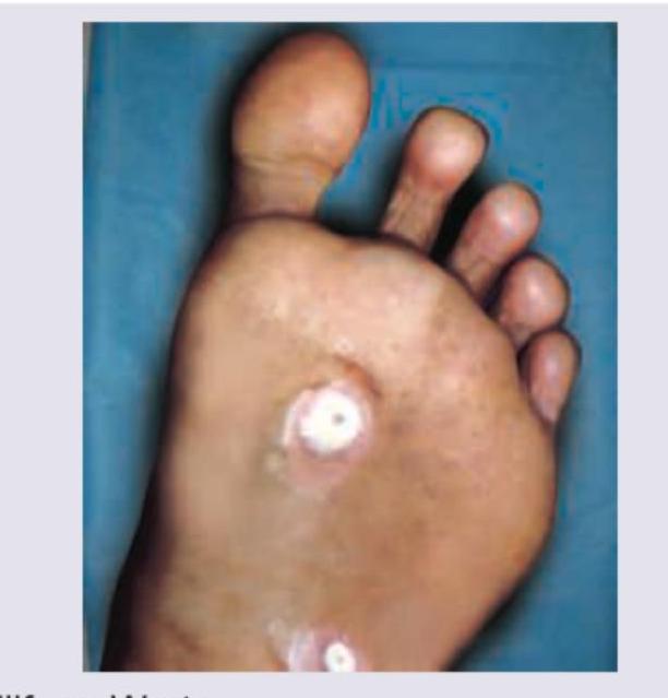
Identify the lesion shown in the image:
Identify the lesions given in the image:
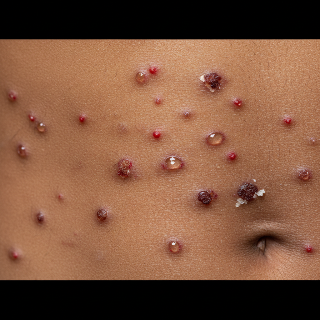
The following image shows:
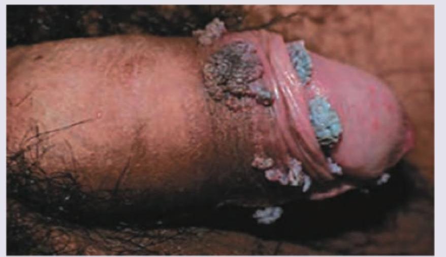
What is the causative agent for the lesion on penis shown below?
A patient presents with painful blisters along the chest wall. All of the following tests are useful for diagnosis except:
Practice by Chapter
Herpes Simplex Virus Infections
Practice Questions
Varicella-Zoster Virus Infections
Practice Questions
Human Papillomavirus Infections
Practice Questions
Molluscum Contagiosum
Practice Questions
Viral Exanthems
Practice Questions
Hand, Foot, and Mouth Disease
Practice Questions
Orf and Milker's Nodule
Practice Questions
Cytomegalovirus Cutaneous Manifestations
Practice Questions
Epstein-Barr Virus Manifestations
Practice Questions
Poxvirus Infections
Practice Questions
HIV-Related Dermatoses
Practice Questions
Viral Infections in Immunocompromised Hosts
Practice Questions
Want unlimited practice?
Get full access to all questions, explanations, and performance tracking.
Scan to download app