What is the most common cause of Kaposi sarcoma?
Which of the following statements is NOT true regarding the skin condition shown in the figure?

Tzank smear is used to diagnose which of the following conditions?
Which of the following conditions is characterized by mucous membrane lesions?
What are Pastia's lines?
Tzank test is positive in the following conditions, except?
Which of the following lesions is/are associated with HPV?
A cutaneous maculopapular rash of the head and neck, preceded by small ulcers in the buccal mucosa, would suggest which of the following?
A patient presents with recurrent palatal pain, multiple punctate ulcers in the hard palate that were preceded by tiny blisters. Her lesions typically heal in about 2 weeks and reappear during stressful times. 
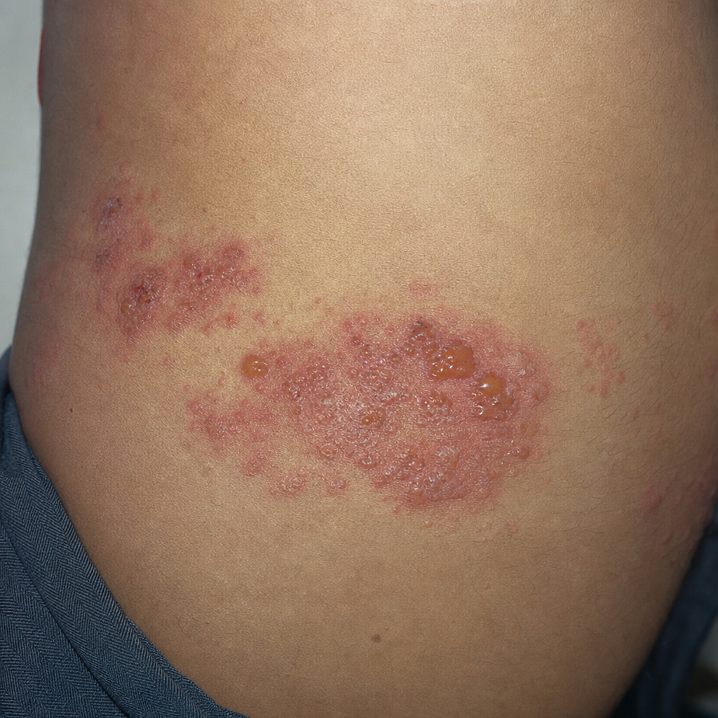
What is the most likely diagnosis?
Herpes zoster is commonly seen in which anatomical region?
Explanation: **Explanation:** **Kaposi Sarcoma (KS)** is a multicentric angioproliferative tumor of the vascular endothelium. The correct answer is **HHV8** (Human Herpesvirus 8), also known as **Kaposi Sarcoma-associated Herpesvirus (KSHV)**. 1. **Why HHV8 is correct:** HHV8 is the primary and necessary etiological agent for all four clinical variants of Kaposi Sarcoma (Classic, Endemic/African, Iatrogenic, and AIDS-associated). The virus encodes proteins like viral cyclin D and v-FLIP that interfere with host cell cycle regulation and apoptosis, leading to the characteristic proliferation of spindle cells and slit-like vascular spaces. 2. **Why other options are incorrect:** * **HHV6:** This virus is the primary cause of **Roseola Infantum** (Exanthem Subitum or Sixth Disease), characterized by high fever followed by a maculopapular rash. * **HHV7:** Similar to HHV6, it is also associated with Roseola Infantum and has been linked to Pityriasis Rosea, but it does not cause KS. * **HHV9:** This is a distractor; there is currently no officially classified human herpesvirus designated as HHV9. **High-Yield Clinical Pearls for NEET-PG:** * **Histopathology:** Look for "Spindle cells," "Slit-like vascular spaces," and "Promontory sign." * **Clinical Presentation:** Presents as palpable purpuric macules, plaques, or nodules, often on the lower limbs or palate. * **Variants:** The **AIDS-associated (Epidemic)** form is the most aggressive and most common variant seen in clinical practice today. * **CD4 Count:** In HIV patients, KS typically occurs when the CD4 count is **<200 cells/mm³**.
Explanation: ***It is common in the elderly.*** - **Molluscum contagiosum** primarily affects **children** and **young adults**, not the elderly population. - The condition is most prevalent in patients under **20 years of age**, making this statement false. *It occurs due to a viral infection.* - **Molluscum contagiosum** is caused by the **Molluscum Contagiosum Virus (MCV)**, a **poxvirus**. - The virus causes characteristic **dome-shaped papules** with **central umbilication**. *It can occur in individuals with a normal immune status.* - **Immunocompetent individuals** frequently develop molluscum contagiosum, especially children. - While **immunocompromised patients** may have more extensive disease, normal immunity does not prevent infection. *It can be transmitted as a sexually transmitted disease.* - In **adults**, molluscum contagiosum is commonly transmitted through **sexual contact**. - **Genital lesions** in adults are typically acquired through intimate skin-to-skin contact.
Explanation: **Explanation:** The **Tzanck smear** is a rapid bedside diagnostic test used primarily for the diagnosis of **vesiculobullous viral infections**, specifically those caused by the Herpesviridae family. **1. Why Option A is Correct:** The Tzanck smear involves taking a scrape from the base of a fresh vesicle. In **Herpes Zoster Virus (HZV)**, Herpes Simplex Virus (HSV-1, HSV-2), and Varicella, the virus causes characteristic cytopathic changes in keratinocytes. Under a microscope (stained with Giemsa, Wright, or Leishman stain), these appear as **multinucleated giant cells** (with "molding" of nuclei) and **acantholytic cells**. **2. Why the Other Options are Incorrect:** * **B. Psittacosis:** Caused by *Chlamydia psittaci*. Diagnosis is typically confirmed via serology (CF test) or PCR, not a Tzanck smear. * **C. Cryptococcosis:** This fungal infection is diagnosed using **India Ink preparation** (to see the capsule) or mucicarmine stain, particularly in CSF samples. * **D. Rickettsial infection:** These are obligate intracellular bacteria diagnosed via the **Weil-Felix reaction** (serology) or skin biopsy with immunostaining. **3. NEET-PG High-Yield Clinical Pearls:** * **Key Finding:** Look for the phrase **"Multinucleated Giant Cells"** in clinical vignettes; this is the hallmark of a positive Tzanck smear for Herpes. * **Other Uses:** Tzanck smear can also be used in **Pemphigus Vulgaris** (to see Tzanck cells/acantholytic cells) and **Molluscum Contagiosum** (to see Henderson-Paterson bodies). * **Limitation:** The Tzanck smear **cannot differentiate** between HSV-1, HSV-2, and HZV; for specific differentiation, viral culture or PCR is required.
Explanation: **Explanation:** **Pemphigus Vulgaris (Option D)** is the correct answer because it is a life-threatening autoimmune blistering disease characterized by the loss of cell-to-cell adhesion (acantholysis). In **80-90% of cases**, it begins with painful oral erosions. The underlying mechanism involves IgG antibodies against **Desmoglein 3** (found primarily in mucosal surfaces) and **Desmoglein 1** (found in the skin). Because Desmoglein 3 is the dominant cadherin in mucous membranes, these sites are almost always involved, often preceding skin lesions by months. **Why the other options are incorrect:** * **Secondary Syphilis (A):** While it can present with "mucous patches" or condyloma lata, it is primarily characterized by a generalized maculopapular rash involving the palms and soles. It is an infectious disease, not a primary blistering disorder. * **Dermatitis Herpetiformis (B):** This is an IgA-mediated condition associated with Celiac disease. It presents as intensely pruritic vesicles on extensor surfaces (elbows, knees). **Mucosal involvement is extremely rare.** * **Psoriasis (C):** This is a chronic inflammatory proliferative disorder. While "geographic tongue" is sometimes associated, classic psoriasis typically spares the mucous membranes. **NEET-PG High-Yield Pearls:** * **Nikolsky Sign:** Positive in Pemphigus (denudation of skin with slight lateral pressure). * **Tzanck Smear:** Shows "Acantholytic cells" or **Tzanck cells** (large, round keratinocytes with hyperchromatic nuclei). * **Immunofluorescence:** Direct Immunofluorescence (DIF) shows a characteristic **"fish-net"** or "lace-like" pattern of IgG/C3 deposits. * **Pemphigoid vs. Pemphigus:** Bullous Pemphigoid is subepidermal and **rarely** involves mucous membranes, whereas Pemphigus is intraepidermal and **frequently** involves them.
Explanation: **Explanation:** **Pastia’s lines** (also known as Thompson’s signs) are a classic clinical sign of **Scarlet Fever**, caused by Group A Beta-hemolytic *Streptococcus pyogenes*. These are pink or red transverse streaks found in the skin folds, most commonly in the antecubital fossa, axillae, and groin. They represent linear petechiae caused by increased capillary fragility due to the circulating erythrogenic toxin. Notably, these lines persist even after the generalized "sandpaper" rash of scarlet fever begins to fade. **Analysis of Options:** * **Option A:** Erythematous streaks in staphylococcal infections (like cellulitis) usually represent lymphangitis, not Pastia’s lines. * **Option C:** White striae in Lichen Planus are known as **Wickham’s striae**, which are caused by hypergranulosis. * **Option D:** The rash in secondary syphilis is typically a generalized maculopapular eruption involving the palms and soles, often described as "copper-colored." **High-Yield Clinical Pearls for NEET-PG:** * **Scarlet Fever Triad:** Exudative pharyngitis, fever, and a generalized "sandpaper" rash. * **Strawberry Tongue:** Initially "White Strawberry Tongue" (coated), followed by "Red Strawberry Tongue" (denuded papillae). * **Schultz-Charlton Reaction:** A diagnostic test where the injection of antitoxin into the skin causes blanching of the scarlet fever rash. * **Dick Test:** Used to determine susceptibility to scarlet fever.
Explanation: The **Tzanck smear** is a rapid bedside diagnostic test used primarily to identify **multinucleated giant cells**, which are characteristic of certain viral infections and autoimmune blistering diseases. ### **Why EBV is the Correct Answer** While the **Epstein-Barr Virus (EBV)** belongs to the *Herpesviridae* family, it primarily infects B-lymphocytes and epithelial cells of the oropharynx. It typically presents as Infectious Mononucleosis or Oral Hairy Leukoplakia. Unlike other herpes viruses, EBV does not cause the formation of intraepidermal vesicles or the specific cytopathic changes (like multinucleation and acantholysis) that a Tzanck smear detects. Therefore, the Tzanck test is **negative** in EBV infections. ### **Analysis of Other Options** * **Herpes Simplex (HSV-1 & 2):** Causes "dew drops on rose petals" vesicles. Tzanck smear shows characteristic multinucleated giant cells with **molding, margination of chromatin, and multinucleation** (the 3 M’s). * **Varicella (Chickenpox):** Caused by the Varicella-Zoster Virus (VZV). It produces intraepidermal vesicles where Tzanck cells are abundant. * **Herpes Zoster (Shingles):** Also caused by VZV (reactivation). Like Varicella and HSV, it shows positive Tzanck smears because these viruses are **epidermotropic**. ### **NEET-PG High-Yield Pearls** * **Tzanck Smear Findings:** Look for **multinucleated giant cells** (Tzanck cells). * **Other Positive Conditions:** Apart from HSV/VZV, the Tzanck test is positive in **Pemphigus Vulgaris** (shows acantholytic cells) and **Cytomegalovirus (CMV)** (rarely, but can show "owl’s eye" inclusions). * **Stains Used:** Giemsa, Wright’s, or Leishman stain. * **Limitation:** The Tzanck test can confirm a Herpes group infection but **cannot differentiate** between HSV-1, HSV-2, and VZV. For differentiation, Viral Culture or PCR is required.
Explanation: Human Papillomavirus (HPV) is a double-stranded DNA virus that infects keratinocytes, leading to various benign and malignant proliferations. **Explanation of Options:** * **Verruca vulgaris (Common Warts):** These are the most common clinical manifestation of HPV. They are typically caused by **HPV types 2 and 4** (and sometimes 1, 27, and 57). They present as firm, periungual, or dorsal hand papules with a "verrucous" (warty) surface. * **Cowden Syndrome (Multiple Hamartoma Syndrome):** While primarily a PTEN gene mutation syndrome, it is classically associated with **multiple acral keratoses** and **verrucous papules** on the skin that are histologically and virologically linked to HPV (often types 6 and 11). * **Focal Epithelial Hyperplasia (Heck’s Disease):** This is a specific oral mucosal condition characterized by multiple asymptomatic, pink-to-white papules on the lips, buccal mucosa, or tongue. It is strongly associated with **HPV types 13 and 32**. **Why "All of the above" is correct:** Each of these conditions represents a distinct clinical morphology (cutaneous, syndromic, and mucosal) triggered by different genotypes of the Human Papillomavirus. **High-Yield Clinical Pearls for NEET-PG:** 1. **Butcher’s Warts:** Associated with HPV-7. 2. **Myrmecia:** Deep palmoplantar warts caused by HPV-1. 3. **Epidermodysplasia Verruciformis (EV):** A genetic susceptibility to HPV; "Tree-man" appearance is associated with **HPV-5 and HPV-8**, which have high malignant potential (Squamous Cell Carcinoma). 4. **Condyloma Acuminatum:** Genital warts caused by HPV-6 and 11 (Low risk) vs. HPV-16 and 18 (High risk for cervical/anal cancer). 5. **Histology Sign:** Look for **Koilocytes** (keratinocytes with pyknotic nuclei and perinuclear halos).
Explanation: ### Explanation The clinical presentation described—a **maculopapular rash** starting on the head and neck, preceded by oral mucosal lesions—is the classic triad of **Rubeola (Measles)**. **1. Why Rubeola is Correct:** Rubeola follows a predictable chronological pattern. The "small ulcers in the buccal mucosa" refer to **Koplik spots**, which are pathognomonic. These are small, bluish-white spots on an erythematous base found opposite the lower molars during the prodromal phase. Following this, a maculopapular (morbilliform) rash appears, typically starting behind the ears and at the hairline, then spreading cephalocaudally (downward) to the trunk and extremities. **2. Why Other Options are Incorrect:** * **Primary Herpes Simplex:** Characterized by grouped vesicles on an erythematous base (e.g., gingivostomatitis), not a generalized maculopapular rash. * **Varicella (Chickenpox):** Presents with a "pleomorphic" rash (macules, papules, and vesicles in different stages of evolution simultaneously) that starts on the trunk and spreads centrifugally. It lacks Koplik spots. * **Primary Syphilis:** Typically presents with a painless, indurated ulcer (chancre) at the site of inoculation. The generalized rash occurs in *secondary* syphilis and characteristically involves the palms and soles. **3. NEET-PG High-Yield Pearls:** * **The 3 C’s of Measles:** Cough, Coryza, and Conjunctivitis (prodromal symptoms). * **Koplik Spots:** Appear 48 hours before the rash and disappear as the rash breaks out. * **Rash Progression:** Cephalocaudal spread; fades in the same order it appeared, often leaving behind "branny" desquamation or brownish staining. * **Vitamin A:** Supplementation is recommended in all children with acute measles to reduce morbidity and mortality. * **Complication:** The most common cause of death in children is pneumonia; the most serious late complication is SSPE (Subacute Sclerosing Panencephalitis).
Explanation: ### Explanation The clinical presentation of recurrent, grouped punctate ulcers on the **keratinized mucosa** (hard palate) preceded by vesicles is pathognomonic for **Recurrent Secondary Herpes (Recurrent Herpes Simplex)**. **1. Why the correct answer is right:** After a primary infection, the Herpes Simplex Virus (HSV-1) remains latent in the sensory ganglia (trigeminal ganglion). Upon reactivation by triggers like **stress**, sunlight, or trauma, the virus travels back to the skin/mucosa. Unlike primary herpes, which is widespread, secondary herpes is localized. A key diagnostic feature is its location: **Recurrent intraoral herpes almost exclusively involves keratinized tissues** (hard palate and gingiva), whereas aphthous ulcers involve non-keratinized tissue. **2. Why other options are wrong:** * **Aphthous ulcers:** These are common but typically occur on **non-keratinized mucosa** (buccal mucosa, labial mucosa, floor of the mouth). They are not preceded by vesicles and do not involve the hard palate. * **Recurrent primary herpes:** This is a contradictory term. "Primary" refers to the initial exposure (Gingivostomatitis), which is usually more severe, involves high fever, and occurs only once. * **Erythema multiforme:** This presents with "target lesions" on the skin and extensive, hemorrhagic crusting of the lips. While it can be triggered by HSV, the oral lesions are typically large, irregular erosions rather than tiny punctate ulcers on the hard palate. **3. NEET-PG High-Yield Pearls:** * **Location Rule:** HSV = Keratinized tissue (Hard palate/Gingiva); Aphthous = Non-keratinized (Soft palate/Cheeks). * **Tzanck Smear:** Look for **Multinucleated Giant Cells** and **Acantholysis**. * **Inclusion Bodies:** Cowdry Type A (Lipschütz bodies) are characteristic of HSV. * **Treatment:** Oral Acyclovir or Valacyclovir is most effective if started during the prodromal phase.
Explanation: **Explanation:** Herpes Zoster (Shingles) is caused by the reactivation of the **Varicella-Zoster Virus (VZV)**, which remains latent in the sensory ganglia following a primary chickenpox infection. While the virus can reactivate in any dermatome, the question asks for the most common site or specific anatomical involvement frequently tested in clinical scenarios. **Why Geniculate Ganglion is the Correct Answer:** In the context of specific named syndromes and high-yield anatomical sites, the **Geniculate Ganglion** of the facial nerve (CN VII) is a classic site of reactivation. This leads to **Ramsay Hunt Syndrome (Herpes Zoster Oticus)**, characterized by a triad of ipsilateral facial palsy, ear pain, and vesicles in the auditory canal or auricle. While thoracic nerves are numerically the most frequent site of involvement overall, in many competitive exams, the geniculate ganglion is highlighted due to its distinct clinical presentation and complications. **Analysis of Incorrect Options:** * **Thoracic region:** Statistically, the thoracic dermatomes (T3–L2) are the most common site for general zoster. However, if "Geniculate Ganglion" is provided as a specific anatomical structure in a single-best-answer format, it often points toward the examiner's focus on cranial nerve involvement. * **Cervical and Lumbar regions:** These are less common than thoracic involvement. Cervical zoster typically affects the neck and arms, while lumbar zoster affects the lower extremities. **NEET-PG High-Yield Pearls:** 1. **Hutchinson’s Sign:** Vesicles on the tip of the nose indicating involvement of the nasociliary branch of the ophthalmic nerve; it predicts a high risk of ocular complications (Herpes Zoster Ophthalmicus). 2. **Tzanck Smear:** Shows **Multinucleated Giant Cells** with Cowdry Type A intranuclear inclusion bodies (common to HSV and VZV). 3. **Post-Herpetic Neuralgia (PHN):** The most common complication, defined as pain persisting >3 months after the rash heals. 4. **Treatment:** Oral Acyclovir (800 mg 5 times/day for 7 days) is the gold standard, ideally started within 72 hours of rash onset.
Herpes Simplex Virus Infections
Practice Questions
Varicella-Zoster Virus Infections
Practice Questions
Human Papillomavirus Infections
Practice Questions
Molluscum Contagiosum
Practice Questions
Viral Exanthems
Practice Questions
Hand, Foot, and Mouth Disease
Practice Questions
Orf and Milker's Nodule
Practice Questions
Cytomegalovirus Cutaneous Manifestations
Practice Questions
Epstein-Barr Virus Manifestations
Practice Questions
Poxvirus Infections
Practice Questions
HIV-Related Dermatoses
Practice Questions
Viral Infections in Immunocompromised Hosts
Practice Questions
Get full access to all questions, explanations, and performance tracking.
Start For Free