
Viral Skin Infections — MCQs

On this page
A 28-year-old woman presents with a "growth" in her genital area, first noticed 3 weeks ago and seemingly grown since then. She has hypothyroidism managed with thyroid hormone replacement. Examination reveals two non-tender, 6 mm, well-circumscribed, flesh-colored, papillated, oval lesions on the labia majora, with no ulceration, erythema, purulence, or inguinal lymphadenopathy. What is the most likely diagnosis?
An 80-year-old female patient complains of a 3-day history of a painful rash extending over the right half of her forehead and down to her right eyelid. There are weeping vesicular lesions on physical examination. Which of the following is the most likely diagnosis?
Which of the following is NOT true about Condyloma acuminata?
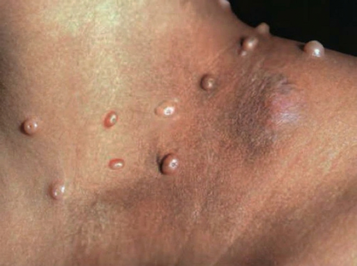
Which of the following statements is NOT true regarding the skin condition shown in the figure?
A patient presents with recurrent palatal pain, multiple punctate ulcers in the hard palate that were preceded by tiny blisters. Her lesions typically heal in about 2 weeks and reappear during stressful times. What is the most likely diagnosis?
A 3-year-old female child develops umbilicated nodules over the face following a trivial viral infection. What is the probable diagnosis?
Genital Warts are seen in:
All of the following are true regarding viral warts except:
Recurrent lesions on the glans that heal with residual hyperpigmentation are suggestive of:
What is the most common cause of erythema multiforme?
Practice by Chapter
Herpes Simplex Virus Infections
Practice Questions
Varicella-Zoster Virus Infections
Practice Questions
Human Papillomavirus Infections
Practice Questions
Molluscum Contagiosum
Practice Questions
Viral Exanthems
Practice Questions
Hand, Foot, and Mouth Disease
Practice Questions
Orf and Milker's Nodule
Practice Questions
Cytomegalovirus Cutaneous Manifestations
Practice Questions
Epstein-Barr Virus Manifestations
Practice Questions
Poxvirus Infections
Practice Questions
HIV-Related Dermatoses
Practice Questions
Viral Infections in Immunocompromised Hosts
Practice Questions
Want unlimited practice?
Get full access to all questions, explanations, and performance tracking.
Scan to download app