
Viral Skin Infections — MCQs

On this page
A patient presents with the skin finding shown in the image. Identify the most likely diagnosis for this lesion.
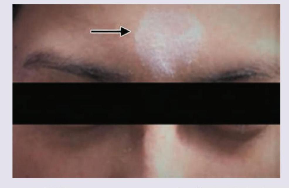
A 28-year-old lady has asymptomatic dome-shaped small lesions on the forehead for the past 2 months. She lives with her 2-year-old daughter who also is having similar lesions. What is the causative agent of these lesions?
Which of the following is INCORRECT regarding genital warts (condyloma acuminata)? 1. It is usually single. 2. It is related to HPV Types 6 and 11. 3. It can be transmitted sexually. 4. It can involve vagina and anus.
Which of the following statements about molluscum contagiosum is FALSE?
What is the primary route of transmission for molluscum contagiosum in adults?
A 30-year-old man with recurrent genital warts has undergone multiple treatments without success. Which of the following treatments would be most appropriate to try next?
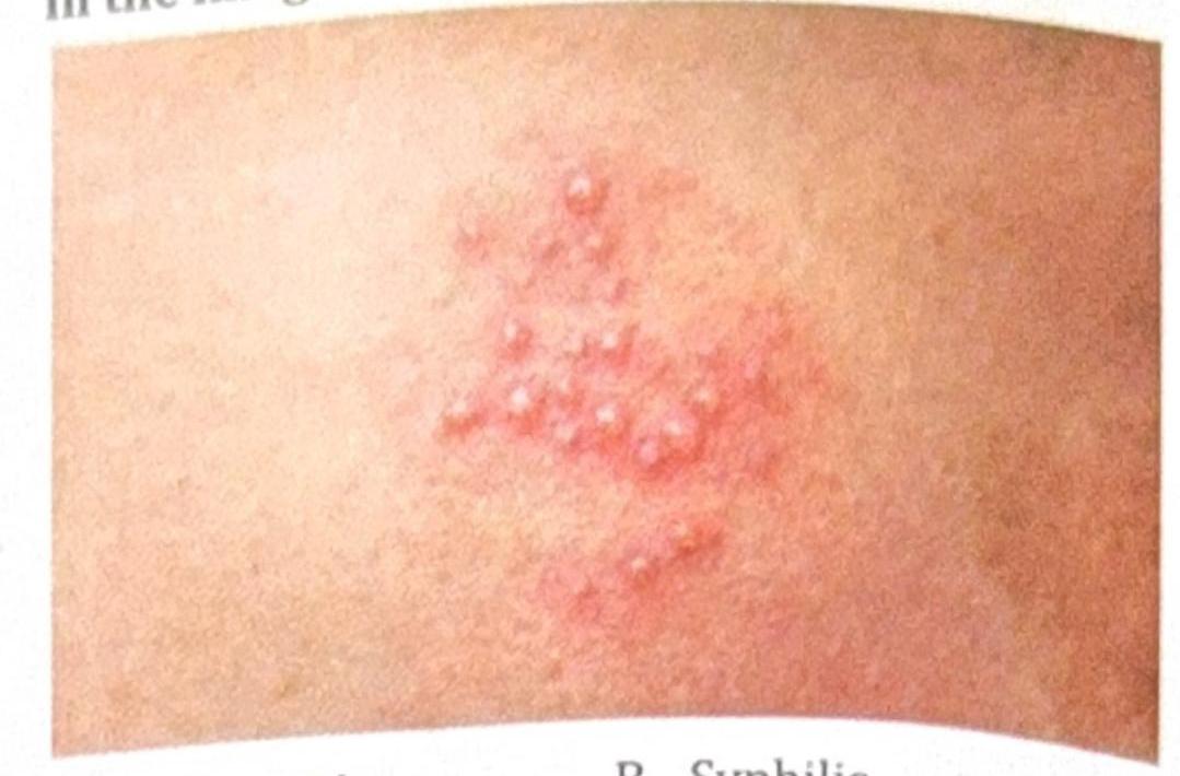
A patient presents with painful vesicles as shown in the image. What is the diagnosis?
A 7-year-old child presents to the dermatology clinic with multiple small, painless, flesh-colored papules on the trunk and arms that have been present for 3 months. The lesions are 2-4 mm in diameter, dome-shaped with a smooth surface, and several have a characteristic central umbilication. The child is otherwise healthy and immunocompetent. The lesions are not pruritic and there is no associated lymphadenopathy. On closer examination, a white, cheesy material can be expressed from the central depression of some lesions. What is the most likely diagnosis?
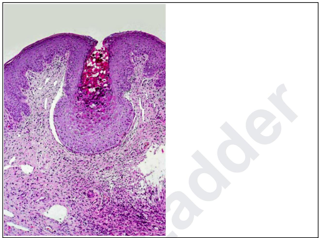
A woman presents with lesions on the inner thighs and peri-anal region. They are nodular, 4-6 mm in size and appear pale. The histopathological image shows multiple intracytoplasmic inclusion bodies consistent with Henderson-Patterson bodies. The diagnosis is:
A 60-year-old patient presents with unilateral vesicular lesions in a dermatomal distribution on the torso. The lesions are painful and appeared over the past 2-3 days, progressing from erythematous patches to fluid-filled vesicles. The patient reports prodromal burning and tingling sensation in the affected area. Which of the following is the most likely diagnosis?
Practice by Chapter
Herpes Simplex Virus Infections
Practice Questions
Varicella-Zoster Virus Infections
Practice Questions
Human Papillomavirus Infections
Practice Questions
Molluscum Contagiosum
Practice Questions
Viral Exanthems
Practice Questions
Hand, Foot, and Mouth Disease
Practice Questions
Orf and Milker's Nodule
Practice Questions
Cytomegalovirus Cutaneous Manifestations
Practice Questions
Epstein-Barr Virus Manifestations
Practice Questions
Poxvirus Infections
Practice Questions
HIV-Related Dermatoses
Practice Questions
Viral Infections in Immunocompromised Hosts
Practice Questions
Want unlimited practice?
Get full access to all questions, explanations, and performance tracking.
Scan to download app