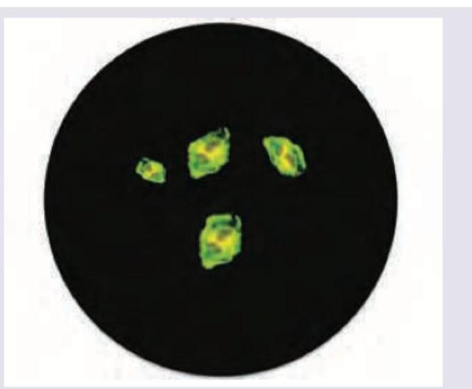
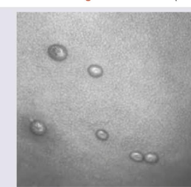
The following findings on Tzanck smear can be seen in:

The following lesion is diagnostic of:

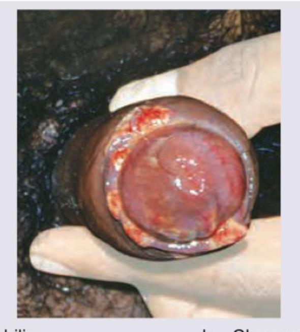
A 20-year-old male with no history of any sexual contact presents with following lesions on his penis. What is the diagnosis?

Identify the lesion shown in the image:

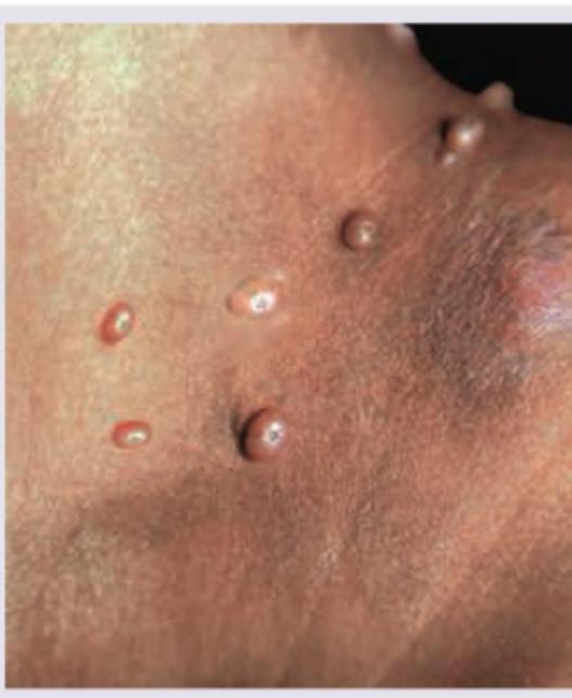
A patient after a trip to Bangkok developed fever and perioral vesicles. What is the diagnosis?

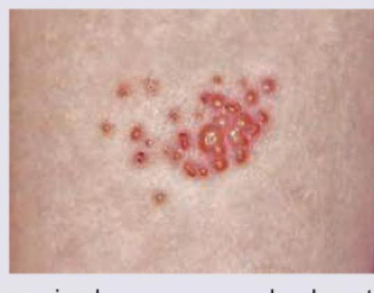
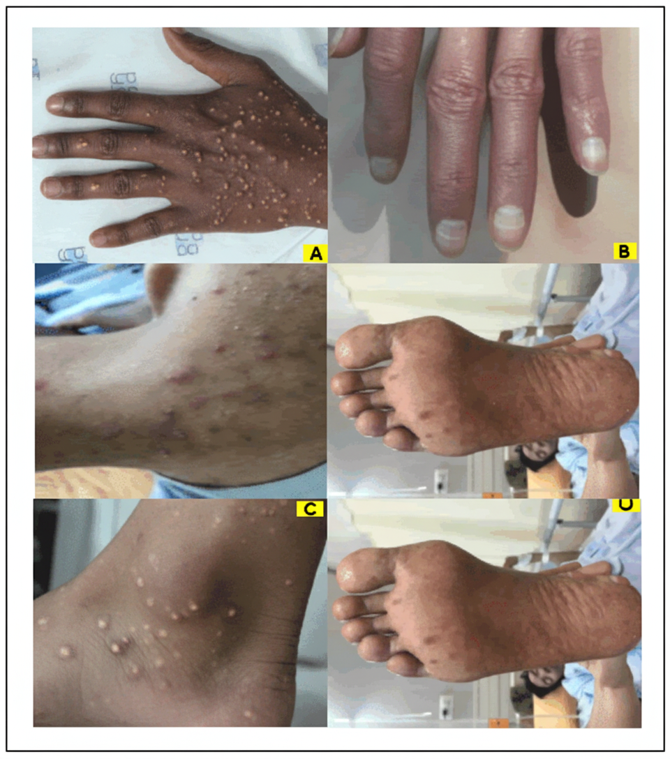
A 28-year-old lady has asymptomatic dome shaped small lesions on the forehead for the last 2 months as shown in the image. She has a 2-year-old daughter with similar lesions. What is the causative agent? (AIIMS May 2016)

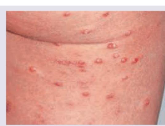
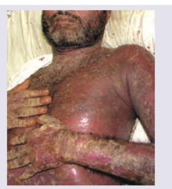
An AIDS positive patient has presented with multiple crusted lesions as shown in the image below. What is the possible diagnosis?

All are true about the lesion shown in the image except:

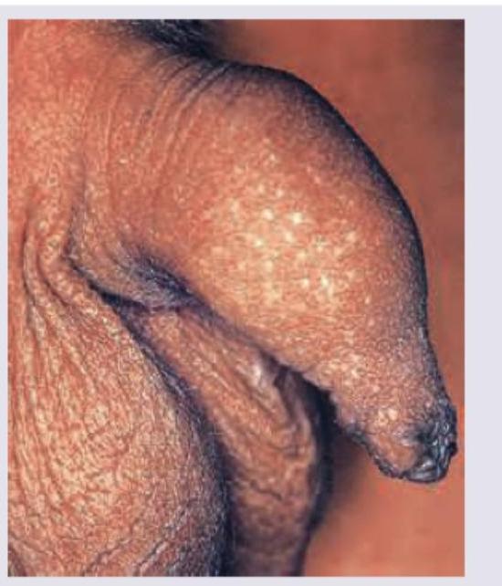
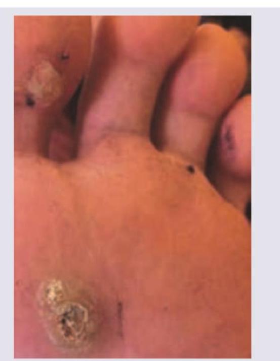
A 25-year-old athlete presents with the following painless lesions in the sole of the foot. The aetiology is:

Comment on the image shown:

Explanation: ***All of the above*** - The image displays multiple **acantholytic cells** (keratinocytes that have lost intercellular connections) with prominent nuclei, which are characteristic findings in several dermatological conditions. - A **Tzanck smear** is a rapid cytological test performed by scraping the base of a fresh blister, staining with Giemsa or Wright stain, and examining under microscopy. **Why all three conditions show similar findings:** *Herpes simplex* - Tzanck smear shows **multinucleated giant cells** with molding of nuclei and **balloon degeneration** of keratinocytes - Acantholytic cells are present due to viral cytopathic effect causing cell separation - These findings are **identical** to those seen in Herpes zoster *Herpes zoster* - Cannot be distinguished from Herpes simplex on Tzanck smear morphology alone - Shows the same **multinucleated giant cells** and **acantholytic keratinocytes** - Viral culture, PCR, or direct fluorescent antibody (DFA) testing needed for definitive differentiation *Paraneoplastic pemphigus* - Shows **acantholytic cells** (rounded keratinocytes with hyperchromatic nuclei) due to autoantibody-mediated destruction of intercellular adhesion - Unlike herpes infections, typically shows acantholytic cells **without** multinucleated giant cells - Definitive diagnosis requires direct immunofluorescence (DIF) on skin biopsy showing intercellular and basement membrane zone IgG/C3 deposition **Note**: While Tzanck smear can show acantholytic cells in all three conditions, the **pattern differs** - herpes shows multinucleated giant cells prominently, while pemphigus shows isolated acantholytic cells. Clinical correlation and confirmatory tests are essential for accurate diagnosis.
Explanation: ***Donovanosis*** - The image shows a **granulomatous, beefy-red ulcer** with rolled borders, which is characteristic of **Donovanosis (granuloma inguinale)**. - This condition is caused by *Klebsiella granulomatis* and typically presents as a painless, progressive ulcerative lesion that can bleed easily. *Syphilis* - Syphilitic chancres are typically **painless, indurated ulcers with clean bases** and well-demarcated borders. - They are usually single, appear about 3 weeks after exposure, and are not beefy-red or friable like the lesion shown. *Chancroid* - Chancroid lesions are typically **painful, ragged ulcers with undermined edges** and often have a gray or yellow exudate. - They tend to be multiple and soft, in contrast to the single, granulomatous lesion seen. *Herpes* - Genital herpes presents as **multiple, painful vesicles** that rupture to form shallow ulcers, which then crust over. - The lesions are usually acute, often recurrent, and do not resemble the chronic, proliferative ulceration shown in the image.
Explanation: ***Lichen nitidus*** - The image shows numerous small, shiny, **pin-head sized papules** on the penis, which are characteristic of lichen nitidus. - This condition is often **asymptomatic** and benign, and it can occur on the penis without any sexual contact history. *Lichenoides keratosis* - This term is broad and often refers to a benign **inflammatory process** with lichenoid features affecting keratinocytes, usually solitary and often in older adults; it does not typically present as widespread, uniform papules on the penis. - Lichenoides keratosis is often a more **solitary lesion** or a reaction pattern, not a diffuse eruption of small papules like those pictured. *Epstein pearls* - **Epstein pearls** are small, white or yellow cysts found in the mouths of newborns, specifically on the gums or palate, and are remnants of epithelial tissue. - They are a normal finding in neonates and are **not found on the penis** or in a 20-year-old male. *Molluscum contagiosum* - Molluscum contagiosum lesions typically present as **dome-shaped, flesh-colored papules with central umbilication**. - While they can appear on the penis and are sexually transmitted, the lesions in the image lack the characteristic **umbilication** of molluscum contagiosum.
Explanation: ***Molluscum contagiosum*** - The image shows **multiple, discrete, flesh-colored to pink, dome-shaped papules** with a characteristic **umbilicated center**. These features are classic for **molluscum contagiosum**. - This viral skin infection is caused by the **molluscum contagiosum virus (MCV)**, a Poxvirus, and is highly contagious, often affecting children or sexually active adults. *Herpes simplex* - Herpes simplex typically presents as **grouped vesicles on an erythematous base**, which later erode and crust. - The lesions seen in the image are **solid papules with central umbilication**, not fluid-filled vesicles or erosions. *Pityriasis rosea* - Pityriasis rosea characteristically begins with a **'herald patch'**, followed by a generalized eruption of smaller, oval, pinkish-red patches with fine scale arranged in a **'Christmas tree' pattern** on the trunk. - The depicted lesions are small, umbilicated papules, not scaly patches with a characteristic distribution. *Gianotti-Crosti syndrome* - Gianotti-Crosti syndrome, also known as papular acrodermatitis of childhood, presents with **monomorphic, flesh-colored to reddish-brown papules** or papulovesicles primarily on the **cheeks, buttocks, and extensor surfaces** of the extremities. - While papular, the lesions in the image have a distinct **umbilicated appearance** not typical for Gianotti-Crosti syndrome, and their distribution is not clearly suggestive of acrodermatitis.
Explanation: ***Herpes simplex*** - The combination of **fever** and **perioral vesicles** after a trip suggests a **viral infection**, with herpes simplex being a classic presentation. - Herpes simplex lesions typically appear as clusters of small, painful **vesicles** on an erythematous base, often affecting the lips and perioral area. *Impetigo* - Impetigo is a **bacterial skin infection** characterized by **honey-colored crusted lesions**, not clear vesicles. - It does not typically present with systemic symptoms like fever unless it is widespread or severe. *Molluscum contagiosum* - Molluscum contagiosum presents as **flesh-colored, dome-shaped papules** with a characteristic **umbilicated center**, not fluid-filled vesicles. - While it's a viral infection, systemic symptoms like fever are rare, and the lesion morphology is distinct. *Bullous pemphigoid* - Bullous pemphigoid is an **autoimmune blistering disease** characterized by large, tense bullae, usually in older adults. - It is not typically associated with acute onset of fever or travel history in this manner and lacks the clustered perioral vesicular appearance.
Explanation: ***Poxvirus*** - The description of **asymptomatic, dome-shaped small lesions** (suggesting **molluscum contagiosum**), especially with transmission from a child to a parent, is classic for **Molluscum contagiosum virus (MCV)**, which belongs to the **Poxviridae family**. - **Molluscum contagiosum** lesions can appear anywhere but are often seen on the face, neck, trunk, and extremities in children, and may be transmitted through close contact. *Papillomavirus* - **Papillomavirus** causes **warts**, which are typically **rough, verrucous** (cauliflower-like) lesions, not smooth, dome-shaped papules. - While warts can also be transmitted through close contact, their morphology is distinct from the lesions described. *Herpes virus* - **Herpes simplex virus (HSV)** causes **vesicular eruptions** (cold sores or fever blisters) that evolve into ulcers or crusts, often associated with pain, burning, or itching. - The lesions described as "asymptomatic dome-shaped" do not fit the typical presentation of herpes infections. *Coxsackie virus* - **Coxsackievirus** most commonly causes **hand-foot-and-mouth disease (HFMD)**, characterized by **fever**, **oral ulcers**, and a **rash of macules or vesicles** on the hands and feet. - This clinical picture is not consistent with the localized, dome-shaped papules on the forehead.
Explanation: ***Norwegian scabies*** - This severe, crusted form of **scabies** is common in immunocompromised individuals like AIDS patients. - The image displays **widespread crusted lesions** consistent with the hyperkeratotic plaques seen in Norwegian scabies, where there are thousands of mites. *Herpes zoster* - **Herpes zoster** typically presents as a painful, vesicular rash in a **dermatomal distribution**, which progresses to crusting. - The lesions in the image are widespread and diffuse, not limited to a dermatome, and appear more crusted and hyperkeratotic than typical herpes zoster. *Bacillary angiomatosis* - **Bacillary angiomatosis** presents as reddish-purple, vascular lesions that can resemble **Kaposi sarcoma** or pyogenic granulomas. - While it can occur in AIDS patients, the lesions are primarily **angiomatous** (blood vessel proliferation) rather than crusted and hyperkeratotic as shown. *Kaposi sarcoma* - **Kaposi sarcoma** in AIDS patients typically manifests as **reddish-purple or brown macules, plaques, and nodules**, often on the skin or mucous membranes. - Although it can be widespread, the image depicts extensive **crusted, hyperkeratotic lesions** rather than the characteristic vascular lesions of Kaposi sarcoma.
Explanation: ***Henderson-Patterson bodies are intranuclear bodies*** - **Henderson-Patterson bodies** are characteristic **eosinophilic intracytoplasmic inclusions** found in keratinocytes infected with Molluscum contagiosum virus, not intranuclear. - This statement is incorrect because the inclusions are cytoplasmic. *Giant extensive lesions in HIV positive patients* - In **immunocompromised individuals**, particularly those with **HIV**, molluscum contagiosum lesions can be unusually large, numerous, and widespread. - This is due to a compromised immune response that prevents the effective clearance of the virus. *Auto-innoculated lesions* - **Molluscum contagiosum** lesions can spread rapidly from one area of the body to another through **scratching** or rubbing, a process known as auto-inoculation. - This characteristic explains the common presentation of multiple lesions in different sites. *Needle extirpation followed by trichloro-acetic acid application* - **Needle extirpation** (pricking the lesion with a sterile needle to express the core) followed by the application of a chemical cauterant like **trichloroacetic acid** is a common and effective treatment method for molluscum contagiosum. - This combination helps destroy the viral particles and prevent recurrence.
Explanation: ***HPV-2*** - The image shows **plantar warts** (verruca plantaris), which are rough, grainy growths on the soles of the feet caused by **human papillomavirus (HPV)**. - **HPV-2** is a well-established cause of plantar warts, along with HPV-1, HPV-4, and HPV-63. - The black dots visible in the lesions are **thrombosed capillaries**, a hallmark feature of warts, distinguishing them from calluses. - Among the given options, **HPV-2 is the only viral etiology** that causes plantar warts. *HSV-2* - **Herpes simplex virus type 2 (HSV-2)** primarily causes **genital herpes**, characterized by painful blisters and ulcers in the genital area. - HSV-2 lesions are typically recurrent, painful, and vesicular, which is distinct from the painless, hyperkeratotic plantar lesions shown. *Candida* - **Candida** infections (candidiasis) can manifest as various skin conditions but rarely as discrete, rough, hyperkeratotic lesions on the sole of the foot. - Fungal infections on the feet typically present as **athlete's foot (tinea pedis)**, characterized by scaling, redness, and itching between toes, not as verrucous lesions. *Neuropathic ulcer* - **Neuropathic ulcers** are common in patients with **peripheral neuropathy** (e.g., from diabetes), often occurring at pressure points of the foot. - These ulcers are typically open sores with surrounding calluses and, while painless due to neuropathy, their appearance is distinct from the verrucous, papillomatous lesions of plantar warts.
Explanation: ***Callosity*** - The image shows **diffuse, hyperkeratotic thickening** spread over a wide area of the palm, characteristic of callosity formation from repeated friction and pressure. - Callosities present as **broad, flat lesions** without a central core, causing painless epidermal thickening over pressure areas. *Corn* - A corn is a **small, localized lesion** with a **painful central core**, unlike the diffuse, widespread thickening seen in this image. - Corns are typically **punctate and well-demarcated**, occurring over bony prominences rather than broad palmar surfaces. *Warts* - **Warts** are caused by **human papillomavirus (HPV)** and show characteristic **black puncta** (thrombosed capillaries) upon paring, which are not visible here. - They present with a **rough, papillomatous surface** with elevated borders, different from the smooth, flat hyperkeratotic appearance shown. *Cutaneous horn* - A **cutaneous horn** appears as a **conical projection** of hyperkeratotic material resembling an animal horn, typically on sun-exposed areas. - It presents as a **solitary, horn-like protrusion** rather than the multiple, flat, diffusely thickened lesions demonstrated in this image.
Herpes Simplex Virus Infections
Practice Questions
Varicella-Zoster Virus Infections
Practice Questions
Human Papillomavirus Infections
Practice Questions
Molluscum Contagiosum
Practice Questions
Viral Exanthems
Practice Questions
Hand, Foot, and Mouth Disease
Practice Questions
Orf and Milker's Nodule
Practice Questions
Cytomegalovirus Cutaneous Manifestations
Practice Questions
Epstein-Barr Virus Manifestations
Practice Questions
Poxvirus Infections
Practice Questions
HIV-Related Dermatoses
Practice Questions
Viral Infections in Immunocompromised Hosts
Practice Questions
Get full access to all questions, explanations, and performance tracking.
Start For Free