
Viral Skin Infections — MCQs

On this page
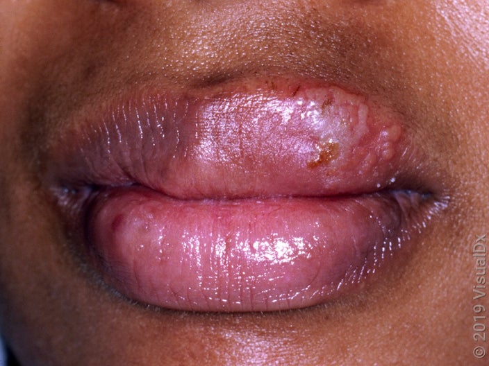
A 10-year-old child had fever for 5 days, along with which he developed multiple fluid filled lesions on the lips as shown below. What is the probable underlying etiology for the skin lesions?
Herpetic Whitlow is seen in
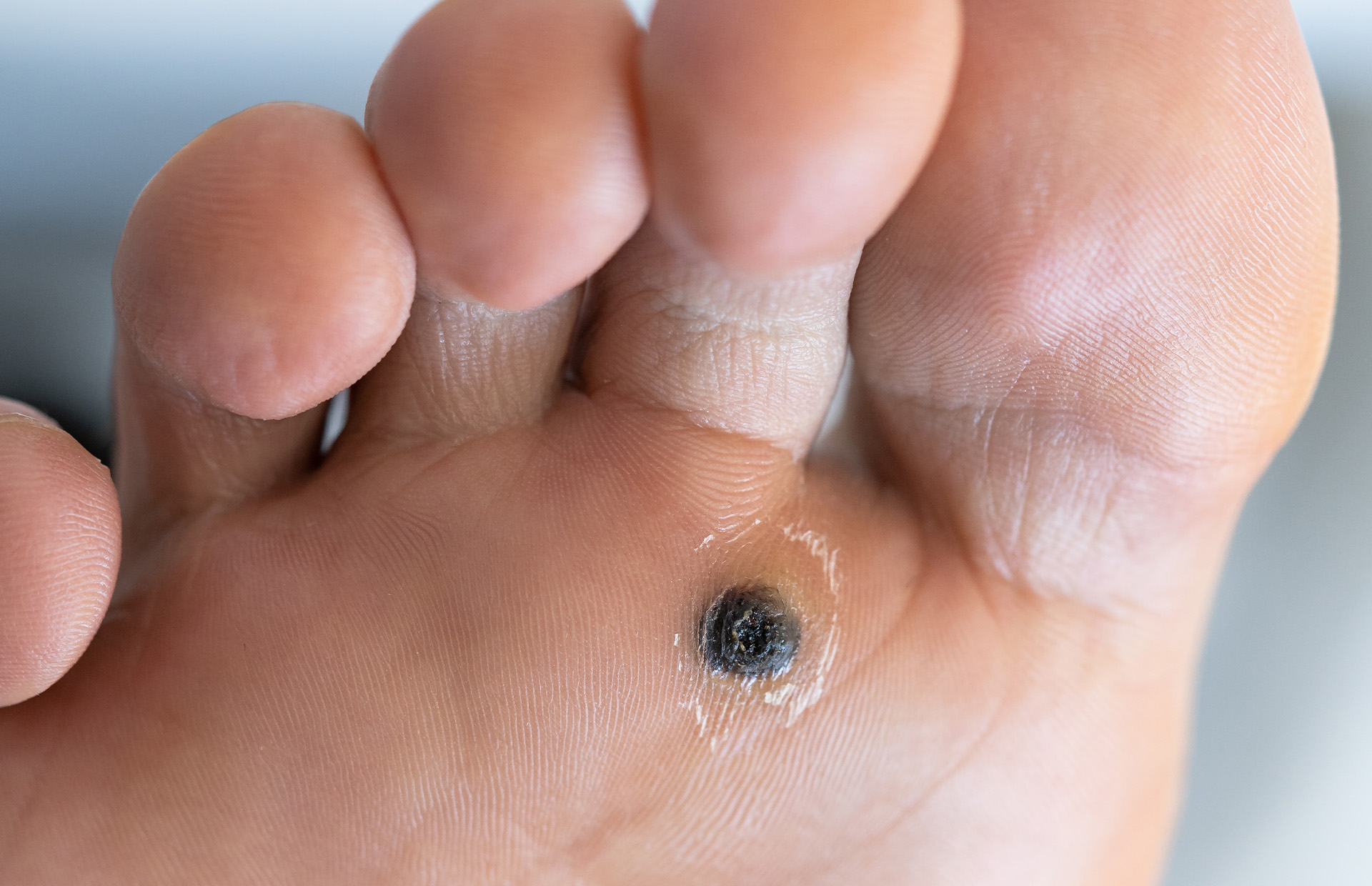
What diagnosis is suggested by the finding on the sole of this patient's foot?
Podophyllin was historically used as treatment of choice in
Myrmecia warts are a type of which wart?
Multinucleated giant cell on Tzanck smear is not seen in?
Most common manifestation of HPV infection in children :
Pityriasis rosea is caused by?
Which of the following conditions is associated with autoinoculation, where lesions spread to other body sites through scratching or direct contact?
A female patient presents with multiple sessile lesions on the vulva that do not bleed on touch. What is the most likely diagnosis?
Practice by Chapter
Herpes Simplex Virus Infections
Practice Questions
Varicella-Zoster Virus Infections
Practice Questions
Human Papillomavirus Infections
Practice Questions
Molluscum Contagiosum
Practice Questions
Viral Exanthems
Practice Questions
Hand, Foot, and Mouth Disease
Practice Questions
Orf and Milker's Nodule
Practice Questions
Cytomegalovirus Cutaneous Manifestations
Practice Questions
Epstein-Barr Virus Manifestations
Practice Questions
Poxvirus Infections
Practice Questions
HIV-Related Dermatoses
Practice Questions
Viral Infections in Immunocompromised Hosts
Practice Questions
Want unlimited practice?
Get full access to all questions, explanations, and performance tracking.
Scan to download app