A child presented with bluish-white spots in the mouth followed by a rash. What is the genome of the most likely causative agent?
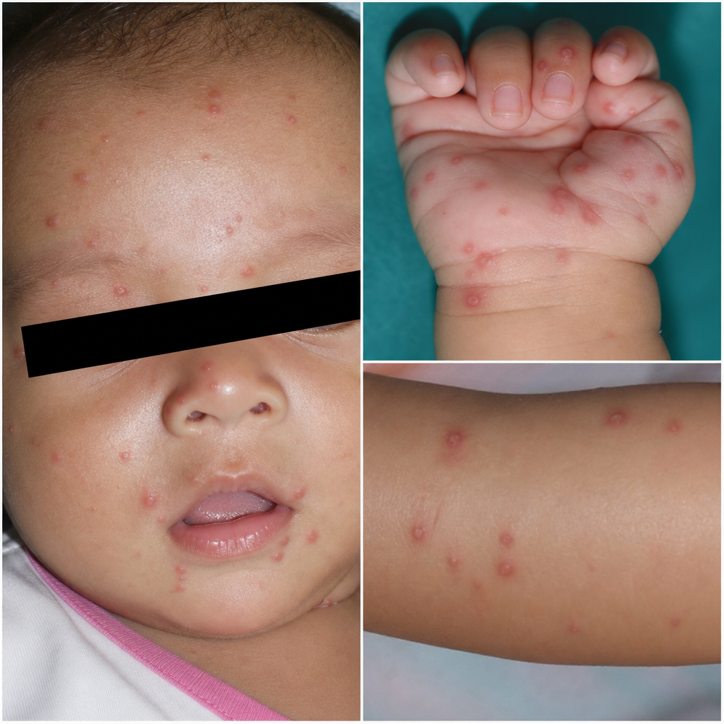
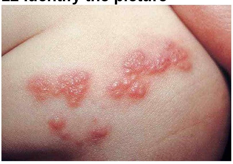
A patient presents with painful vesicular eruptions on one side of the body. What is the most likely diagnosis based on the clinical image?

Fever of unknown origin in a farmer who raises goats would most likely be caused by which of the following organisms?
Which of the following clinical manifestations is LEAST likely in secondary syphilis?
Which of the following does not have a viral etiology?
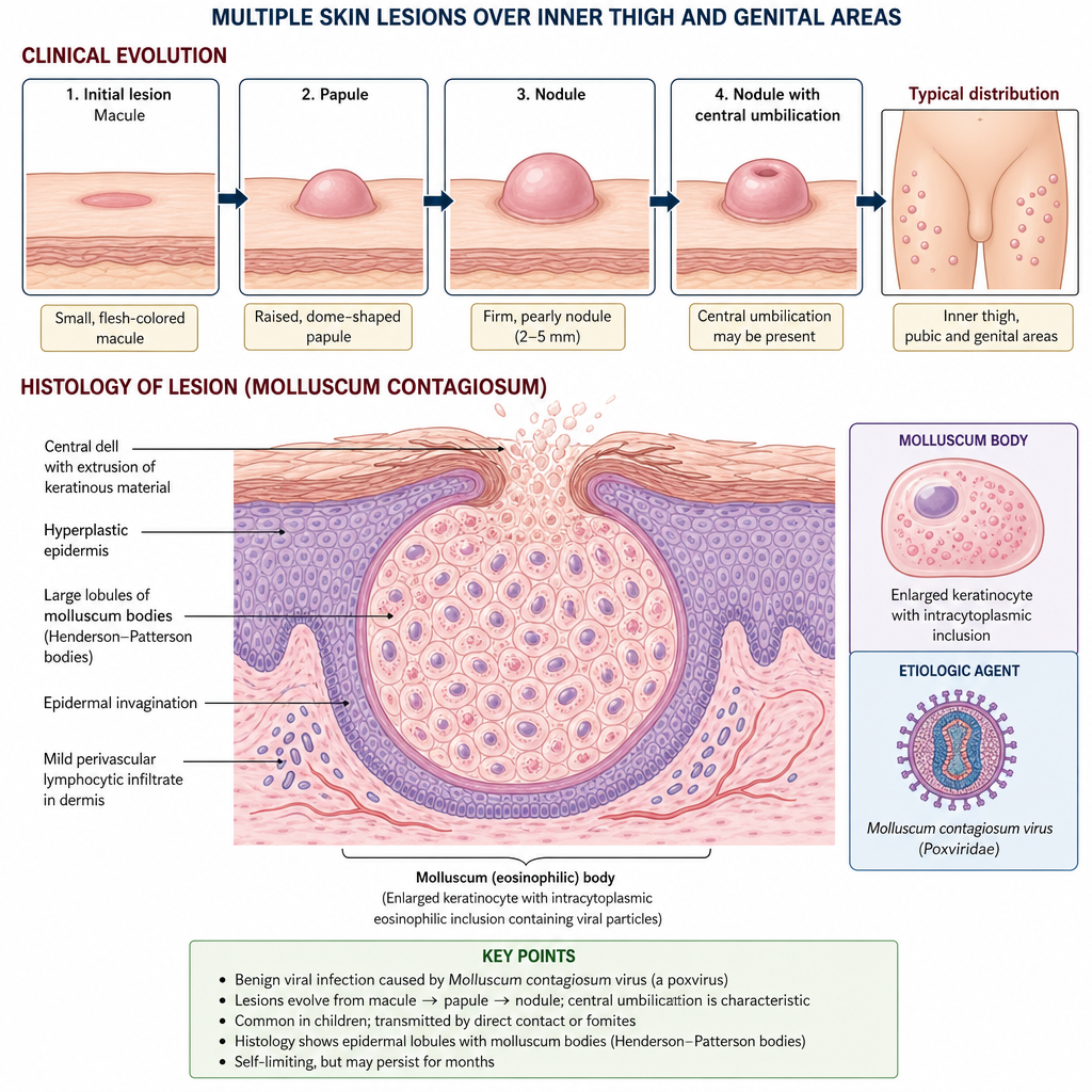
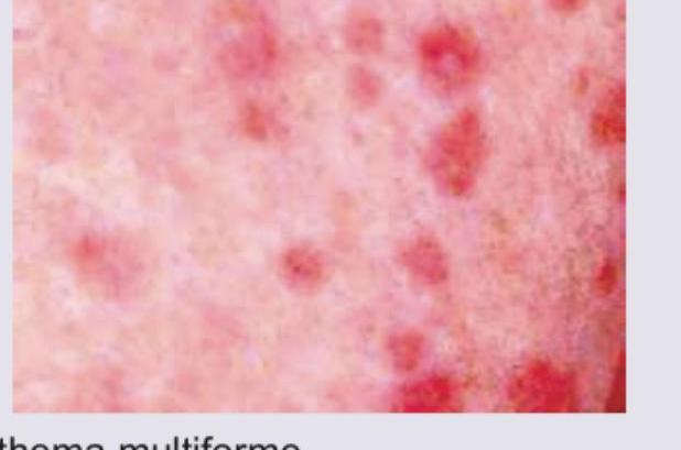
All are true about erythema multiforme except which of the following?
Identify the lesion: (Recent NEET Pattern 2016-17)

Condyloma acuminatum is caused by which virus?
Which HPV type is most commonly implicated in genital warts?
Verrucosa vulgaris is caused by which virus?
Explanation: ***Enveloped virus with single-stranded RNA*** - Bluish-white spots in the mouth (**Koplik spots**) followed by a rash are pathognomonic for **measles**, which is caused by the **measles virus**. - The measles virus is a **paramyxovirus**, characterized as an **enveloped, negative-sense, single-stranded RNA virus**. *Double stranded Naked RNA* - No major human pathogen belongs to this specific genomic and structural classification. - Most **dsRNA viruses** like **rotavirus** are **naked** but cause gastroenteritis, not measles. *Naked virus with single-stranded RNA* - Viruses like **rhinovirus** (common cold) or **poliovirus** fit this description but do not cause Koplik spots or measles. - **Naked viruses** lack a lipid envelope, making them generally more resistant to environmental factors. *Double stranded Enveloped RNA* - There are no known medically significant human viruses that are both **enveloped** and contain **double-stranded RNA**. - Viral genomes are typically either DNA or RNA, and RNA viruses are usually single-stranded (positive or negative sense) or double-stranded, with or without an envelope.
Explanation: ***Herpes zoster*** - The image displays characteristic **vesicular lesions** grouped together on an erythematous base, typically following a **dermatomal distribution**, which is classic for herpes zoster (shingles). - These lesions often cause significant pain and are due to the **reactivation of the varicella-zoster virus**. *Smallpox* - Smallpox lesions are typically **deep-seated, firm, round pustules** that are all in the same stage of development. - While smallpox also features vesicular lesions, their appearance and distribution are distinct from the clustered, dermatomal pattern seen in the image. *Chickenpox* - Chickenpox presents as a generalized rash with lesions at **various stages of development** (macules, papules, vesicles, scabs), often described as a "dewdrop on a rose petal." - Unlike the localized, dermatomal pattern of herpes zoster, chickenpox lesions are typically **widespread** over the body. *Atopic dermatitis* - Atopic dermatitis typically manifests as **erythematous, scaly, intensely itchy patches or plaques**, often in areas like the flexural creases. - It does not present with the characteristic **vesicular, grouped lesions in a dermatomal pattern** seen in the image.
Explanation: ***Brucella melitensis*** - **Brucellosis** is a zoonotic infection commonly acquired through contact with infected animals or consumption of unpasteurized dairy products. - ***Brucella melitensis* is specifically associated with goats and sheep**, making it the most likely organism in this clinical scenario. - It classically presents as **fever of unknown origin (FUO)** due to its varied and non-specific clinical manifestations, including undulating fever, malaise, myalgia, arthralgia, and hepatosplenomegaly. *T. pallidum* - *Treponema pallidum* is the causative agent of **syphilis**, a sexually transmitted infection. - While syphilis can cause fever, its primary modes of transmission (sexual contact, vertical transmission) and clinical manifestations (e.g., chancres, rash, mucous patches) are distinct from the scenario described. - Not associated with occupational goat exposure. *Histoplasma capsulatum* - *Histoplasma capsulatum* causes **histoplasmosis**, a fungal infection associated with exposure to **bird or bat droppings**, particularly in endemic areas (e.g., Ohio and Mississippi River valleys). - Although it can cause disseminated disease and fever, the epidemiological context (exposure to goats, not birds/bats) makes it unlikely in this scenario. *Clostridium novyi* - *Clostridium novyi* is an anaerobic bacterium known to cause **gas gangrene** (clostridial myonecrosis) and occasionally **black disease** in livestock. - It is not typically associated with human fever of unknown origin in the context of occupational animal contact described. - More relevant to wound infections or animal disease rather than systemic FUO in farmers.
Explanation: ***Chancre*** - A **chancre** is the characteristic lesion of **primary syphilis**, appearing at the site of inoculation [1]. - By the time **secondary syphilis** develops (typically weeks to months later), the chancre of primary syphilis has usually healed spontaneously [1]. *Condylomata lata* - **Condylomata lata** are highly infectious, moist, wart-like lesions that occur in intertriginous areas and mucous membranes during **secondary syphilis** [1]. - They are a common manifestation due to the widespread dissemination of **Treponema pallidum** [1]. *Mucous patches* - **Mucous patches** are painless, white to gray lesions found on mucous membranes (e.g., mouth, pharynx, vagina, anus) during **secondary syphilis**. - These are highly infectious and result from the systemic spread of the spirochete. *Palmar and plantar rash* - A diffuse, non-pruritic, maculopapular rash, often involving the **palms and soles**, is a classic and highly characteristic sign of **secondary syphilis** [1]. - This rash indicates the systemic nature of the infection and can vary widely in appearance [1].
Explanation: ***Hodgkin's lymphoma*** - Hodgkin's lymphoma is primarily associated with **genetic factors** and environmental triggers instead of a viral cause [1][2]. - It can arise from **Reed-Sternberg cells**, which are not related to viral infections [1][2]. - EBV is not associated with certain subtypes of Hodgkin lymphoma [3]. *Nasopharyngeal carcinoma* - Strongly linked to **Epstein-Barr virus (EBV)**, which plays a significant role in its pathogenesis. - It is characterized by **nasal obstruction** and symptoms related to tumor invasion, with a clear viral etiology. *Hepatocellular carcinoma* - Often associated with **hepatitis B and C viruses**, which are well-known risk factors for liver cancer. - Chronic infection leads to **cirrhosis**, predisposing individuals to carcinoma development. *Burkitt's lymphoma* - Associated with **EBV**, particularly in endemic forms in Africa, affecting the jaw or abdomen [4]. - It involves **aggressive B-cell proliferation**, strongly linked to viral infection [4]. **References:** [1] Cross SS. Underwood's Pathology: A Clinical Approach. 6th ed. Common Clinical Problems From Diseases Of The Urinary And Male Genital Tracts, pp. 556-557. [2] Kumar V, Abbas AK, et al.. Robbins and Cotran Pathologic Basis of Disease. 9th ed. Diseases of White Blood Cells, Lymph Nodes, Spleen, and Thymus, pp. 614-616. [3] Kumar V, Abbas AK, et al.. Robbins and Cotran Pathologic Basis of Disease. 9th ed. Diseases of White Blood Cells, Lymph Nodes, Spleen, and Thymus, p. 618. [4] Kumar V, Abbas AK, et al.. Robbins and Cotran Pathologic Basis of Disease. 9th ed. Neoplasia, pp. 335-336.
Explanation: ***Does not involve mucosa*** - Erythema multiforme often presents with **mucosal involvement**, particularly in the oral cavity, which can range from mild erosions to severe blistering. - The presence of mucosal lesions, especially oral, ocular, or genital, is a key feature distinguishing more severe forms like **erythema multiforme major**. *Target lesion* - The **target lesion** (or iris lesion) is the hallmark dermatological finding in erythema multiforme, characterized by concentric rings of different colors. - This classic lesion is crucial for the clinical diagnosis of erythema multiforme. *Associated with HSV* - **Herpes Simplex Virus (HSV) infection** is the most common precipitating factor for erythema multiforme, especially for recurrent episodes. - The onset of lesions typically follows an HSV outbreak by several days to weeks. *Extensor involvement* - The rash of erythema multiforme commonly affects the **extensor surfaces of the extremities**, such as the dorsal hands, forearms, and shins. - While it can appear elsewhere, this distribution is a characteristic pattern.
Explanation: ***Erythema multiforme*** - The image displays characteristic **targetoid lesions** with multiple concentric rings of color (erythema, edema, pallor), typical of **erythema multiforme**. - These lesions often appear suddenly, symmetrically, and commonly on the extremities, often triggered by infections (e.g., **herpes simplex virus**) or medications. *Gianotti-Crosti syndrome* - Characterized by **monomorphic, flesh-colored to erythematous papules** and papulovesicles, often on the cheeks, buttocks, and extensor surfaces of the limbs. - This condition is typically observed in **children** after viral infections and does not usually present with target lesions. *Pityriasis rosea* - Starts with a single **"herald patch,"** followed by smaller, oval, pinkish-red patches with fine scales, often arranged in a **"Christmas tree pattern"** on the trunk. - The morphology of the lesions in the image, specifically the targetoid appearance, is not consistent with pityriasis rosea. *Acne rosacea* - Marked by **facial erythema**, papules, pustules, and telangiectasias, primarily affecting the central face. - It does not present with the widespread, distinct target lesions seen in the image.
Explanation: **Explanation:** **Condyloma acuminatum**, commonly known as anogenital warts, is caused by the **Human Papillomavirus (HPV)**. It is a sexually transmitted infection characterized by soft, cauliflower-like growths on the skin and mucous membranes of the genital and perianal regions. The virus infects the basal keratinocytes, leading to epidermal hyperplasia. * **Why HPV is correct:** Specifically, **HPV types 6 and 11** are responsible for approximately 90% of cases. These are considered "low-risk" types because they have a low potential for malignant transformation, unlike "high-risk" types (16 and 18) which are linked to cervical and anal carcinomas. * **Why other options are incorrect:** * **HSV (A):** Causes Herpes Simplex (Type 1: orofacial; Type 2: genital), presenting as painful, grouped vesicles on an erythematous base, not warty growths. * **HIV (C):** While HIV is a sexually transmitted virus that can coexist with HPV, it does not directly cause condyloma. However, HIV-induced immunosuppression can lead to more extensive and recalcitrant HPV lesions. * **VZV (D):** Causes Chickenpox (primary infection) and Herpes Zoster/Shingles (reactivation), characterized by dermatomal vesicular rashes. **High-Yield Clinical Pearls for NEET-PG:** 1. **Histopathology:** Look for **Koilocytes** (keratinocytes with perinuclear halo and wrinkled "raisinoid" nuclei) in the upper epidermis. 2. **Diagnosis:** Usually clinical; application of 5% acetic acid (Acetowhitening) can help visualize subclinical lesions. 3. **Treatment:** Options include Podophyllotoxin, Imiquimod (immunomodulator), Cryotherapy, or Electrocautery. 4. **Note:** Do not confuse *Condyloma acuminatum* (HPV) with *Condyloma lata* (Secondary Syphilis).
Explanation: **Explanation:** **Genital warts (Condyloma acuminatum)** are caused by the Human Papillomavirus (HPV). HPV types are broadly classified into "low-risk" and "high-risk" based on their oncogenic potential. * **Correct Answer (D): HPV-6** (along with HPV-11) is responsible for approximately 90% of all genital warts. These are considered **low-risk types** because they cause benign proliferative lesions rather than invasive squamous cell carcinoma. **Analysis of Incorrect Options:** * **A & B (HPV-16 & 18):** These are the most common **high-risk (oncogenic)** HPV types. They are strongly associated with cervical, anal, and vulvar cancers. While they can be found in genital lesions, they typically cause flat dysplastic changes (intraepithelial neoplasia) rather than the classic cauliflower-like warts. * **C (HPV-31):** This is another high-risk type associated with cervical cancer, though it is less prevalent than types 16 and 18. **High-Yield Clinical Pearls for NEET-PG:** 1. **HPV 6 & 11:** Most common cause of Condyloma acuminatum and Recurrent Respiratory Papillomatosis (RRP). 2. **HPV 16 & 18:** Most common cause of Cervical Carcinoma and Bowenoid papulosis. 3. **HPV 1, 2 & 4:** Associated with common warts (Verruca vulgaris) and plantar warts. 4. **HPV 3 & 10:** Associated with plane warts (Verruca plana). 5. **Histopathology:** The hallmark of HPV infection is the **Koilocyte** (a squamous cell with a perinuclear halo and wrinkled "raisin-like" nucleus). 6. **Treatment:** Podophyllin, Imiquimod (immunomodulator), or destructive methods like cryotherapy and CO2 laser.
Explanation: **Explanation:** **Verruca vulgaris**, commonly known as the common wart, is caused by the **Human Papillomavirus (HPV)**. This is a double-stranded DNA virus that infects the basal layer of the epithelium. Specifically, Verruca vulgaris is most frequently associated with **HPV types 2 and 4** (though types 1, 3, 27, and 57 are also implicated). The virus induces localized proliferation of keratinocytes, leading to the characteristic hyperkeratotic papules with a "verrucous" or cauliflower-like surface. **Analysis of Incorrect Options:** * **Epstein-Barr Virus (EBV):** A herpesvirus associated with Infectious Mononucleosis, Nasopharyngeal carcinoma, and Oral Hairy Leukoplakia (often seen in HIV patients), but not common warts. * **Cytomegalovirus (CMV):** Primarily causes systemic infections in immunocompromised individuals or congenital infections (TORCH); it does not manifest as verrucous skin lesions. * **Human Immunodeficiency Virus (HIV):** While HIV causes immunosuppression that can lead to *extensive* or recalcitrant warts, the direct causative agent of the wart itself remains HPV. **High-Yield Clinical Pearls for NEET-PG:** * **Auspitz Sign vs. Punctate Bleeding:** When the hyperkeratotic surface of a wart is shaved, "black dots" are seen. These represent thrombosed dermal capillaries (a key diagnostic feature). * **Koebner Phenomenon:** Warts can exhibit this (linear spread of lesions along the site of trauma/scratching). * **Histopathology:** Look for **Koilocytes** (keratinocytes with pyknotic nuclei and perinuclear halos) in the upper epidermis, which are pathognomonic for HPV infection. * **Other HPV associations:** Verruca plana (Types 3, 10), Plantar warts (Type 1), and Condyloma acuminatum (Types 6, 11).
Get full access to all questions, explanations, and performance tracking.
Start For Free