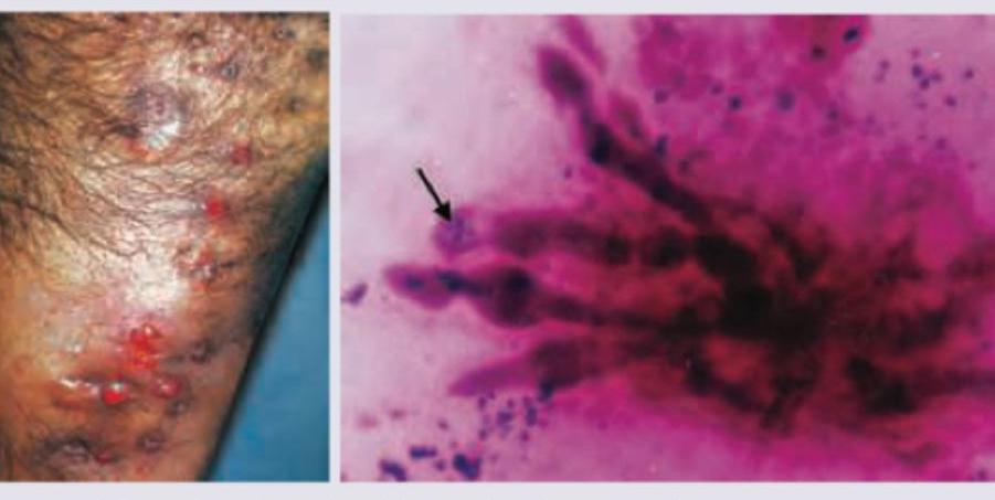
A 65-year-old beggar comes to your OPD with following presentation in upper part of foot Gram stain of discharge was performed. All are true about the condition shown except:

A young male presented with an anesthetic patch on the right forearm. A thickened nerve was palpable on examination. Skin biopsy shows the image below. What is the diagnosis?

All are true about the lesion shown except:

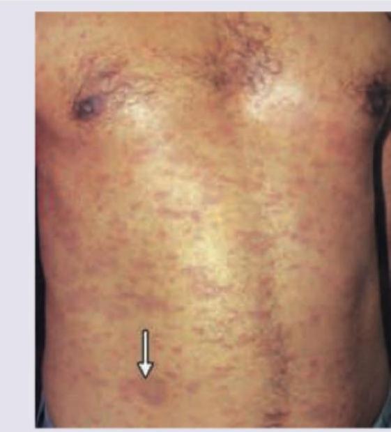
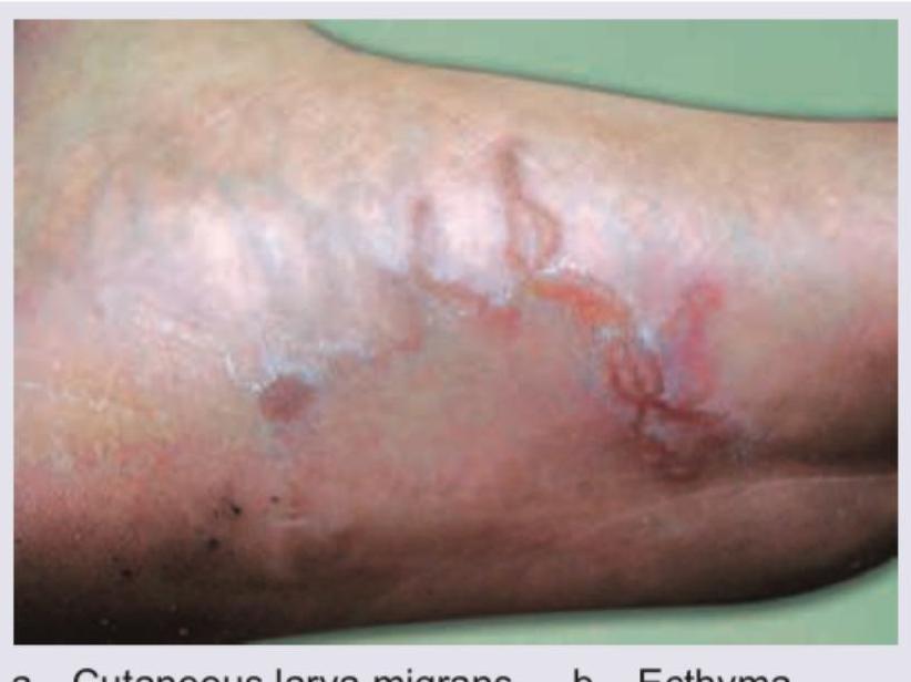
A patient lives in an urban slum and practices open-air defecation. He presented with continuous itching over the sole. The image shows presence of:

The image shows:

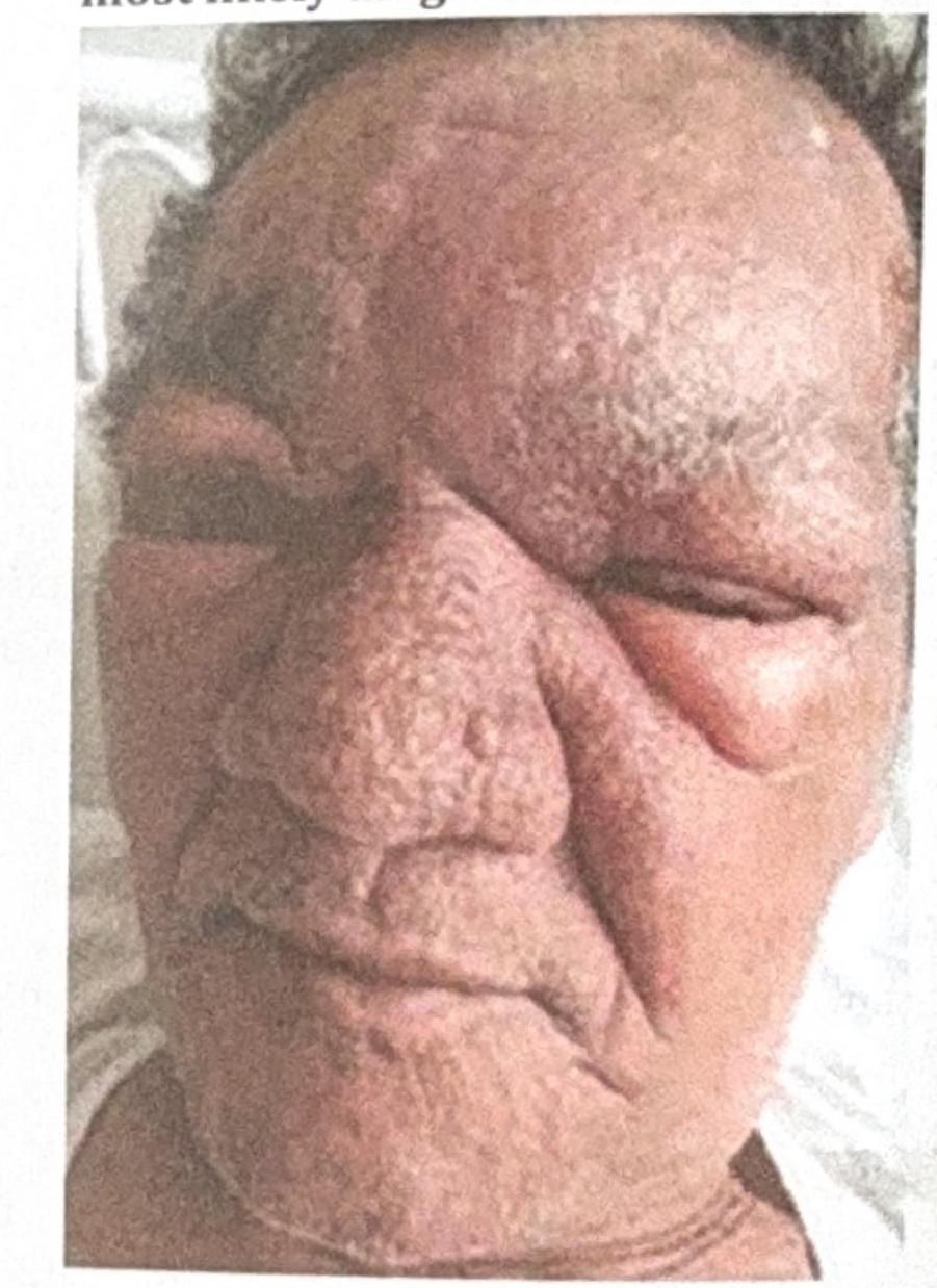
A 53 year-old male presented with erythematous, edematous plaques on his face over pre-existing hypoesthetic patches. He has been experiencing pain for the last 10 days and has been on multibacillary multidrug therapy (MBMDT) for leprosy for the past two months. What is the most likely diagnosis based on the image?

A 12-week pregnant woman on multidrug therapy for leprosy presents with type 2 lepra reaction. What is the treatment of choice for this patient?
Single skin lesion is seen in which type of leprosy -
Multiple hypoaesthetic, hypopigmented macules on right lateral forearm with numerous acid-fast bacilli is indicative of:
A 45-year-old male had multiple hypoesthetic, mildly erythematous large plaques with elevated margins on trunk and extremities. His ulnar and lateral popliteal nerves on both sides were enlarged. The most probable diagnosis is:
Explanation: ***Massive inguinal lymphadenopathy*** - Mycetoma typically has **minimal or no lymph node involvement**, even with extensive lesions. - The lack of significant lymphatic spread is a characteristic feature that differentiates it from other chronic inflammatory conditions. *Most common site is dorsum of foot* - The **foot**, particularly the **dorsum**, is indeed the most common site for mycetoma due to trauma and direct inoculation. - This anatomical location is highly exposed to the environment, increasing the risk of spore implantation. *Granules are aggregates of organized vegetative, septate hyphae* - Mycetoma is characterized by the presence of **"granules"** within the lesions, which are indeed **compact aggregates of fungal hyphae** (for eumycetoma) or bacterial filaments (for actinomycetoma). - These granules can be visualized in discharge or biopsy and are crucial for diagnosis and differentiating between eumycetoma and actinomycetoma. *Amphotericin B has limited role in eumycetoma* - **Amphotericin B** is generally **ineffective** against most causes of eumycetoma, which are difficult to treat with antifungals. - Treatment for eumycetoma often requires more aggressive, **long-term therapy with azoles** (e.g., voriconazole, itraconazole) and surgical debridement, highlighting the limited utility of Amphotericin B.
Explanation: ***LL*** - The image shows a **granuloma with foamy macrophages** (Virchow cells) laden with bacilli, characteristic of **lepromatous leprosy (LL)**. The accompanying clinical features of an anesthetic patch and thickened nerve point towards leprosy. - In LL, there is **poor cell-mediated immunity** with a predominant **Th2 response**, leading to ineffective control of intracellular *Mycobacterium leprae*. While **humoral immunity** is increased, it is ineffective against the intracellular pathogen, resulting in widespread bacterial multiplication and the characteristic **foamy macrophages** (Virchow cells) laden with bacilli. *TT* - **Tuberculoid leprosy (TT)** would typically show a well-formed, epithelioid granuloma with few or no bacilli, reflecting a strong cell-mediated immune response. - Clinical presentation involves well-demarcated, anesthetic patches with significant nerve damage, but the biopsy features would differ from those seen here. *Histiocytosis* - **Histiocytosis** refers to a group of disorders involving abnormal proliferation of histiocytes/macrophages, such as Langerhans cell histiocytosis. - While it involves macrophages, the specific morphology and clinical presentation (anesthetic patch, thickened nerve) are not typical for histiocytosis. *Lymphoma* - **Lymphoma** involves the malignant proliferation of lymphocytes and would present with atypical lymphoid infiltrates, not the macrophage-rich granulomas seen in the image. - The clinical context of an anesthetic patch and thickened nerve is also not characteristic of primary cutaneous lymphoma.
Explanation: ***Predominantly occurs on genitals*** - The image shows a rash consistent with **Pityriasis rosea**, which typically affects the **trunk and proximal extremities**, sparing the face, palms, and soles. - While individual lesions can appear anywhere on the body, the generalized distribution seen here is **not typically genital-predominant**. *HHV-7* - **Pityriasis rosea** is strongly associated with active infection or reactivation of **human herpesvirus 7 (HHV-7)**, and sometimes HHV-6. - Research suggests a causal link, although the exact pathogenic mechanism is still being investigated. *Herald patch* - **Pityriasis rosea** typically begins with a solitary, larger lesion known as a **herald patch** or mother patch, which precedes the generalized eruption by several days or weeks. - This initial lesion is often oval or circular and can be mistaken for other skin conditions. *Spontaneous healing* - **Pityriasis rosea** is a self-limiting rash that usually resolves spontaneously within **6-8 weeks** without specific treatment. - Although the rash can be itchy, the prognosis is excellent, and it rarely recurs.
Explanation: ***Cutaneous larva migrans*** - The image shows **serpiginous, erythematous, and highly pruritic lesions** on the sole, which are characteristic of cutaneous larva migrans. - The patient's history of living in an urban slum and engaging in **open-air defecation** indicates exposure to soil contaminated with **hookworm larvae**, which penetrate the skin and cause these migratory tracks. *Ecthyma* - Ecthyma is a **bacterial skin infection** characterized by **crusted, ulcerative lesions** that extend into the dermis. - It does not present with the characteristic **migratory, linear tracks** seen in the image. *Erysipelas* - Erysipelas is a **superficial bacterial infection** of the dermis with significant lymphatic involvement, presenting as a **sharply demarcated, erythematous, raised rash** with fever and systemic symptoms. - It does not cause the **linear, tunneling appearance** observed in the provided image. *Migratory myiasis* - Myiasis involves infection with **fly larvae** (maggots) that infest living tissue. While some forms of myiasis can be migratory, they typically present as **boil-like lesions** with a central pore, or as subcutaneous nodules, which is different from the **serpiginous tracks** of cutaneous larva migrans. - The source of infection (contaminated soil with hookworm larvae) points away from fly larvae.
Explanation: ***Madura foot*** - The image shows multiple **sinuses** and **abscesses** with evidence of pus discharge on a swollen, deformed foot, which is characteristic of **Madura foot** or **mycetoma**. - This condition is a chronic granulomatous infection of subcutaneous tissues, often caused by **fungi (eumycetoma)** or **bacteria (actinomycetoma)**, and typically affects the foot after minor trauma. *Neuropathic ulcer* - **Neuropathic ulcers** are typically well-demarcated, painless ulcers occurring on pressure points of the foot, often surrounded by calloused skin, and usually present as single lesions. - They do not typically present with the widespread inflammatory swelling, multiple sinuses, and granule discharge seen in the image. *Myrmecia* - **Myrmecia** refers to a specific type of common wart caused by **Human Papillomavirus (HPV)**, characterized by a single, large, deep lesion with surrounding smaller satellite warts. - This condition is a dermal lesion and does not involve the widespread tissue destruction, multiple draining sinuses, and swelling associated with Madura foot. *Sporotrichosis* - **Sporotrichosis** is a subcutaneous fungal infection that often presents as a **lymphocutaneous spread**, with a primary ulcer or nodule and a chain of secondary nodules along lymphatic channels. - While it can cause skin lesions, it typically does not present with the extensive osteomyelitis, soft tissue destruction, and characteristic "grains" or sinus tracts seen in advanced Madura foot.
Explanation: ***Type 1 Lepra reaction*** - The patient presents with **erythematous, edematous plaques on pre-existing hypoesthetic patches** on the face, along with pain and current treatment with **multibacillary multidrug therapy (MBMDT)**. This clinical picture is classic for a type 1 lepra reaction, which is a **delayed-type hypersensitivity reaction** to *Mycobacterium leprae* antigens, often seen during or after treatment. - The image shows significant **facial edema** and **erythema**, particularly around the eyes and nose, consistent with the acute inflammation of a type 1 reaction affecting existing skin lesions and nerves, leading to pain. *Erythema Nodosum Leprosum (ENL)* - ENL is a **Type 2 lepra reaction**, characterized by the appearance of **painful, tender, erythematous nodules** over normal skin, often affecting the limbs and trunk, not typically pre-existing hypoesthetic patches. - It is an **immune complex-mediated reaction** and usually presents more acutely with systemic symptoms like fever and malaise, along with the characteristic nodules, which are not primarily visible in the photograph as widespread edematous plaques. *Cellulitis of the face* - Cellulitis is a **bacterial infection** of the deep dermis and subcutaneous tissue, presenting as a **spreading, warm, red, tender area** with poorly defined borders, often associated with fever and lymphadenopathy. - While there is erythema and edema, the chronic nature of the underlying hypoesthetic patches, the patient's history of leprosy, and the specific distribution suggest a reaction related to leprosy rather than a typical acute bacterial infection. *Erysipelas* - Erysipelas is a **superficial bacterial skin infection**, typically caused by *Streptococcus pyogenes*, characterized by a **sharply demarcated, raised, red, warm, and tender plaque**, often on the face, with characteristic "peau d'orange" texture. - Although it causes facial erythema and edema, the clearly defined borders of erysipelas are not evident, and the association with pre-existing hypoesthetic patches in a leprosy patient points more strongly towards a lepra reaction.
Explanation: ***Continue MDT and add oral steroids*** - **Type 2 lepra reactions (erythema nodosum leprosum)** are inflammatory complications of leprosy and require systemic anti-inflammatory treatment. **Oral corticosteroids** are the mainstay for managing these reactions, particularly in pregnant patients where other immunomodulators are contraindicated. - **Multidrug therapy (MDT)** for leprosy should be continued throughout the reaction, even during pregnancy, to ensure eradication of <b>*Mycobacterium leprae*</b> and prevent drug resistance. Interrupting MDT can lead to relapse and increased neurological damage. *Antibiotics* - This option is incorrect because the type 2 lepra reaction is an **immunological complication** of leprosy, not a bacterial infection requiring additional antibiotics beyond the standard MDT. - The symptoms are due to the immune system's response to dying bacteria, not a new or secondary bacterial infection. *Stop MDT and start oral steroids* - Stopping MDT is inappropriate as the underlying **leprosy infection** still needs to be treated to prevent further progression and drug resistance. - While steroids are crucial for managing the reaction, stopping MDT would compromise the **curative treatment** for leprosy. *Thalidomide* - **Thalidomide** is highly effective in treating **erythema nodosum leprosum (ENL)**. - However, it is an absolute **contraindication** during pregnancy due to its severe **teratogenicity**, causing severe birth defects.
Explanation: ***TT*** - **Tuberculoid leprosy (TT)** is characterized by a strong cell-mediated immune response, leading to a **single, well-demarcated skin lesion** with clear borders and sensory loss. - The few bacilli present are kept localized, preventing widespread dissemination and multiple lesions. *LL* - **Lepromatous leprosy (LL)** is associated with a weak cell-mediated immune response, resulting in **numerous, poorly defined lesions**, nodules, and widespread infiltration. - The high bacillary load leads to systemic involvement and many lesions rather than a single one. *BL* - **Borderline lepromatous (BL)** leprosy is an unstable form with features between borderline tuberculoid and lepromatous, presenting with **multiple, widespread lesions** often with varying sizes and sensory loss. - While skin lesions are present, they are typically numerous and polymorphic, not a single lesion. *BT* - **Borderline tuberculoid (BT)** leprosy presents with **few to several skin lesions**, which are usually smaller, less well-defined, and more numerous than in TT leprosy. - Although closer to TT, it generally involves more than one lesion and exhibits less pronounced sensory loss compared to the single, anesthetic lesion of TT.
Explanation: ***Lepromatous leprosy*** - The hallmark feature is **numerous acid-fast bacilli (AFB)** in skin smears, indicating a **high bacterial load** characteristic of lepromatous leprosy (bacterial index 4-6+). - Lepromatous leprosy is **multibacillary** with **poor cell-mediated immunity**, allowing uncontrolled bacterial multiplication (10⁶-10⁹ bacilli per gram of tissue). - While typically presenting with **widespread, symmetrical lesions**, early lepromatous leprosy can present with **multiple hypopigmented macules** before progressing to diffuse infiltration. - **Sensory loss** may be present but is typically **less pronounced** initially compared to tuberculoid leprosy, as nerve damage occurs gradually. *Borderline leprosy* - Represents the **unstable middle spectrum** (BT, BB, BL) with **moderately impaired immunity** and **variable bacterial load**. - Borderline tuberculoid (BT) has **few AFB**, borderline lepromatous (BL) has **moderate AFB**, but the term "numerous AFB" more specifically indicates lepromatous leprosy. - Lesions are typically **asymmetrical** with variable sensory loss depending on the subtype. *Tuberculoid leprosy* - Characterized by **paucibacillary disease** with **strong cell-mediated immunity** that effectively contains bacterial proliferation. - Skin smears show **few or no detectable AFB** (bacterial index 0-1+) due to robust immune response. - Presents with **few well-defined lesions (1-5)** with **marked sensory loss** and thickened nerves. *Indeterminate leprosy* - The **earliest stage** of leprosy, presenting as a **single hypopigmented or erythematous macule** with minimal sensory changes. - Shows **few or no AFB** on skin smears and may evolve into any form of leprosy or resolve spontaneously. - Not consistent with multiple lesions and numerous bacilli.
Explanation: ***Borderline tuberculoid leprosy*** - The clinical presentation of **multiple hypoesthetic, large plaques with elevated margins** is characteristic of **BT leprosy**, which features **well-demarcated lesions with raised, sloping edges** - **Prominent bilateral nerve enlargement** (ulnar and lateral popliteal nerves) with relatively **few to moderate lesions** is a hallmark of BT leprosy - BT shows **strong cell-mediated immunity** resulting in well-defined plaques with **marked hypoesthesia** and **asymmetric nerve involvement** - The **mildly erythematous** appearance and **elevated margins** indicate active cellular immune response typical of the tuberculoid end of the spectrum *Borderline lepromatous leprosy* - BL presents with **numerous, poorly demarcated lesions** (many more than described) including dome-shaped plaques, papules, and nodules - Lesions in BL have **punched-out centers** rather than elevated margins - Nerve involvement is less prominent relative to the **high number of skin lesions** - The description of well-demarcated plaques with elevated margins points away from the lepromatous pole *Lepromatous leprosy* - LL shows **numerous, diffuse, symmetrical lesions** that are poorly demarcated (macules, papules, nodules, diffuse infiltration) - Lesions are **not hypoesthetic** in early stages due to preserved sensory function until late nerve damage - No elevated margins; lesions are typically smooth, shiny, and dome-shaped *Borderline leprosy* - This is **mid-borderline (BB)** leprosy, which presents with **moderate to numerous lesions** that are more uniform and less well-demarcated than BT - BB has **dimorphous** features without the clear plaques with elevated margins described - The clinical description of elevated margins and prominent nerve involvement with moderate lesion count is more specific for BT
Cutaneous Leishmaniasis
Practice Questions
Leprosy
Practice Questions
Tropical Ulcers
Practice Questions
Onchocerciasis
Practice Questions
Filariasis
Practice Questions
Tropical Mycoses
Practice Questions
Cutaneous Larva Migrans
Practice Questions
Tungiasis
Practice Questions
Myiasis
Practice Questions
Cutaneous Manifestations of Malaria
Practice Questions
Dengue and Other Viral Hemorrhagic Fevers
Practice Questions
Global Health Perspectives in Dermatology
Practice Questions
Get full access to all questions, explanations, and performance tracking.
Start For Free