
Skin Tumors — MCQs

On this page
All are true about squamous cell carcinoma of the skin except?
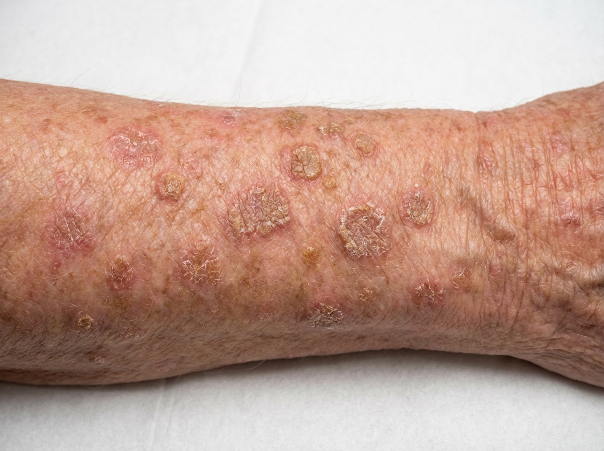
Which of the following is NOT a premalignant skin lesion?
Which of the following is the commonest site for a rodent ulcer?
Which is an aggressive type of cutaneous T-cell lymphoma?
Keratoacanthoma is:
Which skin tumor is known as the "Turban tumor"?
Which of the following melanomas does not have an in situ growth phase?
Prognosis of malignant melanoma depends on which of the following factors?
Which of the following features is most commonly seen?
A 78-year-old woman has multiple long-standing lesions on her face and back. These well-circumscribed lesions are tan to brownish, slightly raised with a rough surface, and typically 0.5 to 1.5 cm in diameter. The clinician examining the patient is able to "peel away" parts of the lesion with the dull side of a scalpel blade. Which of the following diagnoses is most likely?
Practice by Chapter
Benign Epithelial Tumors
Practice Questions
Premalignant Epidermal Tumors
Practice Questions
Basal Cell Carcinoma
Practice Questions
Squamous Cell Carcinoma
Practice Questions
Melanocytic Nevi
Practice Questions
Melanoma
Practice Questions
Merkel Cell Carcinoma
Practice Questions
Vascular Tumors and Malformations
Practice Questions
Cutaneous Lymphomas
Practice Questions
Soft Tissue Tumors
Practice Questions
Metastatic Skin Tumors
Practice Questions
Skin Cancer Prevention and Screening
Practice Questions
Want unlimited practice?
Get full access to all questions, explanations, and performance tracking.
Scan to download app