Which of the following statements about keloids is MOST true?
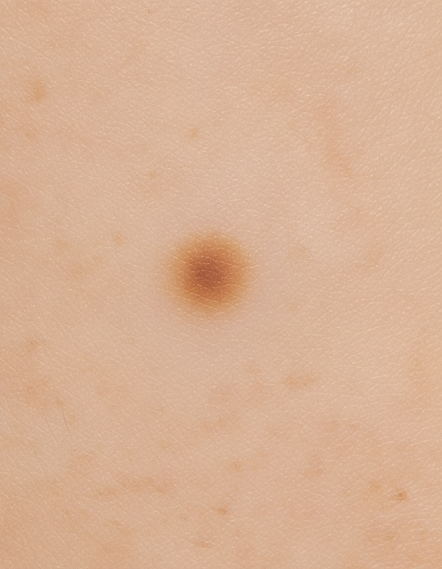
Identify the type of skin lesion shown in the image.

Which of the following statements about Bowen's disease is correct?
All of the following are premalignant conditions except which of the following?
In which part of the body are lesions of Kaposi sarcoma most commonly seen?
A 15cm hyperpigmented macule on an adolescent male undergoes changes such as coarseness, growth of hair & acne. Diagnosis is?
What is the most likely diagnosis for a 15 mm hyperpigmented lesion on the shoulder that is enlarging and has hair growing from it?
Muir–Torre syndrome shows
What is the most common cancer associated with a burn scar?
Which of the following is a common differential diagnosis of verrucous carcinoma?
Explanation: ***Keloids may extend beyond the original wound.*** - Keloids are characterized by their **overgrowth** beyond the boundaries of the original injury. - This distinguishes them from **hypertrophic scars**, which remain confined to the wound edges. *Extended excision is often not the treatment of choice.* - **Excision alone** is usually insufficient for keloids and can even be counterproductive, as the recurring wound often leads to a larger keloid. - While excision can be part of a treatment plan, it is typically combined with supplementary therapies like **steroid injections** or **radiation therapy** to prevent recurrence. *It contains growth factors.* - While keloids involve abnormal fibroblast activity and deposition of **extracellular matrix**, the statement that it "contains growth factors" is too vague and not a defining characteristic that differentiates it from a range of other tissues or conditions. - Many tissues and healing processes involve growth factors, so this statement alone does not provide a specific or most true characteristic of keloids. *None of the options.* - This option is incorrect because the statement that **keloids may extend beyond the original wound** is a hallmark characteristic of keloids and is definitively true.
Explanation: ***Macule*** - The lesion shown is a **macule**, characterized by being **flat and circumscribed** with no elevation above the skin surface. - A macule is defined as a **skin discoloration less than 1 cm in diameter** without any palpable change in texture or thickness. - Classic examples include **freckles, flat nevi, and café-au-lait spots**. - The image demonstrates the typical appearance with clear borders and no vertical component. *Papule* - A papule is an **elevated, solid lesion less than 1 cm** in diameter. - Unlike the lesion shown, papules have a **palpable raised component** above the skin surface. - Examples include acne, warts, and lichen planus. *Patch* - A patch is similar to a macule but is **larger than 1 cm in diameter**. - While also flat, the size criterion differentiates it from a macule. - Examples include vitiligo patches and large birthmarks. *Nodule* - A nodule is a **solid, elevated lesion greater than 1 cm** that extends into deeper skin layers. - It has both elevation and depth, unlike the flat lesion shown. - Examples include dermatofibromas, lipomas, and some cysts.
Explanation: ***It is a form of squamous cell carcinoma in situ.*** - **Bowen's disease** is, by definition, **squamous cell carcinoma in situ (SCC in situ)**. - It is characterized by full-thickness epidermal atypia of keratinocytes **without invasion through the basement membrane**. - This statement is **definitional** and represents the fundamental nature of what Bowen's disease is, making it the **best answer** among the options. *Chronic sun exposure is a known risk factor.* - This statement is **medically accurate**. Chronic UV exposure is indeed a well-established risk factor for Bowen's disease. - However, this describes a **risk factor** rather than defining what the condition is. - Other risk factors include **arsenic exposure**, **ionizing radiation**, **immunosuppression**, and **HPV infection** (particularly in anogenital sites). - While true, this is not as fundamental as the definitional statement. *It is more common in fair-skinned individuals.* - This statement is also **medically accurate**. Bowen's disease occurs more frequently in fair-skinned individuals (Fitzpatrick skin types I-II). - Fair skin provides less melanin protection against UV damage, increasing susceptibility to various forms of skin cancer including Bowen's disease. - However, this describes **epidemiology** rather than defining the condition itself. *There is a link between HSV infection and Bowen's disease.* - This statement is **incorrect**. There is **no established association** between Herpes Simplex Virus (HSV) and Bowen's disease. - **Human Papillomavirus (HPV)**, particularly high-risk types 16 and 18, is associated with Bowen's disease, especially in anogenital locations. - This represents a common confusion between HSV and HPV.
Explanation: ***Pyoderma Gangrenosum*** - This is a **neutrophilic dermatosis** characterized by rapidly enlarging, painful ulcers with undermined, violaceous borders. It is an inflammatory condition, not premalignant. - While often associated with systemic diseases such as **inflammatory bowel disease** or **rheumatoid arthritis**, it does not inherently carry an increased risk of developing into skin cancer. *Bowen's Disease* - This is a form of **squamous cell carcinoma in situ**, meaning the cancerous cells are confined to the epidermis and have not yet invaded the dermis. - It is considered a **premalignant lesion** because it has the potential to progress to invasive squamous cell carcinoma if left untreated. *Actinic Keratosis* - These are **rough, scaly patches** on the skin caused by years of sun exposure, predominantly in fair-skinned individuals. - Actinic keratoses are considered **premalignant lesions** with a risk of transforming into invasive squamous cell carcinoma. *Xeroderma Pigmentosum* - This is a rare, **autosomal recessive genetic disorder** characterized by a defect in DNA repair mechanisms, specifically nucleotide excision repair. - Individuals with xeroderma pigmentosum have an extremely high risk of developing various **skin cancers** (basal cell carcinoma, squamous cell carcinoma, melanoma) at an early age due to their inability to repair UV-induced DNA damage.
Explanation: ***Lower extremities*** - Kaposi sarcoma lesions most frequently appear on the **skin of the lower extremities**, especially the feet and ankles. - This predilection is thought to be due to increased **venous stasis** or other local factors. *Upper extremities* - While Kaposi sarcoma can affect the upper extremities, it is a **less common primary site** compared to the lower limbs. - Lesions here are more likely to appear as the disease **progresses or disseminates**. *Torso* - Kaposi sarcoma lesions can occur on the torso, particularly on the **trunk**, but it is not the most common initial presentation. - Visceral involvement of the **gastrointestinal tract** and lungs can often present without skin lesions on the torso. *Head and neck* - Lesions of Kaposi sarcoma can appear on the head and neck, especially on the **face and oral cavity**, particularly in classic Kaposi sarcoma or in individuals with advanced immunosuppression. - However, this is still **less frequent** than involvement of the lower extremities.
Explanation: ***Becker nevus*** - A Becker nevus is a **hyperpigmented patch** that typically appears during adolescence in males, often on the shoulder or upper trunk. - It characteristically becomes **hairy (hypertrichosis)**, more coarse, and can develop acne within the lesion, particularly during puberty due to androgen sensitivity. *Melanocytic nevus* - While melanocytic nevi are hyperpigmented, they generally do not show the characteristic changes of **coarseness, significant hair growth, or acne** within the lesion during adolescence. - They are typically stable in size and texture after initial development, with changes raising concern for **melanoma**. *Sebaceous nevus* - A sebaceous nevus is a **congenital lesion** often appearing as a yellowish-orange, waxy, or bumpy patch, usually on the scalp or face. - It does not typically present as a large, flat hyperpigmented macule that develops hair and acne in adolescence; instead, it may become verrucous or develop tumors in adulthood. *Sebaceous adenoma* - A sebaceous adenoma is a **benign tumor** of the sebaceous glands, usually appearing as a small, solitary, flesh-colored to yellowish papule or nodule, especially on the face. - It is not typically seen as a large, hyperpigmented macule that grows hair and acne over a broad area, as described in the question.
Explanation: ***Correct: Becker nevus*** This diagnosis is supported by the description of a **hyperpigmented lesion** that is **enlarging** and has **hair growing from it**, typically appearing during adolescence or young adulthood. **Becker nevus** often presents as an **irregular, hyperpigmented patch**, usually on the shoulder or upper trunk, and is characteristically associated with **hypertrichosis** (increased terminal hair growth). The combination of location (shoulder), enlargement, and hair growth in a 15 mm lesion is classic for Becker nevus. *Incorrect: Melanocytic nevus* While **melanocytic nevi** are hyperpigmented, they typically do not continue to **enlarge significantly** after childhood and generally do not develop new onset **hypertrichosis** as a primary feature. The size (15 mm) and progressive growth combined with hair development are more characteristic of a Becker nevus than a common melanocytic nevus. *Incorrect: Sebaceous nevus* **Sebaceous nevi** are typically **yellow-orange to tan, waxy plaques**, often on the scalp or face, with a cobblestone or papillomatous texture. They are not primarily characterized by **hyperpigmentation** and terminal hair growth, but rather by sebaceous gland proliferation. *Incorrect: Comedo nevus* A **comedo nevus** presents as a linear or unilateral group of **dilated follicular openings** filled with keratinous material, resembling blackheads. It is not characterized by diffuse **hyperpigmentation** or the increased terminal hair growth described in this case.
Explanation: ***Sebaceous gland tumors*** - **Muir-Torre syndrome** is a genetic condition characterized by the presence of at least one **sebaceous gland tumor** (adenoma, epithelioma, or carcinoma) and at least one internal malignancy. - It is considered a variant of **Lynch syndrome (hereditary nonpolyposis colorectal cancer - HNPCC)**, stemming from germline mutations in **DNA mismatch repair genes**. *Intestinal polyps (associated with familial adenomatous polyposis)* - While Lynch syndrome (to which Muir-Torre is related) does involve an increased risk of colorectal cancer, **multiple intestinal polyps** are the hallmark of **Familial Adenomatous Polyposis (FAP)**. - FAP is caused by a mutation in the **APC gene**, distinct from the mismatch repair gene mutations seen in Muir-Torre syndrome. *Lisch nodules (associated with Neurofibromatosis type 1)* - **Lisch nodules** are benign **iris hamartomas** typically found in patients with **Neurofibromatosis type 1 (NF1)**. - NF1 is a neurocutaneous disorder caused by a mutation in the **NF1 gene**, presenting with café-au-lait spots, neurofibromas, and optic gliomas, which are unrelated to Muir-Torre syndrome. *Hyperelastic joints (associated with Ehlers-Danlos syndrome)* - **Hyperelasticity of joints** and skin is a characteristic feature of **Ehlers-Danlos syndrome (EDS)**, a group of heritable disorders affecting connective tissue. - EDS is caused by defects in **collagen synthesis or processing**, and its clinical manifestations are distinct from the mucocutaneous and internal malignancies seen in Muir-Torre syndrome.
Explanation: ***Squamous cell carcinoma*** - **Squamous cell carcinoma (SCC)** is the most common malignancy arising in chronic wounds, including **Marjolin's ulcer** (non-healing chronic ulcers), burn scars, and chronic osteomyelitis sinuses. - It often presents as a **non-healing nodular or ulcerative lesion** within the scar tissue. *Fibrosarcoma* - **Fibrosarcoma** is a rare malignant tumor of fibroblasts that can arise in scar tissue, but it is less common than SCC. - It is a type of **soft tissue sarcoma** and typically presents as a firm, rapidly growing mass. *Adenocarcinoma* - **Adenocarcinoma** originates from glandular epithelial cells and is not typically associated with burn scars. - While some rare cutaneous adenocarcinomas exist, they are not the primary malignancy seen in these contexts. *Adeno-squamous Ca* - **Adeno-squamous carcinoma** is a mixed tumor with both glandular and squamous differentiation. - While it has squamous components, it is a less common and more aggressive variant, and SCC without glandular differentiation is the most frequent.
Explanation: ***Condylomata acuminata*** - Both **verrucous carcinoma** and **condylomata acuminata** present as **warty, exophytic lesions**, making differentiation difficult without biopsy. - While verrucous carcinoma is a **well-differentiated squamous cell carcinoma**, condylomata acuminata are benign **genital warts caused by HPV**, highlighting the need for careful diagnosis. *Condylomata lata* - These are **flat, moist, broad-based lesions** associated with **secondary syphilis**, which are distinct from the more exophytic, warty appearance of verrucous carcinoma. - They are typically **painless and highly infectious**, and a diagnosis of syphilis would be confirmed with serological tests. *Adenocarcinoma* - **Adenocarcinomas** originate from **glandular tissue** and typically present as **ulcerative, infiltrative, or polypoid masses**, not wart-like growths. - Their histological features involve **glandular differentiation**, which is fundamentally different from the squamous cell proliferation seen in verrucous carcinoma. *Tuberculosis* - Cutaneous tuberculosis can present in various forms, including **lupus vulgaris** or **scrofuloderma**, but typically does not produce the large, verrucous, cauliflower-like growths seen in verrucous carcinoma. - Diagnosis commonly relies on **histopathology showing granulomas** with caseous necrosis and **acid-fast bacilli**, differing from squamous cell malignancy.
Benign Epithelial Tumors
Practice Questions
Premalignant Epidermal Tumors
Practice Questions
Basal Cell Carcinoma
Practice Questions
Squamous Cell Carcinoma
Practice Questions
Melanocytic Nevi
Practice Questions
Melanoma
Practice Questions
Merkel Cell Carcinoma
Practice Questions
Vascular Tumors and Malformations
Practice Questions
Cutaneous Lymphomas
Practice Questions
Soft Tissue Tumors
Practice Questions
Metastatic Skin Tumors
Practice Questions
Skin Cancer Prevention and Screening
Practice Questions
Get full access to all questions, explanations, and performance tracking.
Start For Free