The image shows presence of:

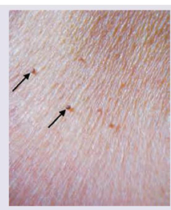
A 50-year-old patient presents with lesion over the nose with rapid growth for last 6 weeks. He has no past history of any skin disease. The image shows presence of?

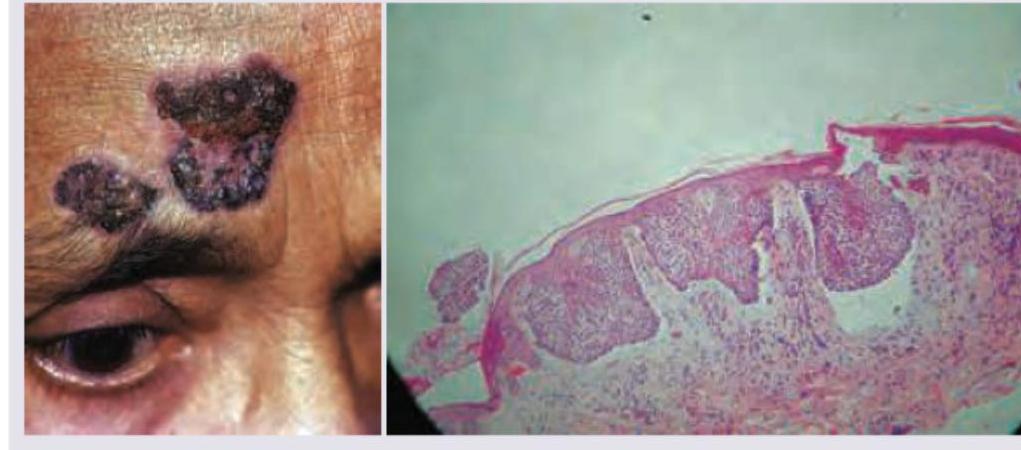
A 70-year-old man presents with an ulcerative lesion over the forehead. A biopsy was performed. All are true about the condition shown except?

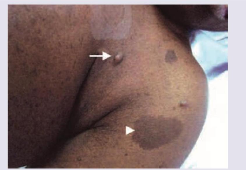
A 25 -year-old patient presents with multiple sebaceous adenomas over the neck and chest. His father too had a similar skin disease and died due to colorectal carcinoma. What is the diagnosis?
The following lesion was noticed in a patient with history of involuntary weight loss. What is the diagnosis?

A 38-year-old man presents with the manifestation shown in the image. He has a number of family members suffering from the same condition, though the severity is different in different members. Which of the following statements is false regarding this condition?

All of the following statements are true for keloids EXCEPT:
A 70 year old man with history of smoking has a 1 cm ulcerative lesion over the vermilion of his upper lip. What is he likely to be suffering from?
A 40 year old man presented with a flat 1x1cm scaly, itchy black mole on the front of thigh. Examination did not reveal any inguinal lymphodenopathy. The best course of management would be:
Which of the following statements is not correct regarding sebaceous cyst?
Explanation: ***Correct: Syringoma*** - Syringomas are **benign adnexal tumors** derived from the eccrine sweat ducts, often appearing as **small, flesh-colored to yellowish papules**, commonly around the eyes or on the face, as seen in the image. - They are typically asymptomatic but can be numerous and persistent. - Histologically, they show **comma-like or tadpole-shaped ducts** in the dermis. *Incorrect: Cutaneous horn* - A cutaneous horn is a **conical projection** of keratin from the skin, often hard and yellowish, resembling an animal's horn, which is not depicted in the image. - It can be associated with underlying benign, premalignant, or malignant lesions. *Incorrect: Acrochordon* - Acrochordons, or **skin tags**, are soft, flesh-colored, pedunculated growths that typically appear in areas of friction like the neck, axilla, or groin. - The lesions in the image are small, firm papules, not soft, pendulous structures. *Incorrect: Keratoacanthoma* - Keratoacanthomas are **rapidly growing, dome-shaped nodules** with a central keratin plug, often appearing on sun-exposed skin. - They have a characteristic crater-like appearance with rolled edges, which is distinct from the lesions in the image.
Explanation: ***Keratoacanthoma*** - Keratoacanthomas are characterized by **rapid growth** over weeks to months, forming a **dome-shaped nodule with a central keratotic plug**, often resembling a volcano. - While generally considered benign, they share morphological and histological features with squamous cell carcinoma, making differentiation crucial. *Seborrheic keratosis* - Seborrheic keratosis typically presents as a **well-demarcated, waxy, 'pasted-on' lesion** that grows slowly over years. - They are usually flat or slightly raised with a finely fissured or verrucous surface, not a rapidly growing nodule with a central crater. *Syringoma* - Syringomas are **benign eccrine sweat duct tumors** that appear as small (1-3 mm) skin-colored or yellowish papules, often found around the eyelids. - They do not exhibit rapid growth or the characteristic central crater seen in the image. *Cutaneous horn* - A cutaneous horn is a **clinical description** of a conical, hyperkeratotic lesion, but it is not a specific diagnosis; rather, it indicates the presence of an underlying pathology like an actinic keratosis, squamous cell carcinoma, or warts. - While it can appear on the nose, the rapid growth and dome shape with a central crater are more indicative of a keratoacanthoma than a generic cutaneous horn.
Explanation: ***Pautrier's micro-abscess formation*** - Pautrier's micro-abscesses are characteristic findings in **Mycosis fungoides (cutaneous T-cell lymphoma)**, which is not depicted here. - The image and description are consistent with basal cell carcinoma, where these micro-abscesses do not occur. *Basaloid cell nests extending into epidermis* - The biopsy image shows nests of **basaloid cells** extending downwards from the epidermis, which is a classic histopathological feature of **basal cell carcinoma**. - These nests often exhibit peripheral palisading and retraction artifact, distinguishing features of this common skin cancer. *Locally destructive* - **Basal cell carcinoma** is known for its **local invasiveness and destructive growth pattern**, often eroding surrounding tissues. - While it rarely metastasizes, its local destruction can cause significant morbidity, especially on the face. *Does not metastasize usually* - **Basal cell carcinoma has a very low metastatic potential**, making it distinct from many other malignancies. - Metastasis is exceedingly rare, occurring in less than 0.1% of cases, though local recurrence is common if not completely excised.
Explanation: ***Muir Torre syndrome*** - This syndrome is characterized by the presence of at least one sebaceous gland tumor (like **sebaceous adenoma**, sebaceous epithelioma, or sebaceous carcinoma) and at least one internal malignancy, most commonly **colorectal carcinoma** or urogenital cancer. - The patient's presentation with **multiple sebaceous adenomas** and a family history of **colorectal carcinoma** (father) is highly indicative of Muir Torre syndrome, which is a variant of **Lynch syndrome**. *Pseudoxanthoma elasticum* - This is an inherited disorder affecting **elastic fibers** in the skin, eyes, and blood vessels, leading to yellowish papules that coalesce into plaques, often in flexural areas. - It is not typically associated with **sebaceous adenomas** or **colorectal carcinoma**. *Bournville disease* - This is a historical term for **tuberous sclerosis complex**, a genetic disorder characterized by tumor growth in multiple organs including the brain, heart, kidneys, and skin. - Skin manifestations include **ash-leaf spots**, facial angiofibromas, and shagreen patches, but not **sebaceous adenomas** in the context of internal malignancy like colorectal cancer. *Sweet syndrome* - Also known as **acute febrile neutrophilic dermatosis**, Sweet syndrome is characterized by the sudden onset of painful, erythematous plaques and nodules accompanied by fever and neutrophilia. - It is often associated with malignancy (especially hematologic), infection, or drugs, but it does not present with **sebaceous adenomas** or a familial link to specific carcinomas like colorectal cancer.
Explanation: ***Leser-Trelat sign*** - The image illustrates numerous rapidly appearing or increasing **seborrheic keratoses**, which, when accompanied by symptoms like **involuntary weight loss**, are highly suggestive of the Leser-Trelat sign. - The Leser-Trelat sign is a **paraneoplastic syndrome** commonly associated with **gastrointestinal adenocarcinomas** or **lymphoid malignancies**. *Acanthosis nigricans* - This condition presents as **dark, velvety patches** on the skin, typically in body folds like the neck, armpits, and groin. - While it can be associated with malignancy (especially gastric adenocarcinoma), the image shows multiple, distinct seborrheic keratoses rather than diffuse hyperpigmentation. *Actinic keratosis* - Actinic keratoses are **premalignant lesions** caused by chronic sun exposure, appearing as rough, scaly patches on sun-exposed areas. - They are typically single or a few scattered lesions and do not usually erupt rapidly or widely as shown, nor are they directly associated with systemic malignancy in the same way as Leser-Trelat. *Intertriginous candida* - Intertriginous candidiasis is a **fungal infection** that occurs in skin folds, characterized by **redness, itching, and satellite lesions**. - The lesions in the image are distinct, raised, and brownish, not consistent with the erythematous and often moist presentation of intertriginous candidiasis.
Explanation: ***Basal cell carcinoma*** - Basal cell carcinoma is a type of skin cancer that is **not typically a primary manifestation** of Neurofibromatosis Type 1 (NF1). - While individuals with NF1 may have an increased risk of certain cancers, basal cell carcinoma is **not one of the characteristic features** of the condition. - **This statement is FALSE**, making it the correct answer to this question. *Autosomal dominant condition* - Neurofibromatosis Type 1 (NF1) is inherited in an **autosomal dominant pattern**, meaning only one copy of the mutated gene is needed to cause the disorder. - The patient's history of **multiple family members** suffering from the same condition with varying severity is consistent with autosomal dominant inheritance. - **This statement is TRUE.** *Chromosomal abnormality seen in chromosome 17* - Neurofibromatosis Type 1 is caused by a mutation in the **NF1 gene**, located on **chromosome 17 (17q11.2)**. - This gene encodes for **neurofibromin**, a tumor suppressor protein, and its dysfunction leads to the characteristic features of NF1. - **This statement is TRUE.** *Café-au-lait spots and neurofibromas are characteristic features* - The image displays characteristic features of NF1, including **café-au-lait spots** (large, hyperpigmented macules) and **neurofibromas** (benign tumors of nerve sheath cells). - These are classic diagnostic findings in Neurofibromatosis Type 1, along with axillary/inguinal freckling and Lisch nodules. - **This statement is TRUE.**
Explanation: ***True keloid does not spread into surrounding tissue*** - This statement is **incorrect** as a defining characteristic of keloids is their tendency to **spread beyond the original wound boundaries**, invading surrounding healthy tissue. - This expansive growth differentiates keloids from hypertrophic scars, which remain confined to the site of injury. *The maturation and stabilization of the collagen fibrils is inhibited* - This statement is **true**. In keloids, there is an impairment in the normal maturation process of collagen, leading to an accumulation of **immature, disorganized collagen fibrils**. - This abnormal collagen synthesis and degradation contribute to the excessive and persistent fibrosis characteristic of keloids. *It is rarely seen in white skinned persons and is more common over the sternum* - This statement is **true**. Keloids are more prevalent in individuals with **skin of color (e.g., African, Hispanic, and Asian descent)** and are less common in Caucasians. - Common locations for keloids include the **sternum**, earlobes, shoulders, and upper back, areas under significant skin tension. *True keloid continues to become worse even after one year* - This statement is **true**. Unlike hypertrophic scars which may regress over time, keloids tend to be **persistent and progressive**, often continuing to grow and worsen in size and appearance even years after the initial injury. - They typically do not resolve spontaneously and may even recur after excision.
Explanation: ***Squamous cell carcinoma*** - **Squamous cell carcinoma (SCC)** is a **common malignancy** of the lip, particularly in older men with a history of **smoking** and **sun exposure**. - **Ulcerative lesions** on the vermilion border are characteristic clinical features of SCC, necessitating biopsy for confirmation. *Spindle cell carcinoma* - **Spindle cell carcinoma** is a rare, aggressive variant of **squamous cell carcinoma**, but it is not the most likely primary diagnosis given the typical presentation. - Diagnosis typically requires **histopathological examination** showing spindle-shaped malignant cells, which cannot be determined clinically. *Basal cell carcinoma* - **Basal cell carcinoma (BCC)** is more common on sun-exposed areas of the face but is rare on the **vermilion border of the lips**. - BCC lesions typically present as **pearly nodules** with **telangiectasia** or **rolled borders**, rather than a purely ulcerative lesion as described. *Adenoid squamous carcinoma* - **Adenoid squamous carcinoma** is also a rare variant of **squamous cell carcinoma** that exhibits glandular differentiation. - This specific subtype is not the most common presentation for an ulcerative lip lesion, making simple **squamous cell carcinoma** a more likely diagnosis.
Explanation: ***Excision biopsy*** - A **flat, scaly, itchy, black mole** is highly suspicious for **melanoma**, and an excision biopsy provides the most accurate histopathological diagnosis and depth assessment. - This procedure removes the entire lesion with a narrow margin of normal-appearing skin, allowing for comprehensive evaluation of its nature and determining further management. *FNAC of lesion* - **Fine needle aspiration cytology (FNAC)** is generally used for evaluating palpable masses or lymph nodes, not primary skin lesions like a suspicious mole. - It provides only cellular samples, making it difficult to assess architectural features, depth of invasion, or determine definitive malignancy in skin lesions. *Incision biopsy* - An **incision biopsy** involves removing only a partial sample of the lesion, which can lead to sampling error and an inaccurate diagnosis if the most aggressive part is missed. - For suspected melanoma, an incomplete biopsy can compromise subsequent staging and definitive treatment planning. *Wide excision with inguinal lymphadenectomy* - This is an **overly aggressive initial approach** before a definitive diagnosis of melanoma and its stage has been established. - **Wide excision** is typically performed after an excision biopsy confirms melanoma and determines its depth, while **lymphadenectomy** is indicated for confirmed lymph node involvement.
Explanation: ***Treatment is incision and drainage*** - The standard treatment for a sebaceous cyst (more accurately an **epidermoid cyst** or **pilar cyst**) is **surgical excision** of the entire cyst wall to prevent recurrence. - **Incision and drainage** only provides temporary relief by emptying the contents but leaves the cyst wall intact, leading to a high chance of the cyst refilling. *Found on hairy areas of the body* - This statement is generally correct as sebaceous cysts often arise from hair follicles and are common in **hair-bearing areas** like the scalp, face, neck, and trunk. - They occur due to the accumulation of **sebum** and keratin within a blocked or damaged sebaceous gland or hair follicle. *Not found on palms and soles* - This statement is correct because **palms and soles** generally **lack sebaceous glands** and hair follicles, hence sebaceous cysts are typically not found in these locations. - Cysts found in these areas are more likely to be **ganglion cysts** or other types of epidermal inclusion cysts. *It has a punctum* - This statement is often correct; many sebaceous cysts (especially epidermoid cysts) have a visible **central punctum** which represents the occluded pore from which the cyst originated. - This punctum is a **key diagnostic feature** and can sometimes exude a cheesy, foul-smelling material.
Benign Epithelial Tumors
Practice Questions
Premalignant Epidermal Tumors
Practice Questions
Basal Cell Carcinoma
Practice Questions
Squamous Cell Carcinoma
Practice Questions
Melanocytic Nevi
Practice Questions
Melanoma
Practice Questions
Merkel Cell Carcinoma
Practice Questions
Vascular Tumors and Malformations
Practice Questions
Cutaneous Lymphomas
Practice Questions
Soft Tissue Tumors
Practice Questions
Metastatic Skin Tumors
Practice Questions
Skin Cancer Prevention and Screening
Practice Questions
Get full access to all questions, explanations, and performance tracking.
Start For Free