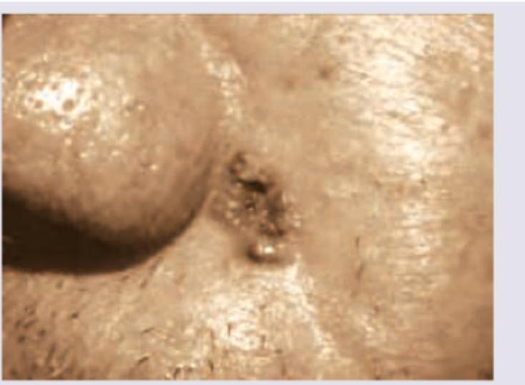
A patient presents with a lesion on the sun-exposed area shown in the image. What is the most likely diagnosis?

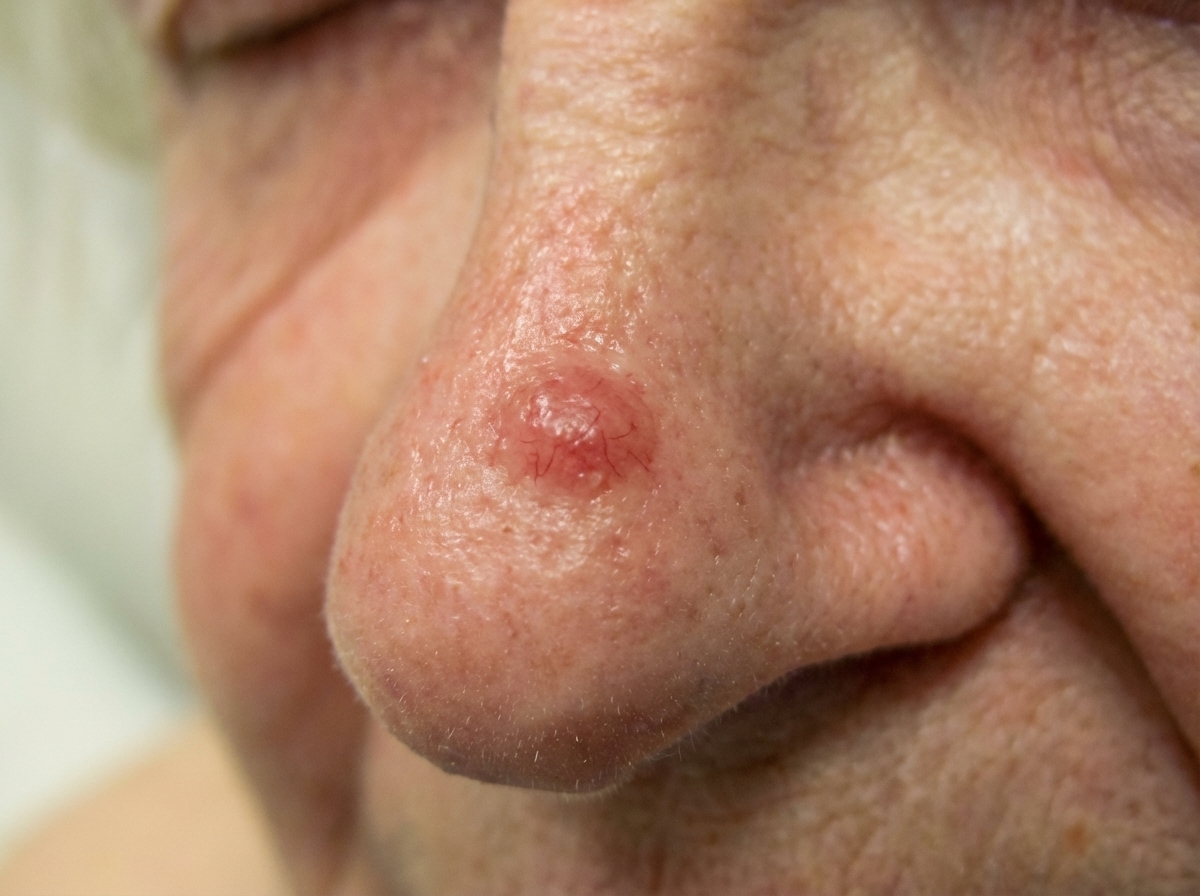
A patient presents with ulcer on the side of the nose, as shown, which bleeds on itching. What is the diagnosis? (AIIMS Nov 2017)

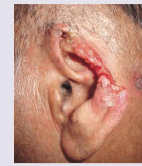
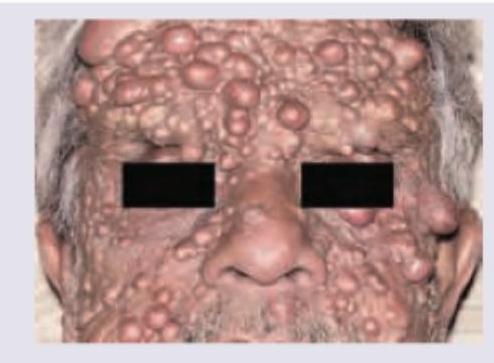
All are true about the lesion shown below except:

What is true regarding the picture shown below?

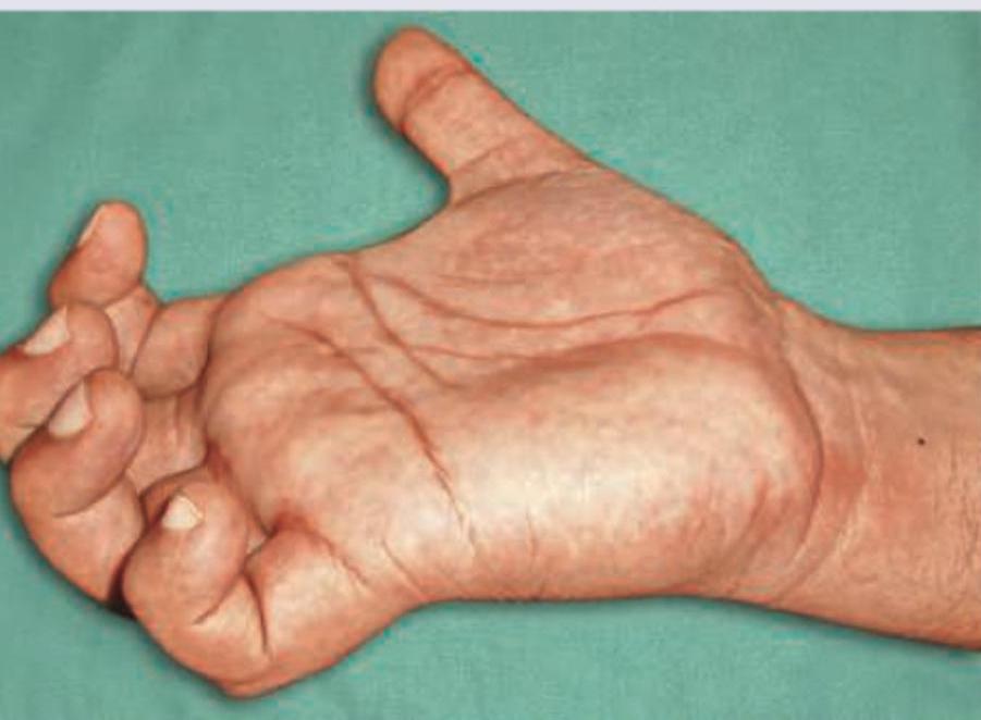
The following clinical presentation is seen in injury to which nerve? (Recent NEET Pattern 2016-17)

The image given below shows:

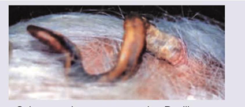
Identify the lesion: (Recent NEET Pattern 2016-17)

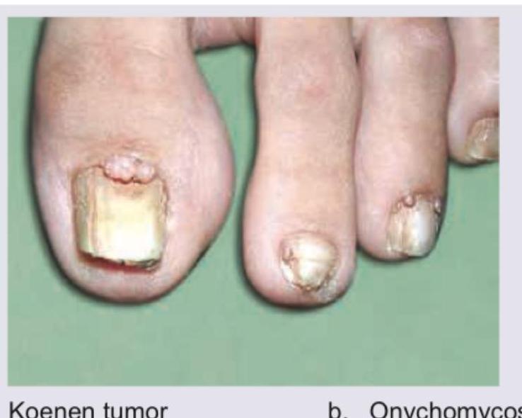
What is the diagnosis based on the clinical image shown?

The following image shows presence of:

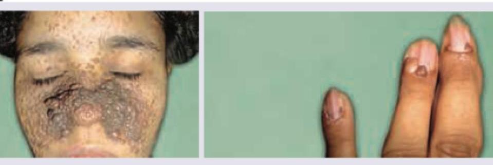
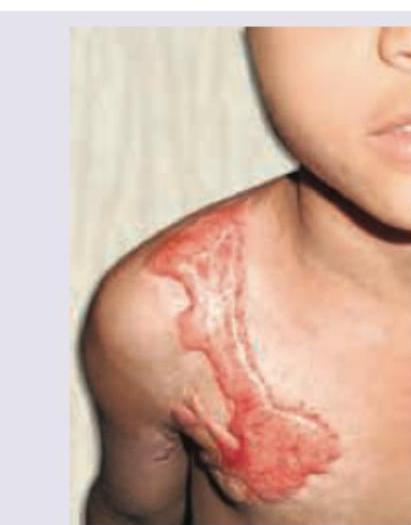
An 8-year-old girl has extreme photosensitivity since birth. She has recently been diagnosed with skin cancer. What is the diagnosis?
Explanation: ***Basal cell carcinoma*** - **Basal cell carcinoma** is the **most common skin cancer**, typically presenting as a **pearly, translucent nodule** with **rolled borders** and **central ulceration** on sun-exposed areas. - It frequently occurs on the **face, scalp, and neck** due to chronic **UV radiation exposure** and has a very low metastatic potential. *Squamous cell carcinoma* - **Squamous cell carcinoma** typically presents as a **red, scaly patch** or **firm, keratotic nodule** that may ulcerate and bleed. - While also common in sun-exposed areas, it has a **higher metastatic potential** than basal cell carcinoma and often arises from **actinic keratoses**. *Melanoma* - **Melanoma** is a less common but highly **aggressive skin cancer** arising from **melanocytes** with significant metastatic potential. - It typically presents as an **atypical mole** with **asymmetry, irregular borders, color variation, and diameter >6mm** (ABCD criteria). *Actinic keratosis* - **Actinic keratosis** is a **precancerous lesion** presenting as **rough, scaly, erythematous patches** on sun-damaged skin. - It represents **dysplastic keratinocytes** that can progress to **squamous cell carcinoma** but is not malignant itself.
Explanation: ***Basal cell carcinoma*** - The image shows a **pearly appearance** with a **central ulceration** and **rolled borders**, which are classic clinical features of **nodular basal cell carcinoma**. - **Bleeding upon minor trauma or itching** (friability) is also a common characteristic of basal cell carcinoma, especially the ulcerated form. *Squamous cell carcinoma* - Squamous cell carcinoma typically presents as a **crusted, firm, red nodule** or a **patch with scales and ulceration**, often in sun-exposed areas. - While it can ulcerate and bleed, it usually lacks the distinct **pearly appearance and rolled borders** seen in basal cell carcinoma. *Marjolin ulcer* - A Marjolin ulcer is a **malignant degeneration of a chronic ulcer**, burn scar, or chronic inflammatory lesion, most commonly an aggressive squamous cell carcinoma. - It would present in the context of a **pre-existing long-standing lesion** or scar, which is not indicated here, and generally has a more irregular, indurated appearance. *Nevus* - A nevus (mole) is a benign growth of pigment-producing cells which typically presents as a **well-demarcated pigmented lesion**. - While some nevi can be raised or irregular, they generally do **not ulcerate spontaneously or bleed easily** on scratching unless traumatized, nor do they typically have the pearly, rolled borders of a basal cell carcinoma.
Explanation: ***Severe itching*** - While keloids can cause **pruritus (itching)**, it is **variable** and not uniformly severe across all cases. - The intensity of itching in keloids varies significantly between patients, and it is **not as consistently present or severe** as the other defining features listed. - The primary **consistent symptoms** of keloids are **abnormal scarring beyond wound boundaries (claw-like extensions), firm raised appearance, and cosmetic concerns**, rather than severe pruritus being a universal hallmark. *Claw like process* - The image shows a **keloid**, which is characterized by its tendency to grow **beyond the boundaries of the original wound**, often forming **claw-like extensions**. - This **exuberant growth** into surrounding normal tissue is a **key differentiating feature** of keloids compared to hypertrophic scars, which remain within the original wound boundaries. *Develops at vaccination sites* - Keloids commonly develop at sites of **skin trauma**, including **vaccination sites**, piercings, acne, and surgical incisions. - The **deltoid region**, where many vaccinations are administered, is a frequent location for keloid formation, especially in **predisposed individuals** (more common in darker skin types and with genetic predisposition). *Collagen bundles are present* - Keloids are composed of **abnormally dense and disorganized collagen bundles** in the dermis, particularly **Type I and Type III collagen**. - Histologically, they show **thick, hyalinized collagen fibers** arranged in a haphazard fashion, extending into the subcutaneous fat, which distinguishes them from normal scars.
Explanation: ***All these options are true*** - The image depicts a **malignant lesion on the ear**, characterized by extensive tissue destruction and ulceration. This appearance is highly consistent with advanced **basal cell carcinoma (BCC)**, especially the aggressive, infiltrative type. - As all the preceding statements describe characteristic features of basal cell carcinoma, and the image is consistent with this diagnosis, this option correctly synthesizes the validity of the individual choices. *This is basal cell carcinoma which arises from the basal layer of keratinocytes* - The image shown, with its destructive ulceration and typical location on the ear (a sun-exposed area), is highly suggestive of advanced **basal cell carcinoma (BCC)**. - **BCC** originates from the **basal keratinocytes of the epidermis**, which are the proliferative cells in the basal layer that give rise to the overlying epidermis. - BCC is the most common skin cancer and typically presents on sun-exposed areas with various morphological patterns. *Rodent ulcer appearance* - The term "**rodent ulcer**" is a classical description for an advanced, ulcerative form of **basal cell carcinoma**. - This appearance is characterized by a **central ulceration** with rolled, pearly borders and often presents with extensive local destruction, as seen in the image. *Exposure to solar UV radiation is a risk factor* - **Ultraviolet (UV) radiation from sun exposure** is the primary and most significant **environmental risk factor** for the development of basal cell carcinoma. - BCC commonly occurs on **sun-exposed areas** such as the face, ears, neck, and hands, reinforcing the link to UV radiation.
Explanation: ***Median nerve*** - The image illustrates **'ape hand' deformity**, characterized by the inability to oppose the thumb and atrophy of the thenar eminence due to paralysis of the **thenar muscles** (abductor pollicis brevis, opponens pollicis, superficial head of flexor pollicis brevis). - These thenar muscles are primarily innervated by the **recurrent branch of the median nerve**. *Ulnar nerve* - Ulnar nerve injury typically causes **'claw hand' deformity** (hyperextension of MCP joints and flexion of IP joints of 4th and 5th digits) due to paralysis of the intrinsic muscles of the hand (interossei and medial two lumbricals). - It would also cause prominent atrophy of the **hypothenar eminence** and interosseous muscles. *Median and ulnar nerve* - Combined median and ulnar nerve injury would result in a **more severe and widespread paralysis** affecting nearly all intrinsic hand muscles, leading to a profound loss of hand function. - This would present with features of both **ape hand** and **claw hand**, commonly referred to as a **'simian hand'** if severe. *Radial nerve* - Radial nerve injury typically causes **'wrist drop'** and inability to extend the fingers and thumb at the metacarpophalangeal joints. - It primarily affects the **extensor muscles of the forearm** and does not directly result in thenar atrophy or the inability to oppose the thumb.
Explanation: ***Cutaneous horn (Cornu cutaneum)*** - The image distinctly shows an **exophytic, conical, or cylindrical lesion** composed of compact keratin, resembling an animal's horn, which is characteristic of a cutaneous horn. - A cutaneous horn is a **clinical descriptive term** for a keratinous projection and is NOT of sebaceous origin; it can arise from various underlying conditions including **seborrheic keratosis, viral warts, actinic keratosis**, or benign lesions. - Importantly, cutaneous horns can rarely **harbor squamous cell carcinoma** or other underlying skin cancers at their base (up to 20% have underlying malignancy), necessitating biopsy and histopathological examination. *Papilloma* - A papilloma is a general term for a **benign epithelial tumor** growing exophytically in a frond-like or papillary pattern, often softer and not typically forming such a dense, hardened projection. - While some papillomas can be keratotic, they usually lack the extreme **horn-like appearance** made of densely packed keratin seen in the image. *Cock's peculiar tumor* - Cock's peculiar tumor, also known as a **calcifying epithelioma of Malherbe** or **pilomatricoma**, is typically a firm, deep-seated nodule arising from hair matrix cells, often with a bluish or reddish hue. - It does not present as a **hard, projecting, horn-like accumulation of keratin** on the skin surface. *Glomus tumor* - A glomus tumor is a rare, **benign neurovascular tumor** typically found in the digits (especially subungual), characterized by exquisite pain, cold sensitivity, and often present as a small, reddish-blue nodule. - Its presentation is distinctly different from the **hyperkeratotic cutaneous projection** shown and does not form a horn-like structure.
Explanation: ***Koenen tumor*** - Koenen tumors, also known as **periungual fibromas**, are small, benign growths that emerge from beneath the nail fold or nail plate. - They are a classic cutaneous manifestation of **tuberous sclerosis complex**, characterized by firm, flesh-colored papules or nodules, often at the base or sides of the nails, as seen in the image. *Onychomycosis* - This is a fungal infection of the nail, typically causing **thickening**, **discoloration (yellow, brown, white)**, and **crumbling of the nail plate**. - While some discoloration is present, the prominent **periungual nodule** is not characteristic of onychomycosis alone. *Onychodystrophy* - This is a general term for any condition that results in abnormal nail appearance or growth, often due to trauma, systemic disease, or local pathology. - It describes the state of the nails, but does not identify the **specific proliferative lesion** at the nail fold. *Onychogryphosis* - Also known as "ram's horn nail," this condition involves **marked thickening and curvature of the nail plate**, often due to chronic trauma or neglect. - The nails in the image show some thickening and discoloration, but the characteristic **overgrowth and extreme curvature** of onychogryphosis are not the primary feature, nor is the periungual growth explained by this term.
Explanation: ***Plexiform neurofibroma*** - The image displays numerous **nodular, flesh-colored to hyperpigmented lesions** predominantly on the face, characteristic of **plexiform neurofibromas**, which are often present in **Neurofibromatosis type 1 (NF1)**. - These lesions are typically soft, sometimes described as feeling like a "bag of worms" on palpation, due to the proliferation of neural tissue, and can cause significant disfigurement. *Proteus syndrome* - Proteus syndrome is characterized by **overgrowth of various tissues**, bones, and organs, leading to asymmetric and disproportionate growth. - While it can involve skin lesions such as **cerebriform connective tissue nevi**, the appearance in the image with diffuse, multiple, distinct neurofibroma-like nodules does not align with the typical presentation of Proteus syndrome. *Cruveilhier-Baumgarten disease* - This is a rare condition characterized by the presence of an **umbilical caput medusae** (dilated veins radiating from the umbilicus), due to portal hypertension with a patent umbilical vein not seen in the image. - It relates to circulatory abnormalities in the abdomen and does not present with cutaneous lesions as shown in the image. *Dermal neurofibroma* - While dermal neurofibromas are benign nerve sheath tumors that can occur on the skin, they are typically **discrete, solitary, or few in number**, often presenting as soft, sessile or pedunculated lesions. - The extensive, diffuse, and nodular appearance across the entire face in the image suggests a more widespread and deeply intertwined growth pattern characteristic of **plexiform neurofibromas**, rather than isolated dermal neurofibromas.
Explanation: ***Angiofibroma, Peri-ungual fibroma*** - The image on the left shows facial **angiofibromas**, characterized by small, reddish-brown papules predominantly on the central face, butterfly distribution on the nose, and cheeks. - The image on the right displays **peri-ungual fibromas** (Koenen tumors), which are flesh-colored growths emerging from under the nail fold, typically seen in tuberous sclerosis. *Angiokeratoma, Peri-ungual warts* - **Angiokeratomas** are usually dark red to blue-black papules with a rough hyperkeratotic surface, often found on the scrotum, vulva, or trunk, which doesn't match the facial lesions. - **Peri-ungual warts** are caused by HPV and appear as cauliflower-like growths, often with black dots (thrombosed capillaries), which differ from the smooth, firm peri-ungual fibromas shown. *Angiosarcoma, Peri-ungual lupus* - **Angiosarcoma** is a rare and aggressive vascular tumor, typically appearing as bruise-like lesions or plaques that can ulcerate; it does not resemble the benign facial growths. - **Peri-ungual lupus** would likely present with signs of inflammation, telangiectases, and nail dystrophy associated with autoimmune disease, not discrete fibrous growths. *Angiolipoma, peri-ungual fibroma* - **Angiolipomas** are benign tumors composed of fat and blood vessels, typically presenting as subcutaneous nodules that can be painful, but they are not characteristic facial lesions like those seen. - While peri-ungual fibromas are correctly identified, the facial lesions are not angiolipomas but rather angiofibromas.
Explanation: ***Xeroderma Pigmentosum*** - This condition is characterized by an extreme sensitivity to **ultraviolet (UV) light** from birth due to defects in **DNA repair mechanisms**, leading to severe sunburns, pigmentary changes (freckles, hypopigmented macules), and a high risk of developing **skin cancers** at a young age. - The history of extreme photosensitivity since birth and the diagnosis of skin cancer in an 8-year-old girl is highly indicative of Xeroderma Pigmentosum. *Bloom syndrome* - Bloom syndrome is an inherited disorder characterized by **stunted growth**, a **photosensitive facial rash (telangiectatic erythema)**, and a predisposition to **various cancers**, including leukemia and lymphomas. - While photosensitivity and cancer risk are present, the extreme skin damage and early onset of specific skin cancers (as opposed to leukemias/lymphomas often seen in Bloom) make Xeroderma Pigmentosum a more fitting diagnosis. *Griscelli syndrome* - Griscelli syndrome is a rare autosomal recessive disorder characterized by **partial albinism**, immunodeficiency, and neurological impairment. - While it involves pigmentary abnormalities, it does not typically present with the extreme photosensitivity or the very early skin cancer development described in the patient. *Chediak Higashi syndrome* - Chediak-Higashi syndrome is an autosomal recessive disorder characterized by **partial albinism**, recurrent pyogenic infections, and neurological abnormalities, due to defective lysosomal trafficking. - This syndrome is not primarily associated with extreme photosensitivity leading to early skin cancers but rather with immunodeficiency and neurological issues.
Benign Epithelial Tumors
Practice Questions
Premalignant Epidermal Tumors
Practice Questions
Basal Cell Carcinoma
Practice Questions
Squamous Cell Carcinoma
Practice Questions
Melanocytic Nevi
Practice Questions
Melanoma
Practice Questions
Merkel Cell Carcinoma
Practice Questions
Vascular Tumors and Malformations
Practice Questions
Cutaneous Lymphomas
Practice Questions
Soft Tissue Tumors
Practice Questions
Metastatic Skin Tumors
Practice Questions
Skin Cancer Prevention and Screening
Practice Questions
Get full access to all questions, explanations, and performance tracking.
Start For Free