
Skin Tumors — MCQs

On this page
What is the most common clinical presentation of basal cell carcinoma?
Which of the following statements associated with Kaposi's sarcoma is FALSE?
What is the most common type of skin carcinoma on the face in light-skinned individuals?
Kaposi sarcoma is commonly seen in which anatomical location?
An 80-year-old man presents with a recently noticed skin lesion. A biopsy was performed, and the histopathology report is available. Which of the following options cannot be used as a management for this clinical scenario?

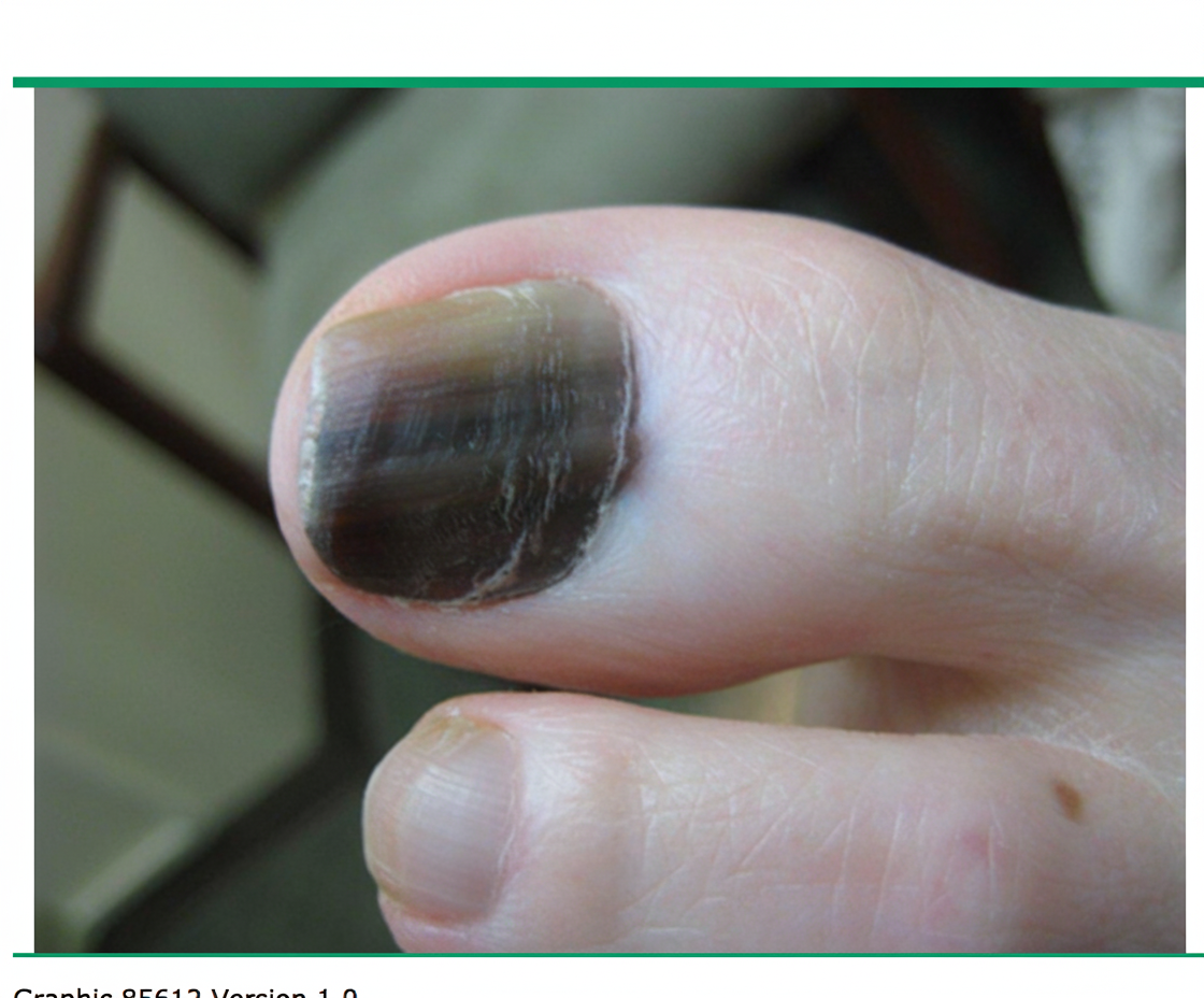
What is the most likely diagnosis in this 50-year-old woman?
Which of the following is NOT a risk factor for squamous cell carcinoma?
Which of the following is NOT a risk factor for melanoma?
Which of the following best describes the characteristic lesion of Kaposi sarcoma?
An elderly man presents with an ulcerative lesion at the inner canthus of his eye with pearly margins. On microscopic examination, it shows a palisading arrangement of cells. Identify the lesion:
Practice by Chapter
Benign Epithelial Tumors
Practice Questions
Premalignant Epidermal Tumors
Practice Questions
Basal Cell Carcinoma
Practice Questions
Squamous Cell Carcinoma
Practice Questions
Melanocytic Nevi
Practice Questions
Melanoma
Practice Questions
Merkel Cell Carcinoma
Practice Questions
Vascular Tumors and Malformations
Practice Questions
Cutaneous Lymphomas
Practice Questions
Soft Tissue Tumors
Practice Questions
Metastatic Skin Tumors
Practice Questions
Skin Cancer Prevention and Screening
Practice Questions
Want unlimited practice?
Get full access to all questions, explanations, and performance tracking.
Scan to download app