
Skin Tumors — MCQs

On this page
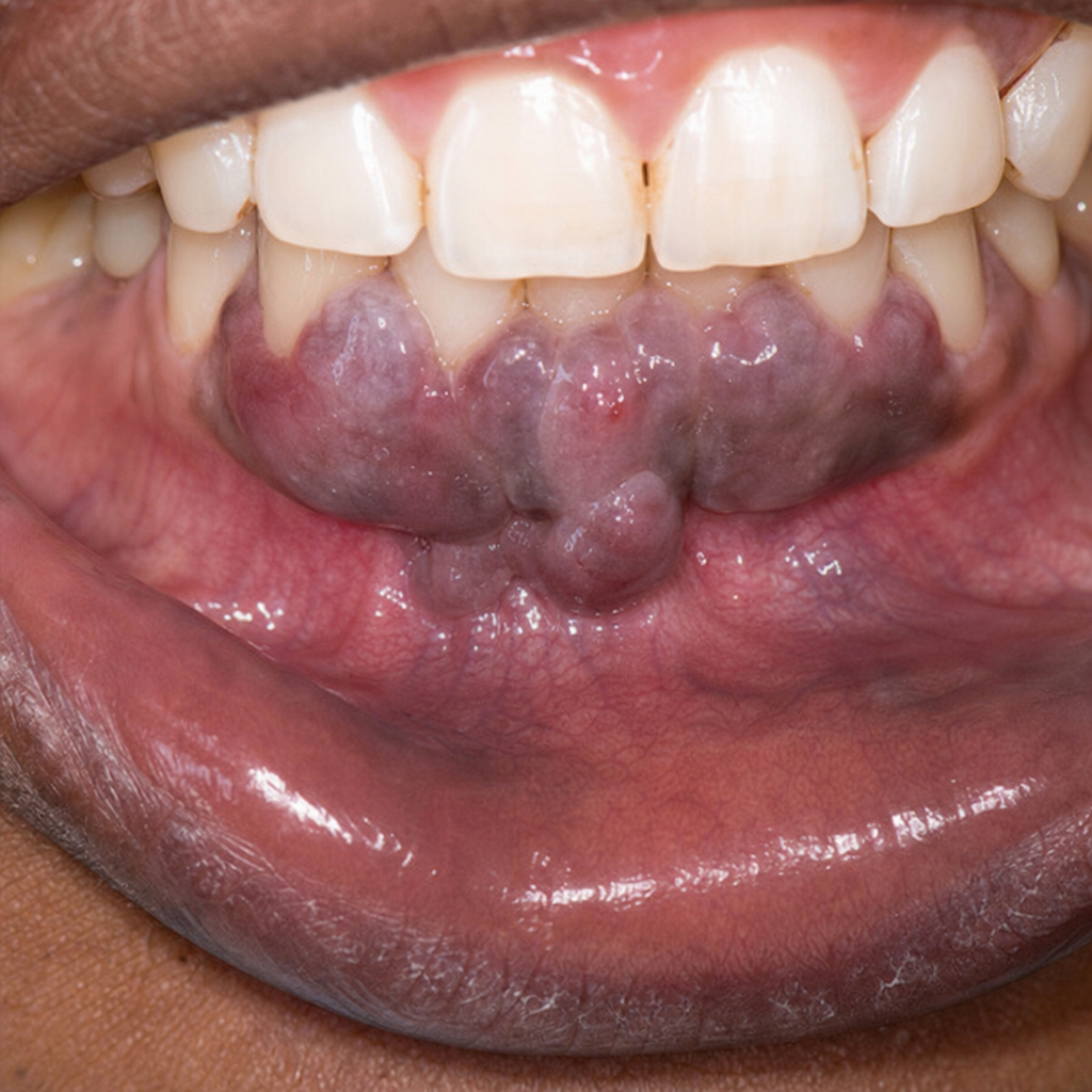
An HIV positive patient presented with the clinical condition as seen in the color plate. Which of the following is the most probable diagnosis?
What is the recommended treatment for Stage 1 cutaneous T-cell lymphoma?
A patient presents with pigmented macules on the palm. Histological examination reveals proliferating melanocytes at the dermoepidermal junction. What is the most likely diagnosis?
What is the treatment for mycosis fungoides syndrome?
A patient presents with diffuse, nonencapsulated fatty deposits. Which of the following is the most likely associated history?
What is true about this condition?
In Alibert-Bazin syndrome, the origin of the lymphoma is from which cell type?
What is the most common presentation of blue rubber bleb nevus syndrome?
What is the most common site for the lentigo maligna subtype of malignant melanoma?
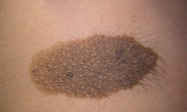
A 45-year-old man is worried about a dark pigmented skin lesion on his arm. The 'mole' is 3 mm wide, symmetric with a regular border and even pigmentation. He reports no change in size or other symptoms. Which of the following is the most appropriate next step in management?
Practice by Chapter
Benign Epithelial Tumors
Practice Questions
Premalignant Epidermal Tumors
Practice Questions
Basal Cell Carcinoma
Practice Questions
Squamous Cell Carcinoma
Practice Questions
Melanocytic Nevi
Practice Questions
Melanoma
Practice Questions
Merkel Cell Carcinoma
Practice Questions
Vascular Tumors and Malformations
Practice Questions
Cutaneous Lymphomas
Practice Questions
Soft Tissue Tumors
Practice Questions
Metastatic Skin Tumors
Practice Questions
Skin Cancer Prevention and Screening
Practice Questions
Want unlimited practice?
Get full access to all questions, explanations, and performance tracking.
Scan to download app