Match the following woods lamp findings: 1. Erythrasma, 2. Pityriasis versicolor, 3. Tinea capitis, 4. Vitiligo || a. Yellow b. Coral red fluorescence c. Pink d. Green e. Milky white
A child comes with a circular 3cm x 3cm scaly patchy hair loss with itching in the lesions. The investigation of choice is
PUVA therapy is used in all except:
Large unilateral hypopigmented lesions on the right trunk and arm in a female are best explained by which of the following?
Koebner's phenomenon is seen in all except
Koebner's phenomenon seen in ?
"Isomorphic response" can be a feature of the following except
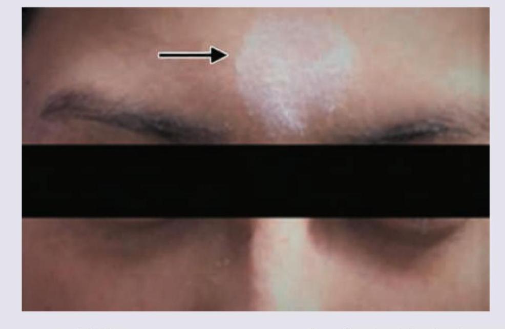
A patient presents with the skin finding shown in the image. Identify the most likely diagnosis for this lesion.

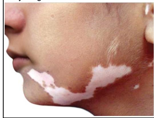
A young girl presents with leukotrichia and lesions as shown in the image. What is the most likely diagnosis?

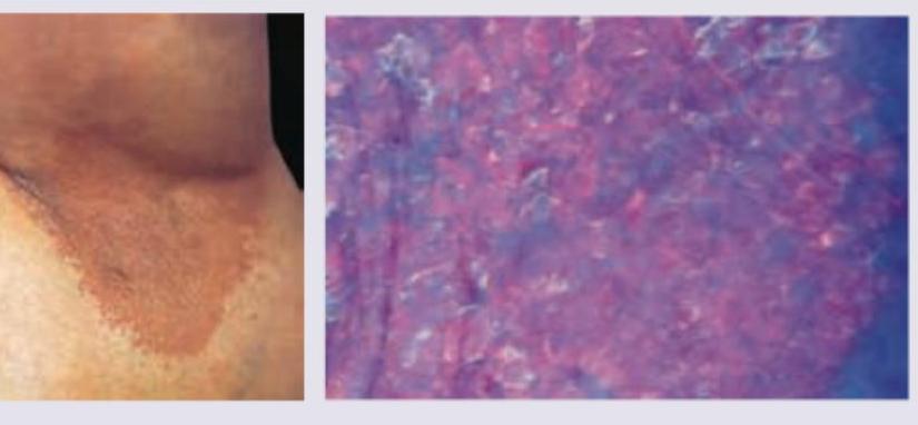
A 35-year-old obese woman presents with recurrent lesions in both axilla in summer season. Wood lamp examination is shown. The diagnosis is:

Explanation: ***1-b, 2-a, 3-d, 4-e*** - **Erythrasma** is caused by *Corynebacterium minutissimum* and produces **porphyrins** that fluoresce **coral red** under a Wood's lamp [1]. - **Pityriasis versicolor** is caused by *Malassezia furfur* and typically fluoresces **yellow to yellowish-green** [2]. - **Tinea capitis** (especially due to *Microsporum* species) shows **green fluorescence** of infected hairs. - **Vitiligo** lesions, due to a complete absence of melanin, appear as **milky white** or bright white areas under a Wood's lamp [3]. *1-d, 2-a, 3-c, 4-e* - This option incorrectly states that Erythrasma fluoresces green. Green fluorescence is characteristic of *Microsporum* species causing **Tinea capitis**. - Additionally, Tinea capitis is incorrectly associated with pink fluorescence, which is not a typical finding. *1-a, 2-c, 3-e, 4-d* - This option incorrectly states that Erythrasma fluoresces yellow. Yellow fluorescence is associated with **Pityriasis versicolor** [2]. - It also incorrectly assigns milky white fluorescence to Tinea capitis and green fluorescence to Vitiligo. *1-b, 2-d, 3-a, 4-c* - This option incorrectly associates Pityriasis versicolor with green fluorescence. While some variations exist, **yellow** is the more characteristic finding [2]. - It also incorrectly links Tinea capitis to yellow fluorescence and Vitiligo to pink, which are not typical Wood's lamp findings for these conditions.
Explanation: ***Correct: KOH mount (Potassium Hydroxide mount)*** - A **KOH mount** is the investigation of choice for suspected **dermatophyte infections** (tinea capitis), which commonly present as circular, scaly patches of hair loss with itching in children. - It involves dissolving keratinous material to visualize **fungal hyphae** and spores directly under a microscope. - This is a quick, cost-effective, and highly specific first-line diagnostic test. *Incorrect: Tzanck smear* - A **Tzanck smear** is primarily used to diagnose **viral infections** like herpes simplex or varicella-zoster by identifying multinucleated giant cells. - It is not effective for detecting fungal elements responsible for scaly hair loss. *Incorrect: Gram stain* - A **Gram stain** is a technique used to classify **bacteria** based on their cell wall properties. - It would not reveal fungal hyphae or spores relevant to the described condition. *Incorrect: Split skin smear* - A **split skin smear** (or slit-skin smear) is typically used in the diagnosis of **leprosy** to identify acid-fast bacilli. - This technique involves scraping the dermis and is not suitable for diagnosing superficial fungal infections.
Explanation: ***Melasma*** - **PUVA (Psoralen plus UVA) therapy** is contraindicated in melasma due to its potential to worsen hyperpigmentation and cause paradoxical darkening. - Melasma is best managed with topical agents like **hydroquinone**, **tretinoin**, and chemical peels, along with strict **sun protection**. *Psoriasis* - **PUVA therapy** is a well-established and effective treatment for moderate to severe psoriasis, especially for patients with widespread plaques. - It works by inhibiting DNA synthesis and cell proliferation in rapidly dividing keratinocytes, leading to a reduction in psoriatic lesions. *Vitiligo* - **PUVA therapy** is a common treatment for vitiligo, stimulating melanocyte activity and promoting repigmentation in affected areas. - Psoralen sensitizes melanocytes to UVA light, which then encourages melanin production. *Mycosis fungoides* - In its early stages, **mycosis fungoides**, a cutaneous T-cell lymphoma, can be effectively treated with **PUVA therapy**. - PUVA induces apoptosis of malignant T-cells in the skin, leading to remission of skin lesions.
Explanation: ***Neurogenic theory*** - This theory posits that **neural mechanisms** play a role in the development of some hypopigmented disorders. The **unilateral distribution** along a dermatome or nerve pathway strongly supports a neurogenic origin. - The **large, unilateral hypopigmented lesions on the right trunk and arm** are characteristic of conditions like **segmental vitiligo** or **hypopigmentation following nerve injury**, where neural factors are implicated in melanocyte dysfunction. *Autoimmune theory* - The autoimmune theory explains **generalized vitiligo**, where the body's immune system attacks melanocytes, leading to widespread depigmentation. - It does not account for the **segmental, unilateral distribution** observed in this case, which is typically not seen in autoimmune conditions. *Genetic predisposition* - While genetics can increase susceptibility to certain pigmentary disorders, it does not explain the **unilateral, segmental pattern** of hypopigmentation. - Genetic factors usually lead to more generalized or bilateral presentations rather than a localized, nerve-distribution pattern. *Lerner's self-destruct theory* - **Lerner's self-destruct theory** suggests that melanocytes may destroy themselves from within due to metabolic defects or oxidative stress. - This theory also fails to explain the **unilateral, dermatomal distribution** of the lesions, as self-destruction would likely occur more randomly or symmetrically.
Explanation: ***Tinea corporis*** - **Koebner's phenomenon**, also known as the isomorphic response, is the appearance of skin lesions characteristic of a **pre-existing dermatosis** at sites of **trauma** to previously uninvolved skin. - **Tinea corporis**, a **superficial fungal infection**, does NOT exhibit true Koebner's phenomenon. - Its spread occurs through **direct fungal contact or autoinoculation**, not through an isomorphic response to non-specific trauma. *Psoriasis* - **Psoriasis** is the **classic example** of Koebner's phenomenon. - New psoriatic plaques can appear at sites of **skin trauma** such as scratches, surgical scars, burns, or tattoos within **10-20 days** of injury. - This occurs in approximately **25-50%** of psoriasis patients. *Warts* - **Warts** (verruca vulgaris), caused by **human papillomavirus (HPV)**, can show what is sometimes called **pseudo-Koebner's phenomenon**. - Trauma facilitates **viral inoculation** and seeding of HPV into the skin, leading to new wart formation along scratch lines. - However, this is technically **viral spread through trauma**, not a true isomorphic response of a pre-existing dermatosis. *Molluscum contagiosum* - **Molluscum contagiosum** can similarly demonstrate **pseudo-Koebner's phenomenon**. - Scratching spreads the **molluscum contagiosum virus** to adjacent areas, creating linear arrays of lesions. - Like warts, this represents **direct viral inoculation** rather than true isomorphic response, but is often grouped with Koebner's phenomenon in clinical practice.
Explanation: ***All of the options*** - **Koebner's phenomenon** (isomorphic response) refers to the development of new lesions at sites of **skin trauma** in patients with pre-existing dermatological conditions. - **All four conditions listed** can exhibit Koebner's phenomenon, making this the correct answer. **Psoriasis** - The **most classic and frequently cited** example of Koebner's phenomenon. - Physical injury triggers characteristic red, scaly plaques at trauma sites. - Seen in approximately **25-50%** of psoriasis patients. **Vitiligo** - Well-documented to exhibit **Koebner's phenomenon**. - New **depigmented patches** appear at sites of trauma, cuts, or friction. - Important diagnostic and prognostic indicator in vitiligo patients. **Lichen planus** - Classic condition showing **Koebner's phenomenon**. - New violaceous, flat-topped papules develop at trauma sites. - One of the hallmark features of this condition. **Lichen nitidus** - Although less commonly emphasized, **Lichen nitidus can exhibit Koebner's phenomenon**. - Tiny, shiny papules may appear in linear distribution following trauma. - Part of the lichenoid reaction group that shows isomorphic response.
Explanation: ***Tinea*** - The **isomorphic response (Koebner phenomenon)** refers to the development of new skin lesions in areas of trauma due to an immunological process. - This phenomenon is **not typically seen in tinea** (fungal infections). - While tinea can spread to new areas, this occurs through **direct fungal inoculation and contact spread**, not through the true Koebner mechanism. *Warts* - **Warts** caused by human papillomavirus (HPV) can exhibit the **isomorphic response**. - Trauma to the skin can lead to **viral inoculation** in that area, resulting in new wart formation along lines of trauma. - This is a well-recognized example of Koebner phenomenon in viral infections. *Molluscum contagiosum* - **Molluscum contagiosum** (poxvirus infection) can demonstrate the **isomorphic response**. - **Scratching or rubbing** can spread the virus to new areas through autoinoculation. - New lesions develop along the lines of trauma, consistent with Koebner phenomenon. *Psoriasis* - **Psoriasis** is the **classic and most well-known** condition exhibiting the isomorphic response or Koebner phenomenon. - New psoriatic plaques appear in areas of **skin injury** (scratches, cuts, burns, surgical incisions, friction). - Seen in approximately **25-50%** of psoriasis patients.
Explanation: ***Piebaldism*** - The image shows a **localized patch of depigmentation** on the forehead, characteristic of **piebaldism**. - **Piebaldism** is a rare, congenital autosomal dominant disorder caused by a defect in melanocyte development and migration, resulting in stable, well-demarcated depigmented areas, often with a **white forelock**. *Vitiligo* - **Vitiligo** typically presents as **progressive, acquired macules and patches of depigmentation** that often enlarge over time. - While it can appear on the face, the sharply demarcated, congenital appearance seen here is more consistent with piebaldism. *Contact leukoderma* - **Contact leukoderma** is an **acquired depigmentation** resulting from exposure to chemicals (e.g., rubber, phenols). - It would usually present in areas of direct contact, and the congenital nature of the lesion in the image rules this out. *Albinism* - **Albinism** is a **generalized hypopigmentation** affecting the skin, hair, and eyes due to a defect in melanin production. - The image shows a localized patch of depigmentation, not a widespread lack of pigment characteristic of albinism.
Explanation: ***Segmental vitiligo*** - Segmental vitiligo characteristically presents as unilateral, **dermatomal** or **quasi-dermatomal depigmentation** with sharply demarcated borders, often including overlying **leukotrichia** (white hairs) in the affected area, as seen in the image. - This form typically has an early onset, rapid progression followed by stabilization, and can be more resistant to conventional treatments than non-segmental vitiligo. *Piebaldism* - Piebaldism is a **congenital leukoderma** characterized by a **white forelock** and symmetrically distributed depigmented patches, primarily on the trunk and extremities, which are usually stable in size and present from birth. - Unlike the progressive nature and unilateral pattern seen in the image, piebaldism is a genetic condition without new lesion development or the characteristic dermatomal distribution. *Focal vitiligo* - Focal vitiligo refers to one or a few localized depigmented macules that do not have a segmental pattern and are not distributed along a specific dermatome. - While it involves localized depigmentation, the clear **segmental distribution** and presence of **leukotrichia** in the image are more indicative of segmental vitiligo. *Nevus depigmentosus* - Nevus depigmentosus is a congenital, **stable hypopigmented lesion** that typically appears as a solitary patch or macule, without subsequent growth or change in size over time. - The lesions shown in the image appear to be multiple and follow a distinct pattern that is not typical of a stable, solitary nevus.
Explanation: ***Erythrasma*** - Erythrasma is a superficial bacterial infection caused by **Corynebacterium minutissimum**, which commonly presents as red-brown patches in intertriginous areas like the axilla, especially in obese individuals and warm, humid conditions (summer season). - The distinctive **coral-red fluorescence under Wood's lamp** is due to porphyrin production by the bacteria, which is a classic diagnostic feature of erythrasma, as shown in the image. *Ecthyma* - Ecthyma is a deeper form of impetigo characterized by **ulcerative lesions with a thick, adherent crust** that extend into the dermis. - It is typically caused by *Streptococcus pyogenes* and sometimes *Staphylococcus aureus*, and would not exhibit coral-red fluorescence under Wood's lamp. *Impetigo contagiosa* - Impetigo contagiosa (non-bullous impetigo) presents with **honey-colored crusted lesions**, usually on the face and extremities. - While also a bacterial skin infection, it is typically caused by *Staphylococcus aureus* or *Streptococcus pyogenes* and does not show coral-red fluorescence under Wood's lamp. *Bullous impetigo* - Bullous impetigo is characterized by **flaccid bullae** (blisters) that rupture to form thin, varnish-like crusts, primarily caused by *Staphylococcus aureus* producing exfoliative toxins. - Similar to other forms of impetigo, it does not produce the coral-red fluorescence under Wood's lamp.
Get full access to all questions, explanations, and performance tracking.
Start For Free