Match the following woods lamp findings: 1. Erythrasma, 2. Pityriasis versicolor, 3. Tinea capitis, 4. Vitiligo || a. Yellow b. Coral red fluorescence c. Pink d. Green e. Milky white
What is the primary condition for which calcitriol is used as a treatment?
Contraindications for skin traction: a) Dermatitis b) Vascularly compromised status of limb c) Abrasions d) Hypopigmentation (vitiligo) e) Bony deformity
How does narrowband UVB therapy work in psoriasis?
Large unilateral hypopigmented lesions on the right trunk and arm in a female are best explained by which of the following?
A 15cm hyperpigmented macule on an adolescent male undergoes changes such as coarseness, growth of hair & acne. Diagnosis is?
A 35-year-old obese woman presents with recurrent lesions in both axilla in summer season. Wood lamp examination is shown. The diagnosis is:

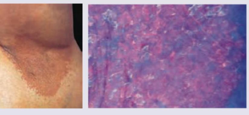
Which of the following is not a part of P. versicolor treatment -
Match the following scale types with their lesions. | Scales | Lesions | | :-- | :-- | | 1. Collarette scales | a. Pityriasis versicolour | | 2. Silvery scales | b. Pityriasis rosea | | 3. Mica-like scales | c. Psoriasis | | 4. Branny scales | d. Pityriasis lichenoides |
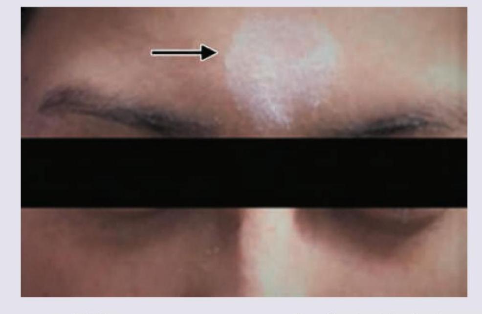
A patient presents with the skin finding shown in the image. Identify the most likely diagnosis for this lesion.

Explanation: ***1-b, 2-a, 3-d, 4-e*** - **Erythrasma** is caused by *Corynebacterium minutissimum* and produces **porphyrins** that fluoresce **coral red** under a Wood's lamp [1]. - **Pityriasis versicolor** is caused by *Malassezia furfur* and typically fluoresces **yellow to yellowish-green** [2]. - **Tinea capitis** (especially due to *Microsporum* species) shows **green fluorescence** of infected hairs. - **Vitiligo** lesions, due to a complete absence of melanin, appear as **milky white** or bright white areas under a Wood's lamp [3]. *1-d, 2-a, 3-c, 4-e* - This option incorrectly states that Erythrasma fluoresces green. Green fluorescence is characteristic of *Microsporum* species causing **Tinea capitis**. - Additionally, Tinea capitis is incorrectly associated with pink fluorescence, which is not a typical finding. *1-a, 2-c, 3-e, 4-d* - This option incorrectly states that Erythrasma fluoresces yellow. Yellow fluorescence is associated with **Pityriasis versicolor** [2]. - It also incorrectly assigns milky white fluorescence to Tinea capitis and green fluorescence to Vitiligo. *1-b, 2-d, 3-a, 4-c* - This option incorrectly associates Pityriasis versicolor with green fluorescence. While some variations exist, **yellow** is the more characteristic finding [2]. - It also incorrectly links Tinea capitis to yellow fluorescence and Vitiligo to pink, which are not typical Wood's lamp findings for these conditions.
Explanation: Secondary hyperparathyroidism - Calcitriol is the active form of vitamin D (1,25-dihydroxyvitamin D₃), and it is crucial for regulating calcium and phosphate levels in the body [1]. - In secondary hyperparathyroidism, often seen in chronic kidney disease (CKD), the kidneys cannot convert vitamin D to its active form, leading to hypocalcemia and increased PTH secretion [1], [2]. - Calcitriol supplementation helps to increase calcium absorption from the gut and suppress the release of parathyroid hormone (PTH), thereby treating the underlying cause of secondary hyperparathyroidism [1], [2]. - This is the primary therapeutic indication for calcitriol in clinical practice. Lichen planus - This is a chronic inflammatory condition affecting the skin, hair, nails, and mucous membranes - Typically treated with corticosteroids or other immunosuppressants - Calcitriol has no primary role in the treatment of lichen planus; its therapeutic applications are predominantly related to calcium and bone metabolism Pemphigus - Pemphigus is a group of rare autoimmune blistering diseases that affect the skin and mucous membranes - Primary treatment involves immunosuppressants like corticosteroids, often in high doses - Calcitriol is not indicated for the treatment of pemphigus, as its mechanism of action is unrelated to the autoimmune processes characteristic of this disease Leprosy - Leprosy is a chronic infectious disease caused by the bacterium Mycobacterium leprae - Treated with multi-drug therapy (MDT), which includes antibiotics like rifampicin, dapsone, and clofazimine - Calcitriol is not an antibiotic and therefore has no role in treating the bacterial infection responsible for leprosy
Explanation: ***ab*** - All conditions listed under 'a' and 'b' (Dermatitis, Vascularly compromised status of limb, Abrasions) are **absolute contraindications** for skin traction as they directly compromise skin integrity or circulation. - Applying skin traction in these situations can lead to **skin breakdown**, infection, or further **ischemic damage**, worsening the patient's condition. *ab* - While **dermatitis**, **vascular compromise**, and **abrasions** are indeed contraindications, the option for 'abc' implies there might be other correct choices included, which is not the case for this option. - This option is incomplete as it misses 'c' (Abrasions) which is also a significant contraindication. *acd* - This option incorrectly includes **hypopigmentation (vitiligo)** as a contraindication, which does not inherently prevent skin traction. - It also omits **vascularly compromised status of limb**, a critical contraindication, while including 'a', 'c', and 'd'. *bcd* - This option incorrectly includes **hypopigmentation (vitiligo)** as a contraindication for skin traction. - It also omits **dermatitis**, a key contraindication, while including 'b', 'c', and 'd'.
Explanation: ***T cell apoptosis*** - Narrowband UVB (NB-UVB) therapy primarily works by inducing **apoptosis (programmed cell death)** of activated **T-lymphocytes** in the psoriatic skin lesions. - By reducing the number of these inflammatory cells, NB-UVB helps to suppress the immune response that drives the **excessive keratinocyte proliferation** in psoriasis. *Melanin synthesis* - While UV radiation does stimulate **melanin synthesis**, leading to tanning, this is a secondary effect and not the primary therapeutic mechanism for psoriasis. - Increased melanin helps protect the skin from UV damage but does not directly treat the underlying pathology of psoriasis. *Collagen breakdown* - UV radiation, especially UVA, can contribute to **collagen breakdown** and photodamage over time, but this is an adverse effect, not a therapeutic mechanism for psoriasis. - Psoriasis treatment aims to normalize skin cell growth and reduce inflammation, not degrade collagen. *Keratinocyte proliferation* - Psoriasis is characterized by **accelerated keratinocyte proliferation**; NB-UVB therapy aims to *reduce* this proliferation, not promote it. - The mechanism by which NB-UVB achieves this reduction is primarily through its effects on immune cells, not by directly enhancing keratinocyte growth.
Explanation: ***Neurogenic theory*** - This theory posits that **neural mechanisms** play a role in the development of some hypopigmented disorders. The **unilateral distribution** along a dermatome or nerve pathway strongly supports a neurogenic origin. - The **large, unilateral hypopigmented lesions on the right trunk and arm** are characteristic of conditions like **segmental vitiligo** or **hypopigmentation following nerve injury**, where neural factors are implicated in melanocyte dysfunction. *Autoimmune theory* - The autoimmune theory explains **generalized vitiligo**, where the body's immune system attacks melanocytes, leading to widespread depigmentation. - It does not account for the **segmental, unilateral distribution** observed in this case, which is typically not seen in autoimmune conditions. *Genetic predisposition* - While genetics can increase susceptibility to certain pigmentary disorders, it does not explain the **unilateral, segmental pattern** of hypopigmentation. - Genetic factors usually lead to more generalized or bilateral presentations rather than a localized, nerve-distribution pattern. *Lerner's self-destruct theory* - **Lerner's self-destruct theory** suggests that melanocytes may destroy themselves from within due to metabolic defects or oxidative stress. - This theory also fails to explain the **unilateral, dermatomal distribution** of the lesions, as self-destruction would likely occur more randomly or symmetrically.
Explanation: ***Becker nevus*** - A Becker nevus is a **hyperpigmented patch** that typically appears during adolescence in males, often on the shoulder or upper trunk. - It characteristically becomes **hairy (hypertrichosis)**, more coarse, and can develop acne within the lesion, particularly during puberty due to androgen sensitivity. *Melanocytic nevus* - While melanocytic nevi are hyperpigmented, they generally do not show the characteristic changes of **coarseness, significant hair growth, or acne** within the lesion during adolescence. - They are typically stable in size and texture after initial development, with changes raising concern for **melanoma**. *Sebaceous nevus* - A sebaceous nevus is a **congenital lesion** often appearing as a yellowish-orange, waxy, or bumpy patch, usually on the scalp or face. - It does not typically present as a large, flat hyperpigmented macule that develops hair and acne in adolescence; instead, it may become verrucous or develop tumors in adulthood. *Sebaceous adenoma* - A sebaceous adenoma is a **benign tumor** of the sebaceous glands, usually appearing as a small, solitary, flesh-colored to yellowish papule or nodule, especially on the face. - It is not typically seen as a large, hyperpigmented macule that grows hair and acne over a broad area, as described in the question.
Explanation: ***Erythrasma*** - Erythrasma is a superficial bacterial infection caused by **Corynebacterium minutissimum**, which commonly presents as red-brown patches in intertriginous areas like the axilla, especially in obese individuals and warm, humid conditions (summer season). - The distinctive **coral-red fluorescence under Wood's lamp** is due to porphyrin production by the bacteria, which is a classic diagnostic feature of erythrasma, as shown in the image. *Ecthyma* - Ecthyma is a deeper form of impetigo characterized by **ulcerative lesions with a thick, adherent crust** that extend into the dermis. - It is typically caused by *Streptococcus pyogenes* and sometimes *Staphylococcus aureus*, and would not exhibit coral-red fluorescence under Wood's lamp. *Impetigo contagiosa* - Impetigo contagiosa (non-bullous impetigo) presents with **honey-colored crusted lesions**, usually on the face and extremities. - While also a bacterial skin infection, it is typically caused by *Staphylococcus aureus* or *Streptococcus pyogenes* and does not show coral-red fluorescence under Wood's lamp. *Bullous impetigo* - Bullous impetigo is characterized by **flaccid bullae** (blisters) that rupture to form thin, varnish-like crusts, primarily caused by *Staphylococcus aureus* producing exfoliative toxins. - Similar to other forms of impetigo, it does not produce the coral-red fluorescence under Wood's lamp.
Explanation: **Griseofulvin (Correct - NOT used for P. versicolor)** - **Griseofulvin** interferes with fungal cell division and is primarily used for dermatophyte infections of the skin, hair, and nails, not superficial yeast infections like *P. versicolor*. - It is systemically absorbed and incorporated into **keratin precursor cells**, offering protection against dermatophytes in newly formed tissue. - *Malassezia* species (causing P. versicolor) are **yeasts**, not dermatophytes, making griseofulvin ineffective. *Selenium sulfide (Incorrect - IS used)* - **Selenium sulfide** is an effective topical antifungal agent commonly used in shampoos and lotions to treat *P. versicolor* by inhibiting the growth of *Malassezia* species. - It works by reducing **sebum production** and having a direct fungistatic effect on the yeast. *Clotrimazole (Incorrect - IS used)* - **Clotrimazole** is a broad-spectrum azole antifungal that is very effective as a topical treatment for *P. versicolor* by inhibiting ergosterol synthesis in the fungal cell membrane. - It works well for localized patches of the infection. *Ketoconazole (Incorrect - IS used)* - **Ketoconazole**, another azole antifungal, is highly effective for *P. versicolor* and can be used topically (shampoos, creams) or orally in more extensive or recalcitrant cases. - It disrupts the fungal cell membrane by inhibiting the synthesis of **ergosterol**.
Explanation: ***1-b, 2-c, 3-d, 4-a*** - **Collarette scales** are pathognomonic of **Pityriasis rosea**, appearing as fine, trailing scales around the periphery of oval lesions in a "Christmas tree" distribution. - **Silvery scales** are the classic hallmark of **Psoriasis**, presenting as thick, adherent, silvery-white scales overlying well-demarcated erythematous plaques. - **Mica-like scales** are characteristic of **Pityriasis lichenoides**, appearing as thick, shiny, adherent scales that can be peeled off like mica sheets. - **Branny scales** are typical of **Pityriasis versicolor**, presenting as fine, powdery scales caused by **Malassezia** yeast overgrowth. *1-d, 2-c, 3-a, 4-b* - Incorrectly matches **collarette scales with Pityriasis lichenoides**, which typically presents with mica-like scales, not collarette scales. - Misassociates **mica-like scales with Pityriasis versicolor**, which characteristically has branny (fine, powdery) scales. *1-c, 2-b, 3-d, 4-a* - Wrongly pairs **collarette scales with Psoriasis**, which is known for thick silvery scales, not peripheral collarette scales. - Incorrectly matches **silvery scales with Pityriasis rosea**, which has collarette scales at lesion periphery, not silvery scales. *1-a, 2-b, 3-d, 4-c* - Falsely associates **collarette scales with Pityriasis versicolor**, which has branny scales from yeast infection, not collarette scales. - Mismatches **branny scales with Psoriasis**, which has characteristic thick silvery scales, not fine powdery scales.
Explanation: ***Piebaldism*** - The image shows a **localized patch of depigmentation** on the forehead, characteristic of **piebaldism**. - **Piebaldism** is a rare, congenital autosomal dominant disorder caused by a defect in melanocyte development and migration, resulting in stable, well-demarcated depigmented areas, often with a **white forelock**. *Vitiligo* - **Vitiligo** typically presents as **progressive, acquired macules and patches of depigmentation** that often enlarge over time. - While it can appear on the face, the sharply demarcated, congenital appearance seen here is more consistent with piebaldism. *Contact leukoderma* - **Contact leukoderma** is an **acquired depigmentation** resulting from exposure to chemicals (e.g., rubber, phenols). - It would usually present in areas of direct contact, and the congenital nature of the lesion in the image rules this out. *Albinism* - **Albinism** is a **generalized hypopigmentation** affecting the skin, hair, and eyes due to a defect in melanin production. - The image shows a localized patch of depigmentation, not a widespread lack of pigment characteristic of albinism.
Get full access to all questions, explanations, and performance tracking.
Start For Free