Mycosis fungoides primarily involves which type of immune cell?
False about Tinea versicolor
Recalcitrant acne is treated by:
A 35 years old female presented with acne. She was treated for her acne but after the treatment, she developed pigmentation. Which drug is responsible for hyperpigmentation?
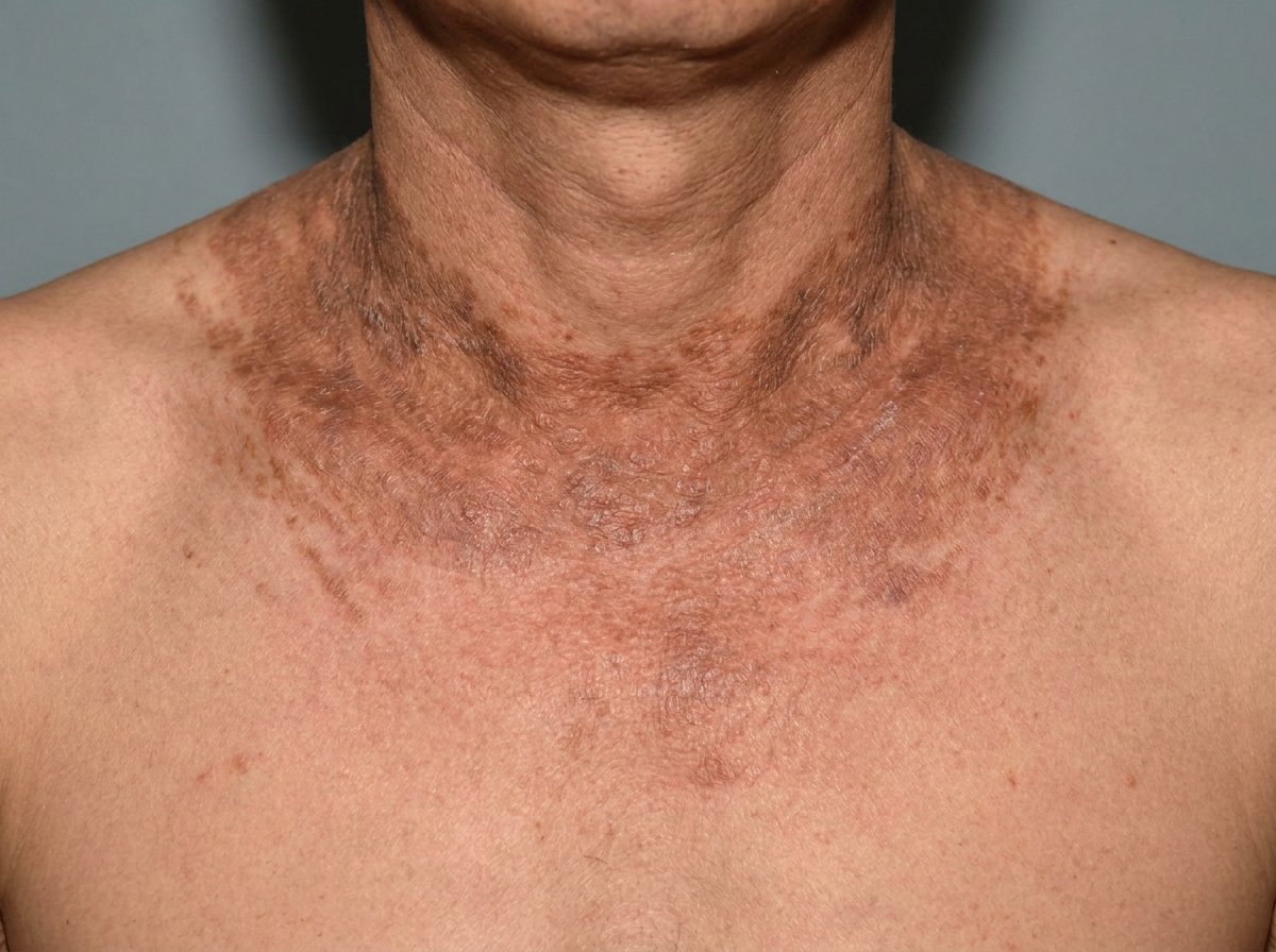
Dermatological manifestation of which of the following diseases?

Which of the following is not true about hydroquinone?
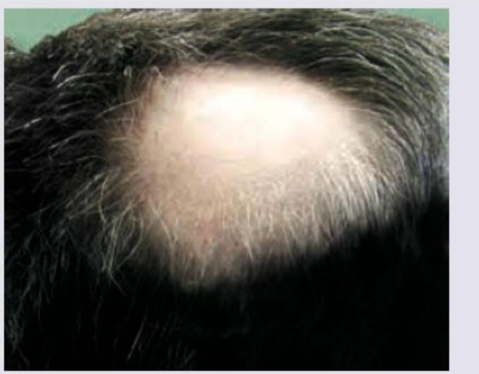
In a patient with the following lesion on scalp, what changes are seen in the nails?

In which of the following conditions is the Koebner phenomenon most commonly observed?
What is the most common association with Acanthosis nigricans?
Defect seen in Vitiligo is:
Explanation: ***CD4+ T Cells*** - Mycosis fungoides is a type of **cutaneous T-cell lymphoma**, primarily involving **CD4+ T cells** which infiltrate the skin [1][2]. - The disease is characterized by **pleomorphic** skin lesions caused by **malignant T-cell proliferation** [3]. *K Cells (not primarily involved in mycosis fungoides)* - K Cells are involved in **immunological responses** but are not specifically linked to mycosis fungoides. - They do not play a primary role in **cutaneous lymphoproliferative disorders**. *B Cells (involved in humoral immunity)* - B Cells are mainly responsible for **antibody production**, which is not the primary mechanism in mycosis fungoides. - The condition involves **T cell malignancy**, rather than abnormalities in B cell function. *NK Cells (part of innate immunity)* - NK Cells are important for **innate immunity** and target viral and tumor cells but are not primarily involved in this lymphoma. - Mycosis fungoides is characterized by **T cell-mediated responses**, not NK cell activity. **References:** [1] Kumar V, Abbas AK, et al.. Robbins and Cotran Pathologic Basis of Disease. 9th ed. Diseases of White Blood Cells, Lymph Nodes, Spleen, and Thymus, pp. 613-614. [2] Kumar V, Abbas AK, et al.. Robbins and Cotran Pathologic Basis of Disease. 9th ed. The Skin, p. 1162. [3] Cross SS. Underwood's Pathology: A Clinical Approach. 6th ed. Common Clinical Problems From Diseases Of The Urinary And Male Genital Tracts, pp. 564-565.
Explanation: ***Wood's lamp examination gives Apple green Fluorescence*** - **Tinea versicolor** typically exhibits a **yellow-green or yellowish-orange fluorescence** under Wood's lamp, not an apple-green fluorescence. - **Apple-green fluorescence** is characteristic of certain bacterial infections, such as those caused by *Pseudomonas aeruginosa*, but not for *Malassezia* species in Tinea versicolor. *Lesions can be both hypo & hyperpigmented* - This statement is true; **Tinea versicolor** lesions can indeed present as **hypopigmented (lighter)** or **hyperpigmented (darker)** patches. - The color variation is due to *Malassezia's* interference with melanin production or its direct pigment production. *It is superficial fungal infection caused by Malassezia* - This statement is true; **Tinea versicolor** is a **superficial fungal infection** of the skin caused by species of the yeast **Malassezia**. - The most common causative agent is **Malassezia globosa**, which is a normal commensal of the skin but can become pathogenic under certain conditions. *Scratch sign is positive* - This statement is true; the **scratch sign** (also known as the Besnier's sign) is positive in **Tinea versicolor**. - When the lesions are gently scraped, fine, **powdery scales** become more apparent, which is a characteristic finding.
Explanation: ***Retinoids*** - **Oral retinoids**, particularly **isotretinoin**, are highly effective for **recalcitrant, severe acne** that has not responded to conventional therapies. - They work by reducing **sebum production**, inhibiting **Propionibacterium acnes**, normalizing **follicular keratinization**, and possessing **anti-inflammatory** properties. *Steroids* - **Systemic steroids** are generally not used for long-term acne treatment due to significant side effects and the potential for **steroid-induced acne**. - They may be used short-term for **severe nodulocystic acne** with significant inflammation, but not as a primary treatment for recalcitrance. *Oral erythromycin* - **Oral erythromycin** is an antibiotic sometimes used for acne, but resistance is common, limiting its effectiveness, especially in **recalcitrant cases**. - It primarily targets **Propionibacterium acnes** and has some **anti-inflammatory** effects, but is less potent than retinoids for severe, persistent acne. *Oral tetracycline* - **Oral tetracyclines** (e.g., doxycycline, minocycline) are commonly used for moderate to severe acne, but if acne is **recalcitrant**, it indicates a lack of response to these antibiotics. - Their mechanism involves reducing **bacterial growth** and inflammation, but they do not address the underlying pathogenesis of severe acne as comprehensively as retinoids.
Explanation: ***Minocycline*** - **Minocycline** is known to cause different types of hyperpigmentation, including blue-grey discoloration of the skin, scars, mucosa, eyes, and teeth, especially with long-term use. - This pigmentation can be due to the accumulation of **iron oxide** and **minocycline degradation products** in tissues. *Doxycycline (a tetracycline antibiotic)* - While doxycycline is a tetracycline, it is **less commonly associated with significant hyperpigmentation** compared to minocycline. - It can cause photosensitivity, which might lead to hyperpigmentation in sun-exposed areas, but direct drug-induced blue-grey discoloration is rare. *Tetracycline (a tetracycline antibiotic)* - **Tetracycline** can cause tooth discoloration, especially in children, and photosensitivity, but direct drug-induced skin hyperpigmentation as described is **less common** than with minocycline. - Other side effects like gastrointestinal upset are more prominent. *Erythromycin (a macrolide antibiotic)* - **Erythromycin** is a macrolide antibiotic and is **not typically associated with significant skin hyperpigmentation** as a side effect. - Common side effects include gastrointestinal disturbances like nausea, vomiting, and diarrhea.
Explanation: ***Pellagra*** - The image shows a classic "butterfly" rash on the face, specifically a photosensitive dermatitis, which is a hallmark of **pellagra**. - Pellagra is caused by a deficiency of **niacin (vitamin B3)**, characterized by the "3 D's": **dermatitis**, **diarrhea**, and **dementia**. *Photo dermatitis* - While pellagra often presents with photosensitive dermatitis, "photo dermatitis" is a general term for **skin inflammation caused by light exposure** and not a specific disease itself. - It could be caused by various factors, including medication, immune reactions, or other underlying conditions, but the pattern seen here is highly suggestive of pellagra. *Acrodermatitis enteropathica* - This condition is a **hereditary zinc deficiency** that typically presents with a periorificial and acral dermatitis. - The skin lesions are typically **vesicular-pustular or eczematous** and do not usually have the distinct butterfly pattern of photosensitive dermatitis seen in the image. *Vitamin B deficiency* - While pellagra is a vitamin B **(niacin, B3)** deficiency, this option is too broad. - Other vitamin B deficiencies, such as **riboflavin (B2)** or **pyridoxine (B6)** deficiency, have different dermatological manifestations like angular cheilitis, glossitis, or seborrheic dermatitis, but not the characteristic facial rash seen here.
Explanation: ***It should not be used for melasma or chloasma of pregnancy*** - This statement is **NOT TRUE** - hydroquinone is actually a **first-line treatment for melasma** including chloasma (melasma of pregnancy) - Hydroquinone 2-4% is one of the **most effective topical agents** for treating melasma and is widely recommended in dermatological guidelines - While hydroquinone use during **active pregnancy** is approached with caution (FDA Category C), it is definitely indicated for treating melasma/chloasma **after pregnancy** and for general melasma in non-pregnant patients - The condition (melasma/chloasma) is appropriately treated with hydroquinone; only the **timing during pregnancy** requires consideration *Response is incomplete and pigmentation may recur* - This is a **TRUE statement** about hydroquinone therapy - Treatment response is often **incomplete** with partial lightening of hyperpigmentation - **Recurrence is common** after discontinuation, especially with continued sun exposure or hormonal triggers - Maintenance therapy is often needed to sustain results *It inhibits tyrosinase* - This is a **TRUE statement** - hydroquinone's primary mechanism of action - Acts as a **competitive inhibitor of tyrosinase**, the rate-limiting enzyme in melanin synthesis - This inhibition reduces melanin production in melanocytes, leading to depigmentation *It requires prescription strength concentrations above 2%* - This is a **TRUE statement** in most countries including India and the USA - Hydroquinone concentrations **≤2%** are available over-the-counter (OTC) - Concentrations **>2% (typically 3-4%)** require a prescription - Higher concentrations provide greater efficacy but also increased risk of side effects like ochronosis
Explanation: ***Pitting of nails*** - The image shows a patch of **alopecia areata** on the scalp. **Nail pitting** is the most common and characteristic nail change associated with alopecia areata, occurring in **10-66% of cases**. - Pitting appears as small depressions or **"ice-pick" marks** on the nail surface, resulting from defective nail matrix keratinization. - Other nail changes in alopecia areata include **trachyonychia (rough nails), red spotted lunulae, onycholysis**, and **Beau's lines**. *Dorsal pterygium of nails* - **Dorsal pterygium** occurs when the proximal nail fold fuses with and extends over the nail plate, creating a wing-like scar. - This is classically associated with **lichen planus, trauma, burns, vasculitis**, and **graft-versus-host disease** — **NOT alopecia areata**. - It can lead to permanent nail dystrophy or nail loss. *Azure nails* - **Azure nails** (blue nails) are typically associated with **Wilson's disease** (copper accumulation) or **minocycline use**, not alopecia areata. - They represent a blue-gray discoloration of the nail bed or lunula. *Yellow nail discolouration* - **Yellow nail syndrome** is a rare condition characterized by slow-growing, thickened, yellow nails, often associated with **lymphedema** and **respiratory problems** (pleural effusions, chronic bronchitis). - It is not linked to alopecia areata.
Explanation: ***Correct: Psoriasis*** - **Psoriasis** is the **most classic and commonly cited example** of the Koebner phenomenon (isomorphic response) - New psoriatic plaques characteristically develop at sites of cutaneous trauma, scratches, or surgical incisions in 25-50% of psoriasis patients - This is a **pathognomonic feature** frequently tested in competitive exams and considered the prototype condition for demonstrating this phenomenon - The mechanism involves inflammatory cascades triggered by trauma in genetically predisposed skin *Incorrect: Lichen planus* - While lichen planus does exhibit the Koebner phenomenon with purplish polygonal papules appearing along scratch lines, it is **less commonly observed** compared to psoriasis - Seen in approximately 10-25% of lichen planus cases - Not considered the primary example when teaching about Koebner phenomenon *Incorrect: Viral warts* - Viral warts can demonstrate **pseudo-Koebner phenomenon** where new warts form along trauma lines due to viral inoculation - This is more accurately described as **autoinoculation** rather than true isomorphic response - Less commonly discussed in the context of classic Koebner phenomenon compared to psoriasis *Incorrect: All of the options* - While all three conditions can show Koebner-like responses, the question asks for "**most commonly observed**" - Psoriasis remains the **gold standard** and most frequently encountered example in clinical practice and medical literature
Explanation: **Explanation:** **Acanthosis Nigricans (AN)** is a common dermatological condition characterized by hyperpigmented, velvety plaques, typically found in intertriginous areas like the axilla and neck. **Why Obesity is the Correct Answer:** Obesity is the **most common** association and cause of Acanthosis Nigricans (Pseudo-acanthosis nigricans). The underlying mechanism is **Insulin Resistance**. In obese individuals, high levels of circulating insulin bind to **Insulin-like Growth Factor-1 (IGF-1) receptors** on keratinocytes and fibroblasts. This stimulates excessive proliferation of these cells, leading to the characteristic epidermal thickening and hyperpigmentation. **Analysis of Incorrect Options:** * **Diabetes Mellitus (B):** While AN is a strong cutaneous marker for Type 2 Diabetes, it usually precedes the clinical onset of diabetes. Obesity remains the primary driver and more frequent association. * **Hypertension (A) & Hypothyroidism (D):** These are often part of the "Metabolic Syndrome" or associated endocrinopathies (like PCOS), but they are not the primary or most common cause of the skin changes seen in AN. **High-Yield Clinical Pearls for NEET-PG:** * **Malignant Acanthosis Nigricans:** If AN appears suddenly, is very extensive, or involves the palms (**Tripe Palms**) and oral mucosa, it is highly suggestive of internal malignancy, most commonly **Gastric Adenocarcinoma**. * **Histopathology:** Shows hyperkeratosis and papillomatosis. Note that "acanthosis" (thickening of the stratum spinosum) is actually minimal despite the name. * **Common Sites:** Neck (most common), axilla, groins, and knuckles. * **Drug-induced AN:** Can be caused by Nicotinic acid, systemic corticosteroids, and OCPs.
Explanation: **Explanation:** **Vitiligo** is an acquired, chronic pigmentary disorder characterized by the selective destruction of melanocytes. **1. Why Option B is correct:** The hallmark of vitiligo is the **complete absence of functional melanocytes** in the affected skin. This is primarily due to an autoimmune-mediated destruction where T-cells target melanocyte-specific antigens. Histopathologically, a skin biopsy of a stable vitiligo lesion shows a total lack of melanocytes (DOPA-negative) and a consequent absence of melanin in the epidermis. **2. Why other options are incorrect:** * **Option A (Absent melanosomes):** This is seen in **Chediak-Higashi syndrome** or specific trafficking defects. In vitiligo, the "factory" (melanocyte) is gone, so melanosomes are naturally absent, but the primary defect is the cell loss itself. * **Option C (Reduction in melanin synthesis):** This describes **Albinism**, where melanocytes are present in normal numbers, but there is a genetic defect in the enzyme tyrosinase, leading to decreased melanin production. * **Option D (Reduction in number of melanocytes):** This describes **Nevus Depigmentosus** or **Pityriasis Alba**, where melanocytes are present but decreased in number or activity. In vitiligo, the loss is absolute in the lesion. **High-Yield Clinical Pearls for NEET-PG:** * **Most common association:** Autoimmune thyroid disease (Hashimoto’s). * **Koebner Phenomenon:** Vitiligo is Koebner positive (new lesions at sites of trauma). * **Segmental Vitiligo:** Does not follow the Koebner phenomenon and has a dermatomal distribution. * **Treatment of Choice:** Narrowband UVB (NB-UVB) is the gold standard for generalized vitiligo. * **Wood’s Lamp:** Lesions show a characteristic **"milky white"** fluorescence.
Get full access to all questions, explanations, and performance tracking.
Start For Free