
Pigmentary Disorders — MCQs

On this page
What is a common skin finding in patients with Fanconi's anemia?
A melanocytic nevus surrounded by a depigmented halo is called:
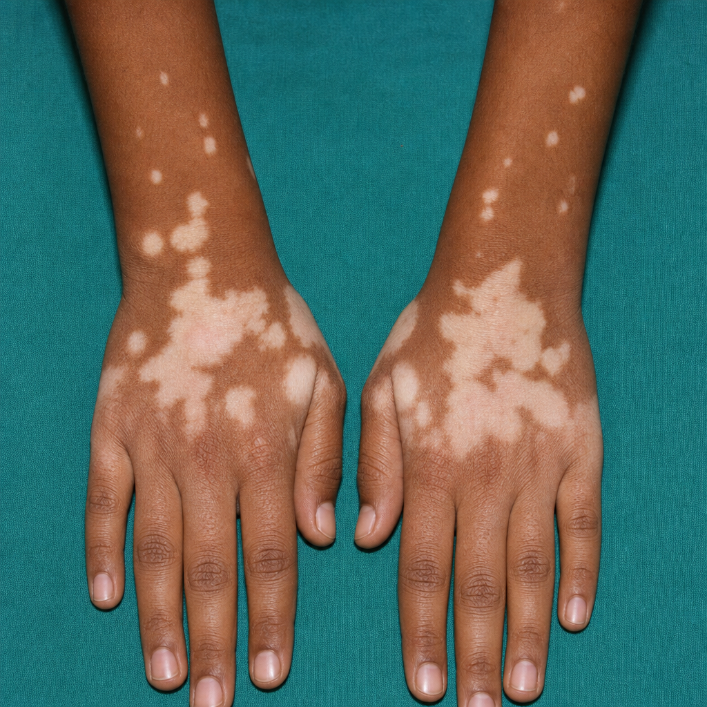
Identify the skin condition based on the provided image.
Brown macular pigmentation in malar area in a pregnant female is due to ?
Schamberg's purpura is seen on?
Which of the following skin lesions is not classified as a nevus of melanocytes?
Raindrop pigmentation is caused by?
Which of the following conditions is not typically associated with vitiligo?
What is the first-line treatment for melasma?
Large unilateral hypopigmented lesions on the right trunk and arm in a female are best explained by which of the following?
Practice by Chapter
Melanocyte Biology
Practice Questions
Vitiligo
Practice Questions
Melasma
Practice Questions
Post-inflammatory Hyperpigmentation
Practice Questions
Post-inflammatory Hypopigmentation
Practice Questions
Albinism
Practice Questions
Drug-Induced Pigmentary Changes
Practice Questions
Pityriasis Alba
Practice Questions
Pigmentary Demarcation Lines
Practice Questions
Nevi of Ota and Ito
Practice Questions
Management of Hyperpigmentation
Practice Questions
Management of Hypopigmentation
Practice Questions
Want unlimited practice?
Get full access to all questions, explanations, and performance tracking.
Scan to download app