
Pigmentary Disorders — MCQs

On this page
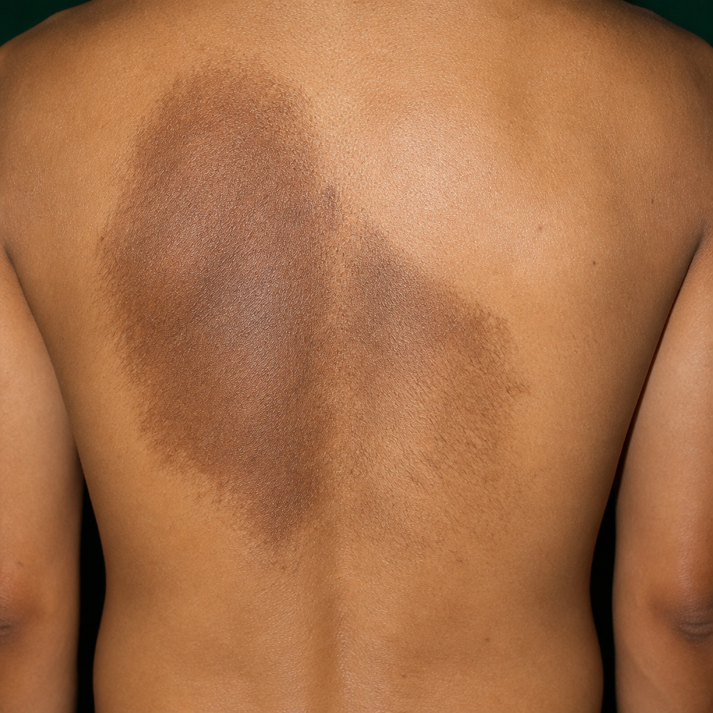
A child presents with a hyperpigmented patch over the back since birth. The level of melanin in this patch on biopsy is likely to be?
A newborn child presents with a solitary white well-defined hypopigmented patch on their right thigh. What is the diagnosis?
Psoralen-A is used in the treatment of:
A 39-year-old woman presents with a thickened, hyperpigmented, velvety lesion over the axillae. This condition is associated with which of the following?
A female presents with hypopigmented lesions on the central forehead. Which drug is responsible?
A large, dark brown patch is noted on the back. What is the most likely diagnosis?
In an elderly patient, acanthosis nigricans usually indicates what?
A 28-year-old pregnant female presents with complaints of brownish pigmentation on the bridge of her nose and cheeks, noticed after returning from a beach vacation. There is no pain or itching at the affected site. What is your most likely diagnosis?
LEOPARD syndrome does not include which of the following?
A 19-year-old girl presents with light brown pigmentation over the malar eminence. What is the likely diagnosis?
Practice by Chapter
Melanocyte Biology
Practice Questions
Vitiligo
Practice Questions
Melasma
Practice Questions
Post-inflammatory Hyperpigmentation
Practice Questions
Post-inflammatory Hypopigmentation
Practice Questions
Albinism
Practice Questions
Drug-Induced Pigmentary Changes
Practice Questions
Pityriasis Alba
Practice Questions
Pigmentary Demarcation Lines
Practice Questions
Nevi of Ota and Ito
Practice Questions
Management of Hyperpigmentation
Practice Questions
Management of Hypopigmentation
Practice Questions
Want unlimited practice?
Get full access to all questions, explanations, and performance tracking.
Scan to download app