Which drug is generally contraindicated in the management of traumatic hyphema in a patient with sickle cell disease?
An adult presents with oval, scaly, hypopigmented macules over the chest and back. The diagnosis is
Which of the following strategies are part of the National Leprosy Control Programme?
All of the following are used in systemic therapy of psoriasis except
Multiple hypoaesthetic, hypopigmented macules on right lateral forearm with numerous acid-fast bacilli is indicative of:
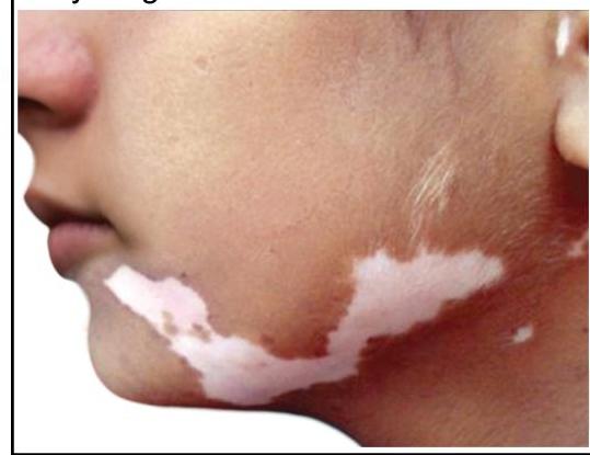
A young girl presents with leukotrichia and lesions as shown in the image. What is the most likely diagnosis?

A young male presented with an anesthetic patch on the right forearm. A thickened nerve was palpable on examination. Skin biopsy shows the image below. What is the diagnosis?

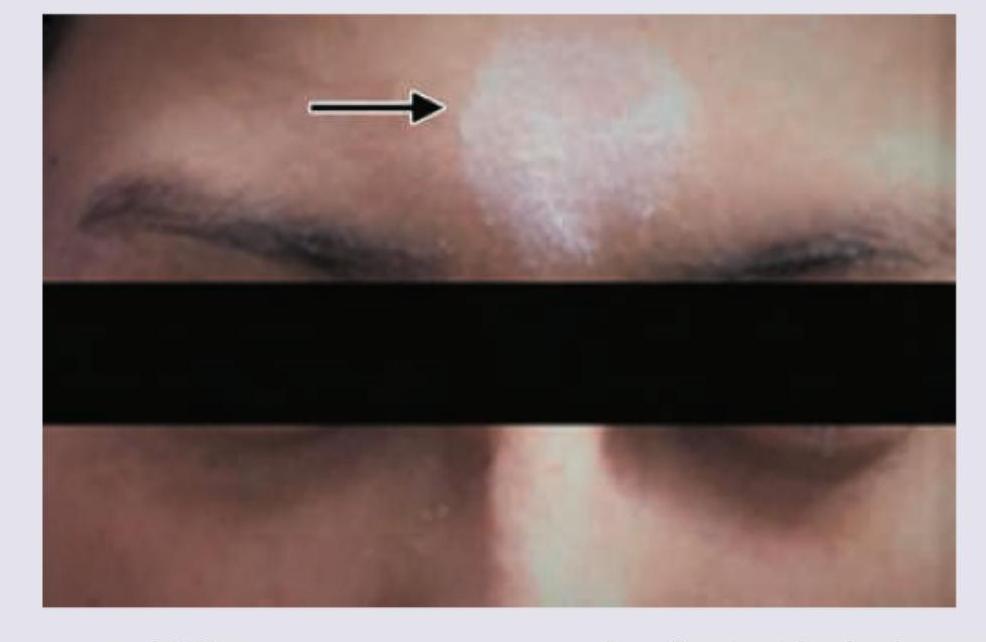
A patient presents with the skin finding shown in the image. Identify the most likely diagnosis for this lesion.

What is the most common association with Acanthosis nigricans?
Defect seen in Vitiligo is:
Explanation: ***Acetazolamide*** - **Acetazolamide** is a **carbonic anhydrase inhibitor** that is **generally contraindicated** in patients with **sickle cell disease or trait**. - It causes **systemic acidosis** by increasing renal bicarbonate excretion, which lowers blood pH. - **Acidosis promotes sickling** of red blood cells, which can lead to **vaso-occlusion**, increased blood viscosity, and potential complications including **anterior chamber obstruction** and **secondary glaucoma**. - Despite its usefulness in lowering intraocular pressure in other settings, this risk makes it contraindicated in sickle cell patients with hyphema. *Timolol* - **Timolol** is a **beta-blocker** that reduces aqueous humor production and is generally **safe and effective** for reducing **intraocular pressure** in traumatic hyphema. - It does not cause systemic acidosis or affect red blood cell sickling. - Commonly used in hyphema management regardless of sickle cell status. *Steroids* - **Topical or systemic steroids** are often used to reduce **inflammation** and anterior chamber reaction in traumatic hyphema. - They help prevent **secondary hemorrhage** and reduce complications. - They do not contribute to red blood cell sickling or systemic acidosis and are safe in sickle cell disease. *Atropine* - **Atropine** is a **cycloplegic agent** used to paralyze the ciliary body and dilate the pupil, which helps **relieve pain** and prevent **posterior synechiae** in hyphema. - It has no adverse effects related to **sickle cell disease** or red blood cell sickling. - Routinely used in hyphema management.
Explanation: ***Pityriasis Versicolor*** - This condition is caused by a **Malassezia species** yeast infection, presenting as characteristic **oval, scaly, hypopigmented or hyperpigmented macules** primarily on the trunk. - The hypopigmentation is often due to the production of **azelaic acid** by the yeast, which inhibits melanin synthesis. *Tuberculosis Cutis* - This refers to various skin manifestations of **tuberculosis**, which can include papules, nodules, ulcers, or verrucous lesions, often with significant inflammation or necrosis. - It does not typically present as widespread, oval, scaly, hypopigmented macules without other systemic signs of tuberculosis. *Psoriasis* - Characterized by **well-demarcated, erythematous plaques** covered with silvery scales, often found on extensor surfaces, scalp, and nails. - **Psoriasis** lesions are typically red and inflamed, not hypopigmented, and do not usually have the fine, scaly appearance described. *Granuloma Annulare* - Presents as **annular (ring-shaped) papules or plaques**, often flesh-colored, red, or pink, most commonly on the hands, feet, and ankles. - It is typically a **non-scaly eruption** and does not cause hypopigmented macules over the trunk.
Explanation: ***Early detection of cases and short course multi-drug therapy*** - The primary strategy of the National Leprosy Control Programme (NLCP) is to actively identify new cases early to prevent disabilities. - **Multi-drug therapy (MDT)**, a short-course regimen, is administered to cure the disease and interrupt transmission. - This combination represents the **core control strategy** of NLCP. *Chemoprophylaxis with dapsone and rehabilitation* - **Chemoprophylaxis with dapsone** is not a standard strategy under NLCP due to concerns about resistance and limited effectiveness for mass prevention. - **Rehabilitation** is important for disability management but is a secondary/tertiary prevention strategy, not a primary control measure. *Short course multi-drug therapy and chemoprophylaxis* - While **MDT** is a cornerstone of NLCP, **chemoprophylaxis** is not included as a widespread intervention. - The focus remains on treating diagnosed cases rather than mass preventive drug administration. *Rehabilitation and early detection of cases* - **Early detection** is indeed a key component, but pairing it with **rehabilitation** alone misses the critical treatment component. - The core control strategy requires both early detection **and MDT treatment**, not just detection and rehabilitation.
Explanation: ***Oral glucocorticoids*** - **Oral glucocorticoids** are generally avoided in psoriasis because they can precipitate severe **rebound flares** upon discontinuation or during dose tapering. - While they can temporarily suppress inflammation, the risk of worsening psoriasis and other systemic side effects makes them unsuitable for long-term systemic therapy. *Methotrexate* - **Methotrexate** is a commonly used systemic agent for psoriasis due to its **immunosuppressive** and **anti-proliferative effects**, targeting rapidly dividing cells. - It works by inhibiting dihydrofolate reductase and is typically given once weekly for chronic plaque psoriasis. *Cyclosporine* - **Cyclosporine** is an effective systemic immunosuppressant used for severe, resistant psoriasis, particularly when rapid control is needed. - It primarily acts by inhibiting **T-cell activation** and proliferation, thereby reducing the inflammatory response in psoriasis. *Acitretin* - **Acitretin** is an oral retinoid derivative of vitamin A, used in severe forms of psoriasis, especially **pustular** and **erythrodermic** types. - It works by modulating **keratinocyte differentiation** and proliferation, helping to normalize skin cell growth.
Explanation: ***Lepromatous leprosy*** - The hallmark feature is **numerous acid-fast bacilli (AFB)** in skin smears, indicating a **high bacterial load** characteristic of lepromatous leprosy (bacterial index 4-6+). - Lepromatous leprosy is **multibacillary** with **poor cell-mediated immunity**, allowing uncontrolled bacterial multiplication (10⁶-10⁹ bacilli per gram of tissue). - While typically presenting with **widespread, symmetrical lesions**, early lepromatous leprosy can present with **multiple hypopigmented macules** before progressing to diffuse infiltration. - **Sensory loss** may be present but is typically **less pronounced** initially compared to tuberculoid leprosy, as nerve damage occurs gradually. *Borderline leprosy* - Represents the **unstable middle spectrum** (BT, BB, BL) with **moderately impaired immunity** and **variable bacterial load**. - Borderline tuberculoid (BT) has **few AFB**, borderline lepromatous (BL) has **moderate AFB**, but the term "numerous AFB" more specifically indicates lepromatous leprosy. - Lesions are typically **asymmetrical** with variable sensory loss depending on the subtype. *Tuberculoid leprosy* - Characterized by **paucibacillary disease** with **strong cell-mediated immunity** that effectively contains bacterial proliferation. - Skin smears show **few or no detectable AFB** (bacterial index 0-1+) due to robust immune response. - Presents with **few well-defined lesions (1-5)** with **marked sensory loss** and thickened nerves. *Indeterminate leprosy* - The **earliest stage** of leprosy, presenting as a **single hypopigmented or erythematous macule** with minimal sensory changes. - Shows **few or no AFB** on skin smears and may evolve into any form of leprosy or resolve spontaneously. - Not consistent with multiple lesions and numerous bacilli.
Explanation: ***Segmental vitiligo*** - Segmental vitiligo characteristically presents as unilateral, **dermatomal** or **quasi-dermatomal depigmentation** with sharply demarcated borders, often including overlying **leukotrichia** (white hairs) in the affected area, as seen in the image. - This form typically has an early onset, rapid progression followed by stabilization, and can be more resistant to conventional treatments than non-segmental vitiligo. *Piebaldism* - Piebaldism is a **congenital leukoderma** characterized by a **white forelock** and symmetrically distributed depigmented patches, primarily on the trunk and extremities, which are usually stable in size and present from birth. - Unlike the progressive nature and unilateral pattern seen in the image, piebaldism is a genetic condition without new lesion development or the characteristic dermatomal distribution. *Focal vitiligo* - Focal vitiligo refers to one or a few localized depigmented macules that do not have a segmental pattern and are not distributed along a specific dermatome. - While it involves localized depigmentation, the clear **segmental distribution** and presence of **leukotrichia** in the image are more indicative of segmental vitiligo. *Nevus depigmentosus* - Nevus depigmentosus is a congenital, **stable hypopigmented lesion** that typically appears as a solitary patch or macule, without subsequent growth or change in size over time. - The lesions shown in the image appear to be multiple and follow a distinct pattern that is not typical of a stable, solitary nevus.
Explanation: ***LL*** - The image shows a **granuloma with foamy macrophages** (Virchow cells) laden with bacilli, characteristic of **lepromatous leprosy (LL)**. The accompanying clinical features of an anesthetic patch and thickened nerve point towards leprosy. - In LL, there is **poor cell-mediated immunity** with a predominant **Th2 response**, leading to ineffective control of intracellular *Mycobacterium leprae*. While **humoral immunity** is increased, it is ineffective against the intracellular pathogen, resulting in widespread bacterial multiplication and the characteristic **foamy macrophages** (Virchow cells) laden with bacilli. *TT* - **Tuberculoid leprosy (TT)** would typically show a well-formed, epithelioid granuloma with few or no bacilli, reflecting a strong cell-mediated immune response. - Clinical presentation involves well-demarcated, anesthetic patches with significant nerve damage, but the biopsy features would differ from those seen here. *Histiocytosis* - **Histiocytosis** refers to a group of disorders involving abnormal proliferation of histiocytes/macrophages, such as Langerhans cell histiocytosis. - While it involves macrophages, the specific morphology and clinical presentation (anesthetic patch, thickened nerve) are not typical for histiocytosis. *Lymphoma* - **Lymphoma** involves the malignant proliferation of lymphocytes and would present with atypical lymphoid infiltrates, not the macrophage-rich granulomas seen in the image. - The clinical context of an anesthetic patch and thickened nerve is also not characteristic of primary cutaneous lymphoma.
Explanation: ***Piebaldism*** - The image shows a **localized patch of depigmentation** on the forehead, characteristic of **piebaldism**. - **Piebaldism** is a rare, congenital autosomal dominant disorder caused by a defect in melanocyte development and migration, resulting in stable, well-demarcated depigmented areas, often with a **white forelock**. *Vitiligo* - **Vitiligo** typically presents as **progressive, acquired macules and patches of depigmentation** that often enlarge over time. - While it can appear on the face, the sharply demarcated, congenital appearance seen here is more consistent with piebaldism. *Contact leukoderma* - **Contact leukoderma** is an **acquired depigmentation** resulting from exposure to chemicals (e.g., rubber, phenols). - It would usually present in areas of direct contact, and the congenital nature of the lesion in the image rules this out. *Albinism* - **Albinism** is a **generalized hypopigmentation** affecting the skin, hair, and eyes due to a defect in melanin production. - The image shows a localized patch of depigmentation, not a widespread lack of pigment characteristic of albinism.
Explanation: **Explanation:** **Acanthosis Nigricans (AN)** is a common dermatological condition characterized by hyperpigmented, velvety plaques, typically found in intertriginous areas like the axilla and neck. **Why Obesity is the Correct Answer:** Obesity is the **most common** association and cause of Acanthosis Nigricans (Pseudo-acanthosis nigricans). The underlying mechanism is **Insulin Resistance**. In obese individuals, high levels of circulating insulin bind to **Insulin-like Growth Factor-1 (IGF-1) receptors** on keratinocytes and fibroblasts. This stimulates excessive proliferation of these cells, leading to the characteristic epidermal thickening and hyperpigmentation. **Analysis of Incorrect Options:** * **Diabetes Mellitus (B):** While AN is a strong cutaneous marker for Type 2 Diabetes, it usually precedes the clinical onset of diabetes. Obesity remains the primary driver and more frequent association. * **Hypertension (A) & Hypothyroidism (D):** These are often part of the "Metabolic Syndrome" or associated endocrinopathies (like PCOS), but they are not the primary or most common cause of the skin changes seen in AN. **High-Yield Clinical Pearls for NEET-PG:** * **Malignant Acanthosis Nigricans:** If AN appears suddenly, is very extensive, or involves the palms (**Tripe Palms**) and oral mucosa, it is highly suggestive of internal malignancy, most commonly **Gastric Adenocarcinoma**. * **Histopathology:** Shows hyperkeratosis and papillomatosis. Note that "acanthosis" (thickening of the stratum spinosum) is actually minimal despite the name. * **Common Sites:** Neck (most common), axilla, groins, and knuckles. * **Drug-induced AN:** Can be caused by Nicotinic acid, systemic corticosteroids, and OCPs.
Explanation: **Explanation:** **Vitiligo** is an acquired, chronic pigmentary disorder characterized by the selective destruction of melanocytes. **1. Why Option B is correct:** The hallmark of vitiligo is the **complete absence of functional melanocytes** in the affected skin. This is primarily due to an autoimmune-mediated destruction where T-cells target melanocyte-specific antigens. Histopathologically, a skin biopsy of a stable vitiligo lesion shows a total lack of melanocytes (DOPA-negative) and a consequent absence of melanin in the epidermis. **2. Why other options are incorrect:** * **Option A (Absent melanosomes):** This is seen in **Chediak-Higashi syndrome** or specific trafficking defects. In vitiligo, the "factory" (melanocyte) is gone, so melanosomes are naturally absent, but the primary defect is the cell loss itself. * **Option C (Reduction in melanin synthesis):** This describes **Albinism**, where melanocytes are present in normal numbers, but there is a genetic defect in the enzyme tyrosinase, leading to decreased melanin production. * **Option D (Reduction in number of melanocytes):** This describes **Nevus Depigmentosus** or **Pityriasis Alba**, where melanocytes are present but decreased in number or activity. In vitiligo, the loss is absolute in the lesion. **High-Yield Clinical Pearls for NEET-PG:** * **Most common association:** Autoimmune thyroid disease (Hashimoto’s). * **Koebner Phenomenon:** Vitiligo is Koebner positive (new lesions at sites of trauma). * **Segmental Vitiligo:** Does not follow the Koebner phenomenon and has a dermatomal distribution. * **Treatment of Choice:** Narrowband UVB (NB-UVB) is the gold standard for generalized vitiligo. * **Wood’s Lamp:** Lesions show a characteristic **"milky white"** fluorescence.
Get full access to all questions, explanations, and performance tracking.
Start For Free