
Hair and Nail Disorders — MCQs

On this page
Pitting of nails is seen in:
A male patient presents with patchy loss of hair involving the scalp, eyebrows, and beard with presence of grey hair in the affected areas. What is the most likely diagnosis?
Pterygium of nail is seen in?
Nail is involved in all except-
A female presented with complaints of hair fall. Her delivery was 2 months ago. Physician diagnosed her condition as Telogen Effluvium. All of the following are true regarding telogen effluvium, EXCEPT:
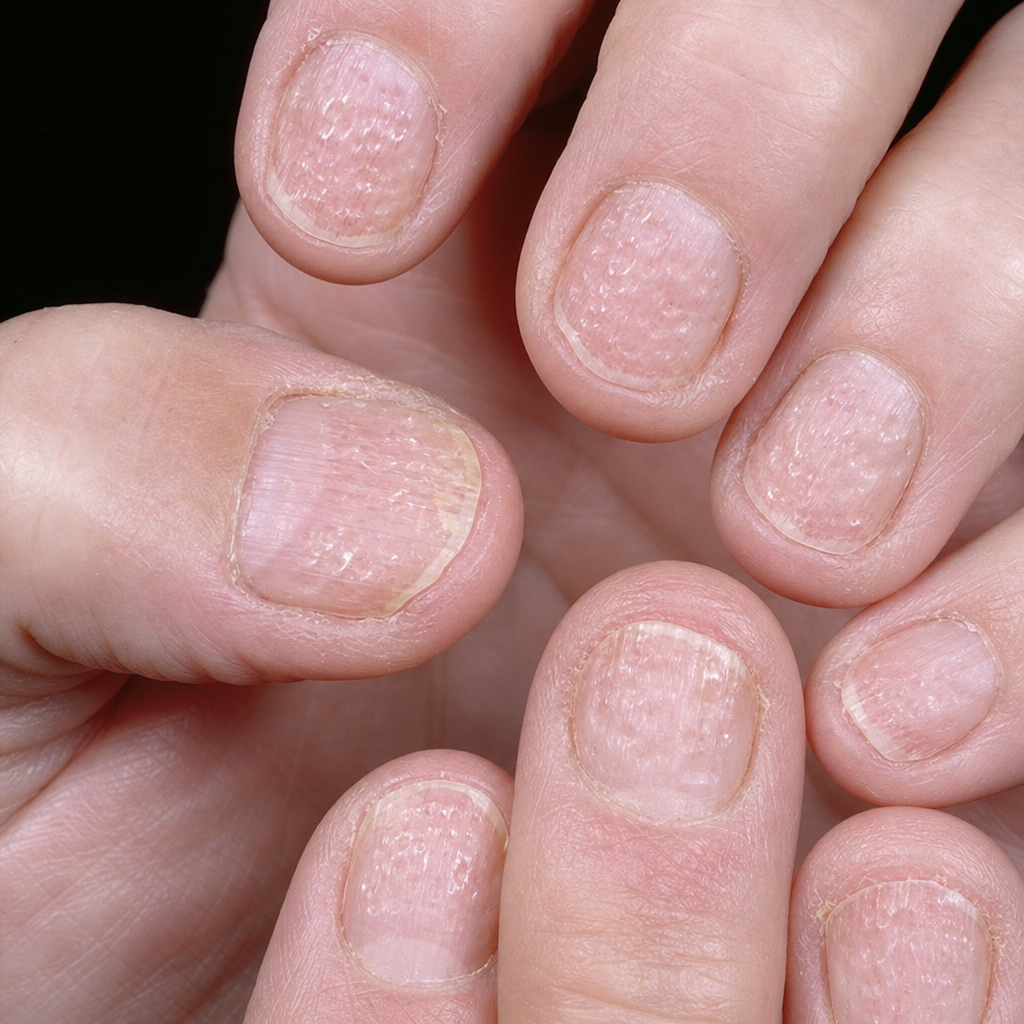
The above nail changes are seen in:
A child presenting with localized patches of complete hair loss with normal appearance of scalp. The diagnosis is:
Pterygium of nail is characteristically seen in -
Which of the following is characteristically seen in alopecia areata?
What is the characteristic nail finding in lichen planus?
Practice by Chapter
Hair Growth Cycle and Anatomy
Practice Questions
Alopecia Areata
Practice Questions
Androgenetic Alopecia
Practice Questions
Telogen Effluvium
Practice Questions
Scarring Alopecias
Practice Questions
Hair Shaft Abnormalities
Practice Questions
Hirsutism and Hypertrichosis
Practice Questions
Nail Anatomy and Growth
Practice Questions
Nail Infections
Practice Questions
Nail Psoriasis and Other Inflammatory Nail Disorders
Practice Questions
Nail Tumors
Practice Questions
Management of Hair and Nail Disorders
Practice Questions
Want unlimited practice?
Get full access to all questions, explanations, and performance tracking.
Scan to download app