What is the best range of UV light used for treatment of skin diseases?
Which of the following burn cases requires IMMEDIATE referral to a specialized burn center?
Which of the following events does NOT occur in rods in response to light
The most specific test to detect blood stains is:
Which of the following are correct for managing hypertrophic scars? 1. Silicone gel sheeting 2. Intralesional steroid injections 3. Vitamin A gel applications 4. Laser treatment Select the answer using the code given below.
What is the first-line treatment for melasma?
What is the best method to treat a large port-wine stain?
Which of the following statements about conjunctival lesions is NOT true?
A 40 year old man presented with a flat 1x1cm scaly, itchy black mole on the front of thigh. Examination did not reveal any inguinal lymphodenopathy. The best course of management would be:
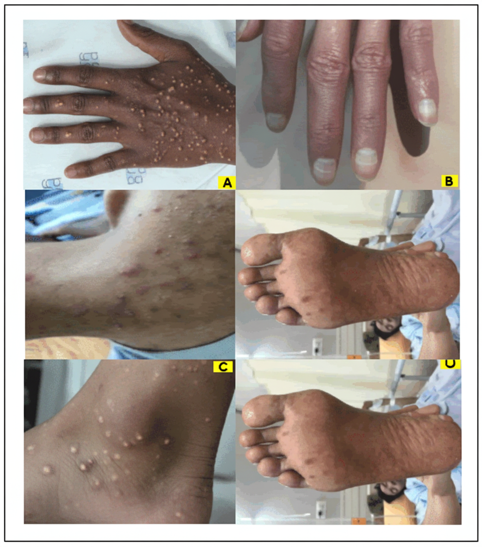
Comment on the image shown:

Explanation: ***200 – 400 nm*** - This range encompasses **UVA (320-400 nm)** and **UVB (290-320 nm)**, which are the most commonly used portions of the **UV spectrum** for treating various skin conditions like psoriasis and eczema. - Specifically, **narrowband UVB (311-313 nm)** is highly effective due to its therapeutic benefits with reduced side effects compared to broadband UVB or UVA. *100 – 200 nm* - This range falls into the **vacuum UV (VUV)** spectrum, which is largely absorbed by air and is not practical for dermatological phototherapy due to its limited penetration and potential for significant cellular damage. - It is known for its germicidal properties but is not used for treating skin diseases in living tissue due to its **high energy** and **low penetration** depth. *> 700 nm* - Wavelengths above 700 nm fall into the **infrared (IR) spectrum** or visible light, which primarily produces heat and has different therapeutic applications. - While IR light can be used for therapies like **pain relief** and **wound healing**, it does not have the immunomodulatory effects on skin cells needed for conditions traditionally treated by UV. *400 – 700 nm* - This range represents the **visible light spectrum**, which is used in some dermatological treatments like **photodynamic therapy (PDT)** or for certain **pigmentary disorders**. - However, visible light does not possess the same **immunomodulatory** and **antiproliferative effects** on keratinocytes and T-cells that make UV light effective for conditions like psoriasis.
Explanation: ***25% deep burn in adult*** - A **deep burn** (full thickness or deep partial thickness) covering **greater than 10% TBSA** is an **absolute criterion** for immediate referral to a specialized burn center per ABA guidelines. - This is due to the high risk of **complications**, need for specialized **wound care**, and potential for **surgical intervention** like skin grafting. - The **combination of depth and extent** makes this the most urgent scenario requiring immediate specialized care. *25% superficial burn in adult* - **Superficial burns** (first-degree) involve only the epidermis and typically heal within days without scarring. - While 25% TBSA is extensive, **superficial burns** can often be managed with supportive care and do not meet the depth criterion for mandatory burn center referral. *Burn in palm* - **Burns involving hands** are considered **special areas** and typically require burn center evaluation for optimal functional outcomes. - However, without specification of **depth and extent**, a small superficial palm burn may be managed locally initially, whereas the question asks for IMMEDIATE referral. - The **25% deep burn** takes precedence due to its life-threatening nature and clear-cut indication. *10% superficial burn in child* - For children, burns greater than **10% TBSA** warrant consideration for burn center referral due to higher morbidity risk. - However, **superficial burns** (first-degree) in children, while concerning, are less urgent than deep burns of significant extent. - The depth of injury is a critical factor; superficial burns may be managed with close monitoring if appropriate expertise is available locally. *5% superficial scald in adult* - A **5% TBSA superficial burn** in an adult does not meet the threshold for mandatory burn center referral (typically >10% for partial thickness burns). - **Superficial scalds** can usually be managed with outpatient care, wound dressing, and pain control. - This would only require referral if other complicating factors were present (e.g., involvement of special areas, inhalation injury).
Explanation: ***Opening of Na+ channels*** - In response to light, **rods hyperpolarize** due to the **closure of Na+ channels**, which reduces the influx of positive ions. - The opening of Na+ channels would lead to depolarization, which is the opposite of what occurs during light detection in rods. *Activation of transducin* - Light causes **conformational changes in rhodopsin**, which in turn activates the G-protein **transducin**. - Activated transducin then goes on to activate **phosphodiesterase (PDE)** as part of the phototransduction cascade. *Structural changes in rhodopsin* - When light strikes the rhodopsin molecule, the **11-cis-retinal chromophore** isomerizes to **all-trans-retinal**. - This **conformational change** in rhodopsin is the initial step that triggers the entire phototransduction pathway. *Decreased intracellular cGMP* - Activated **phosphodiesterase (PDE)**, stimulated by transducin, hydrolyzes **cGMP to GMP**. - The reduction in **cGMP levels** leads to the closure of cGMP-gated Na+ channels, causing hyperpolarization.
Explanation: ***Spectroscopic test*** - The **spectroscopic test** is considered the most specific for detecting blood stains because it identifies the characteristic absorption bands of **hemoglobin** and its derivatives. - This test is highly definitive due to the unique **light absorption properties** of blood components, making it less prone to false positives compared to chemical tests. *Benzidine test* - The **benzidine test** is a sensitive preliminary test for blood but is **not specific**, as it reacts with other oxidizing agents (e.g., rust, certain plant peroxidases). - It works by detecting the **peroxidase-like activity of hemoglobin**, leading to color changes but lacks confirmation of blood origin. *Teichmann's test* - **Teichmann's test** (hemin crystal test) is a moderately specific confirmatory test that produces **rhombic crystals of hemin** when heated with glacial acetic acid and a halide salt. - While more specific than presumptive tests, it can sometimes produce **false-negative results** with old or degraded bloodstains and may be less sensitive than spectroscopy. *Orthotoluidine test* - Similar to the benzidine test, the **orthotoluidine test** is another **presumptive test** that detects the peroxidase-like activity of hemoglobin, resulting in a blue-green color change. - It is **highly sensitive but not specific**, meaning it can also give positive reactions with other substances that have similar peroxidase activity, leading to potential false positives.
Explanation: ***1, 2 and 4*** - **Silicone gel sheeting**, **intralesional steroid injections**, and **laser treatment** are all established and effective methods for managing hypertrophic scars. - Silicone gel helps to hydrate the scar, reduce collagen synthesis, and decrease itching, while steroids reduce inflammation and collagen production, and lasers can help to improve scar texture and color. *1, 3 and 4* - This option incorrectly includes **Vitamin A gel applications** as a primary treatment. While retinoids can have some skin benefits, they are not a first-line or well-established treatment for hypertrophic scars. - **Silicone gel sheeting** and **laser treatment** are indeed effective, but the inclusion of Vitamin A makes this option less accurate. *1, 2 and 3* - This option also incorrectly includes **Vitamin A gel applications**. While **silicone gel sheeting** and **intralesional steroid injections** are effective, Vitamin A is not a standard treatment for hypertrophic scars. - The primary methods for managing hypertrophic scars focus on reducing collagen production and inflammation, which Vitamin A gel does not effectively address in this context. *2, 3 and 4* - This option correctly includes **intralesional steroid injections** and **laser treatment**, but it again incorrectly includes **Vitamin A gel applications** and omits **silicone gel sheeting**, which is a widely recommended and often first-line treatment. - Omitting **silicone gel sheeting** significantly weakens the effectiveness of this combination as a comprehensive management strategy.
Explanation: ***Topical hydroquinone*** - **Topical hydroquinone** (2-4%) is the **most effective single-agent treatment** among the given options, serving as the gold standard for melasma by inhibiting **tyrosinase enzyme** and melanin production. - Demonstrates **highest efficacy rates** (60-80% improvement) when combined with strict sun protection, making it both first-line and most effective monotherapy choice. *Laser therapy* - Carries **high risk of paradoxical darkening** and post-inflammatory hyperpigmentation, especially in darker skin types common in melasma patients. - Requires **specialized expertise** and should only be considered as adjunctive therapy after optimizing topical treatments, not as primary treatment. *Chemical peels* - Provide **variable and inconsistent results** as monotherapy, typically requiring multiple sessions with unpredictable outcomes. - Risk of **post-inflammatory hyperpigmentation** particularly in Fitzpatrick skin types IV-VI, making them less reliable than hydroquinone. *Microdermabrasion* - Offers only **superficial exfoliation** with minimal clinical improvement in melasma pigmentation. - May actually **worsen pigmentation** through mechanical irritation and is not recommended in evidence-based treatment guidelines.
Explanation: ***Pulsed dye laser*** - The **pulsed dye laser (PDL)** is considered the **gold standard** for treating port-wine stains due to its specific targeting of hemoglobin in the dilated capillaries without damaging surrounding tissue. - This treatment involves multiple sessions to progressively lighten the stain and prevent complications such as **nodularity** and **tissue hypertrophy**. *Radiotherapy* - **Radiotherapy** is generally not recommended for port-wine stains due to its potential for **scarring**, **pigment changes**, and risk of **malignancy**. - It is an aggressive treatment typically reserved for **cancerous conditions** or severe proliferative vascular lesions not amenable to other treatments. *Tattooing* - **Tattooing** involves injecting skin-colored pigments into the lesion to camouflage it, but it does not treat the underlying vascular abnormality. - This method can result in an **artificial appearance**, **uneven coverage**, and potential for **allergic reactions** or infections. *Excision with skin grafting* - **Surgical excision** of a large port-wine stain would result in a **significant scar** and require **skin grafting**, which carries risks of graft failure, poor aesthetic outcome, and color mismatch. - This method is generally reserved for very small, localized lesions or those with significant **nodular hypertrophy** that cannot be effectively managed by laser therapy.
Explanation: ***Surgery is treatment of choice*** - While surgery can be used to treat conjunctival lesions, it is not always the **treatment of choice**, especially for smaller, asymptomatic lesions like **pinguecula** which may only require observation and lubrication. - Many conjunctival lesions, such as uncomplicated **pterygium** or **pinguecula**, are managed conservatively unless they cause significant symptoms, vision impairment, or cosmetic concerns. *Arise from any part of conjunctiva* - **Conjunctival lesions** can indeed arise from various parts of the conjunctiva, including the palpebral, bulbar, and forniceal conjunctiva. - For example, **pterygium** typically arises from the bulbar conjunctiva, while **pinguecula** also originates in the bulbar conjunctiva, specifically in the interpalpebral fissure. *Can cause Astigmatism* - Larger **conjunctival lesions**, particularly a **pterygium** that encroaches onto the cornea, can induce or alter astigmatism. - The growth of the lesion can change the **curvature of the cornea**, leading to optical distortion and astigmatism. *UV exposure is risk factor* - **Ultraviolet (UV) light exposure** is a well-established risk factor for the development of many conjunctival lesions, including **pterygium** and **pinguecula**. - Chronic UV exposure leads to **elastotic degeneration** of the conjunctival collagen and is thought to play a key role in the pathogenesis of these growths.
Explanation: ***Excision biopsy*** - A **flat, scaly, itchy, black mole** is highly suspicious for **melanoma**, and an excision biopsy provides the most accurate histopathological diagnosis and depth assessment. - This procedure removes the entire lesion with a narrow margin of normal-appearing skin, allowing for comprehensive evaluation of its nature and determining further management. *FNAC of lesion* - **Fine needle aspiration cytology (FNAC)** is generally used for evaluating palpable masses or lymph nodes, not primary skin lesions like a suspicious mole. - It provides only cellular samples, making it difficult to assess architectural features, depth of invasion, or determine definitive malignancy in skin lesions. *Incision biopsy* - An **incision biopsy** involves removing only a partial sample of the lesion, which can lead to sampling error and an inaccurate diagnosis if the most aggressive part is missed. - For suspected melanoma, an incomplete biopsy can compromise subsequent staging and definitive treatment planning. *Wide excision with inguinal lymphadenectomy* - This is an **overly aggressive initial approach** before a definitive diagnosis of melanoma and its stage has been established. - **Wide excision** is typically performed after an excision biopsy confirms melanoma and determines its depth, while **lymphadenectomy** is indicated for confirmed lymph node involvement.
Explanation: ***Callosity*** - The image displays several **thickened, hyperkeratotic patches** on the palm, characteristic of callosities. - Callosities are caused by repeated friction and pressure, leading to **diffuse epidermal thickening** without a central core. *Corn* - A **corn** is a small, well-demarcated lesion with a **central core** that causes localized pain, unlike the diffuse thickening seen here. - They typically occur over bony prominences and are less spread out than the lesions in the image. *Warts* - **Warts** are caused by the **human papillomavirus (HPV)** and present as rough, elevated lesions with characteristic **black puncta** (thrombosed capillaries) upon paring, which are not visible in the image. - They often have a **papillomatous** or verrucous surface, different from the relatively smooth, thickened appearance here. *Cutaneous horn* - A **cutaneous horn** is a conical projection of **hyperkeratotic material** resembling an animal horn, typically developing on sun-exposed areas. - It is usually a solitary lesion and has a different morphology than the multiple, flat, thickened lesions shown.
Get full access to all questions, explanations, and performance tracking.
Start For Free