A child presenting with localized patches of complete hair loss with normal appearance of scalp. The diagnosis is:
A 25-year-old presents with silvery scales on elbows and knees. Likely diagnosis?
Match the following scale types with their lesions. | Scales | Lesions | | :-- | :-- | | 1. Collarette scales | a. Pityriasis versicolour | | 2. Silvery scales | b. Pityriasis rosea | | 3. Mica-like scales | c. Psoriasis | | 4. Branny scales | d. Pityriasis lichenoides |
Skin biopsy shows psoriasiform hyperplasia with neutrophilic microabscesses in stratum corneum. Most likely diagnosis?
Which of the following is used in the treatment of Pityriasis versicolor?
An eleven-year-old boy has Tinea capitis on his scalp. Which of the following is the most appropriate line of treatment for this condition?
An 8-year-old child has localized non-cicatricial alopecia over scalp with itching and scales. The diagnosis is :
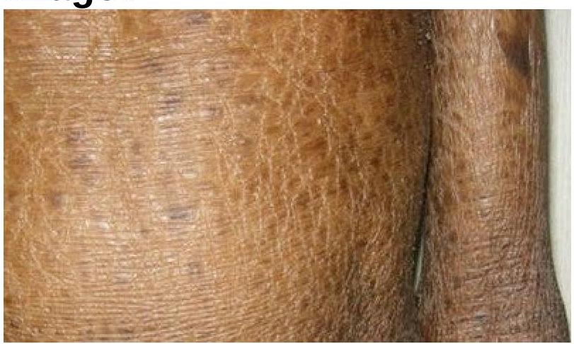
Identify the skin condition depicted in the image.

Which of the following organisms has a role to play in Seborrheic dermatitis?
Pompholyx affects:
Explanation: ***Alopecia areata*** - This condition is characterized by **localized, well-demarcated patches of complete hair loss** on the scalp. - The underlying skin typically appears **normal, smooth, and healthy**, without inflammation or scaling. *Tinea capitis* - This fungal infection usually presents with **scaly patches**, inflammation, pustules, or "black dots" where hairs have broken off. - The scalp appearance is typically **abnormal** due to scaling and inflammation, unlike the normal scalp seen here. *Cradle cap* - Also known as **seborrheic dermatitis in infants**, it presents as greasy, yellowish, scaly patches on the scalp. - It does not cause **complete hair loss** in localized patches, but rather diffuse scaling and sometimes mild thinning. *Telogen effluvium* - This condition involves **diffuse hair shedding** (increased number of hairs falling out), often triggered by stress, illness, or medications. - It does not present as **localized patches of complete hair loss**, and the hair thinning is generally widespread.
Explanation: ***Psoriasis*** - **Silvery scales** on **extensor surfaces** like elbows and knees are classic presentations of plaque psoriasis. - This chronic inflammatory skin condition is characterized by **accelerated epidermal turnover**. *Atopic dermatitis* - Typically presents as **eczematous lesions** characterized by **red, itchy, and often oozing or crusted patches**, mainly on flexural surfaces in adults. - It is strongly associated with a history of **allergies, asthma, or hay fever**. *Pityriasis rosea* - Usually starts with a **herald patch** followed by smaller, oval, pinkish-orange macules and patches with fine scales in a **Christmas tree pattern** on the trunk. - It is distinguished from psoriasis by its distribution and appearance of scales. *Lichen planus* - Characterized by **pruritic, purple, polygonal, planar papules and plaques** (the "6 P's") often with fine, white lacy streaks called **Wickham's striae**, typically affecting flexor surfaces, wrists, and oral mucosa. - It does not present with silvery scales on extensor surfaces.
Explanation: ***1-b, 2-c, 3-d, 4-a*** - **Collarette scales** are pathognomonic of **Pityriasis rosea**, appearing as fine, trailing scales around the periphery of oval lesions in a "Christmas tree" distribution. - **Silvery scales** are the classic hallmark of **Psoriasis**, presenting as thick, adherent, silvery-white scales overlying well-demarcated erythematous plaques. - **Mica-like scales** are characteristic of **Pityriasis lichenoides**, appearing as thick, shiny, adherent scales that can be peeled off like mica sheets. - **Branny scales** are typical of **Pityriasis versicolor**, presenting as fine, powdery scales caused by **Malassezia** yeast overgrowth. *1-d, 2-c, 3-a, 4-b* - Incorrectly matches **collarette scales with Pityriasis lichenoides**, which typically presents with mica-like scales, not collarette scales. - Misassociates **mica-like scales with Pityriasis versicolor**, which characteristically has branny (fine, powdery) scales. *1-c, 2-b, 3-d, 4-a* - Wrongly pairs **collarette scales with Psoriasis**, which is known for thick silvery scales, not peripheral collarette scales. - Incorrectly matches **silvery scales with Pityriasis rosea**, which has collarette scales at lesion periphery, not silvery scales. *1-a, 2-b, 3-d, 4-c* - Falsely associates **collarette scales with Pityriasis versicolor**, which has branny scales from yeast infection, not collarette scales. - Mismatches **branny scales with Psoriasis**, which has characteristic thick silvery scales, not fine powdery scales.
Explanation: ***Psoriasis*** - **Psoriasiform hyperplasia**, characterized by regular epidermal acanthosis and elongated rete ridges, is a classic histological feature of psoriasis. - The presence of **neutrophilic microabscesses (Munro microabscesses)** in the stratum corneum is a pathognomonic finding for psoriasis. *Seborrheic dermatitis* - Histologically, seborrheic dermatitis typically shows **irregular acanthosis** with parakeratosis and a **perivascular lymphocytic infiltrate**, but not regular psoriasiform hyperplasia or Munro microabscesses. - There may be *spongiosis* and neutrophils in the stratum corneum, but not the distinct microabscesses seen in psoriasis. *Pityriasis rosea* - Pityriasis rosea histology often reveals **focal parakeratosis**, **spongiosis**, and a **perivascular lymphocytic infiltrate** with extravasated red blood cells. - It does not demonstrate the characteristic regular psoriasiform hyperplasia or neutrophilic microabscesses of psoriasis. *Lichen planus* - Lichen planus is characterized by a **"sawtooth" rete ridge pattern**, a **band-like lymphocytic infiltrate** at the dermo-epidermal junction, and **colloid bodies (Civatte bodies)**. - It does not exhibit psoriasiform hyperplasia or neutrophilic microabscesses in the stratum corneum.
Explanation: ***Itraconazole*** - **Itraconazole** is an effective oral antifungal agent commonly used to treat Pityriasis versicolor, particularly in widespread or recurrent cases. - It works by inhibiting fungal cytochrome P450-dependent 14α-lanosterol demethylase, thereby disrupting ergosterol synthesis and fungal cell membrane integrity. - Standard regimen: 200 mg once daily for 5-7 days or 200 mg twice daily for 1 day. *Griseofulvin* - **Griseofulvin** is an oral antifungal primarily used for dermatophyte infections (e.g., tinea capitis, tinea corporis), not Pityriasis versicolor, which is caused by *Malassezia* species (a yeast). - It acts by disrupting fungal mitosis and is concentrated in keratinocytes, but has **no activity against yeasts** like *Malassezia*. *Terbinafine* - **Terbinafine** is an allylamin antifungal that primarily targets dermatophytes by inhibiting squalene epoxidase, an enzyme involved in ergosterol synthesis. - While it has **limited activity** against *Malassezia* species, it is not considered a first-line or preferred systemic treatment for Pityriasis versicolor. - Azoles (itraconazole, fluconazole) are far more effective for this condition. *All of the options* - This option is incorrect because **griseofulvin** has no antifungal activity against *Malassezia* species and **terbinafine** is not a preferred treatment. - Only **itraconazole** among the listed options is an appropriate and effective systemic treatment for Pityriasis versicolor.
Explanation: ***Oral griseofulvin therapy*** - **Systemic antifungal agents** are essential for treating **Tinea capitis**, as the fungal infection is deep within the hair follicles and cannot be reached effectively by topical treatments alone. - **Griseofulvin** is a well-established and effective oral antifungal for **Tinea capitis** in children. *Shaving of the scalp* - While shaving the scalp might reduce some fungal load and facilitate topical treatment, it is **not a definitive treatment** for **Tinea capitis** on its own, as the infection remains deep in the hair follicles. - It does not address the underlying systemic nature of the infection within the hair shaft. *Topical griseofulvin therapy* - **Topical griseofulvin** is generally **ineffective** for **Tinea capitis** because the fungus resides deep within the hair follicle and hair shaft, where topical preparations cannot penetrate sufficiently. - **Systemic absorption** is required to deliver adequate drug concentrations to the site of infection. *Selenium sulphide shampoo* - **Selenium sulfide shampoo** can be used as an **adjunctive therapy** to reduce shedding of spores and prevent spread, but it is **not curative** for **Tinea capitis**. - It helps to reduce skin scaling and fungal burden on the surface but does not eradicate the infection deep within the hair follicles.
Explanation: ***Tinea Capitis*** - **Tinea capitis** typically presents as **localized, non-cicatricial alopecia** with features like **scaling**, **itching**, and broken hairs, which are consistent with the child's symptoms. - It's a common **dermatophyte infection** of the scalp, particularly in children, caused by fungi like *Trichophyton* or *Microsporum*. *Lichen planus* - **Lichen planus** can cause alopecia, but it is typically a **cicatricial (scarring)** alopecia, unlike the non-cicatricial finding described. - It is more commonly associated with **purplish, polygonal, pruritic papules** on the skin and mucous membranes. *Tinea Barbae* - **Tinea barbae** specifically affects the **beard and mustache area** in adult males and would not present as alopecia on the scalp in an 8-year-old child. - It usually involves deep follicular inflammation with **pustules and nodules**. *Alopecia areata* - **Alopecia areata** is characterized by **smooth, circular patches of non-scarring hair loss** without associated scaling or significant itching. - The presence of **scaling and itching** in this case makes alopecia areata less likely.
Explanation: ***Ichthyosis*** - The image clearly displays widespread **dry, scaling, and thickened skin**, consistent with the characteristic presentation of ichthyosis. - This condition is characterized by a defect in **skin barrier function** leading to excessive dryness and accumulation of scales. *Syndromic ichthyosis* - While syndromic ichthyosis also involves skin scaling, it is associated with **additional systemic symptoms** or **organ involvement**, which cannot be determined from this image alone. - The term "ichthyosis" broadly covers this appearance, and without more clinical information, specifying it as syndromic is not the most direct identification. *Leprosy* - Leprosy typically presents with **hypopigmented, anesthetic skin patches** or **nodules**, which are not seen in the image. - The texture and color changes in the image are not characteristic of the primarily neurological and dermatological manifestations of leprosy. *Cutaneous sarcoidosis* - Cutaneous sarcoidosis manifests as **reddish-brown papules, plaques, or nodules**, often on the face, neck, or extremities. - The widespread, fine scaling and dryness seen in the image do not align with the typical granulomatous lesions of sarcoidosis.
Explanation: ***Pityrosporum ovale*** - **Pityrosporum ovale**, now known as **Malassezia furfur**, is a lipophilic yeast that colonizes the skin and plays a significant role in the pathogenesis of **seborrheic dermatitis**. - Its presence is commonly associated with the inflammatory response seen in seborrheic dermatitis, though the exact mechanism is not fully understood. *Candida albicans* - **Candida albicans** is a common cause of **mucocutaneous candidiasis** and **intertrigo**, but it is not directly implicated in the etiology of seborrheic dermatitis. - While it can cause skin infections, its typical presentation involves erythematous, macerated rashes with satellite lesions in skin folds. *Propionibacterium* - **Propionibacterium acnes**, now known as **Cutibacterium acnes**, is primarily associated with the pathogenesis of **acne vulgaris**. - It plays a role in the inflammation and comedone formation characteristic of acne, not the scaling and erythema of seborrheic dermatitis. *None of the above* - This option is incorrect because **Pityrosporum ovale (Malassezia furfur)** is a well-recognized organism involved in seborrheic dermatitis. - The other organisms listed are associated with different dermatological conditions.
Explanation: ***Palms and soles*** - **Pompholyx**, also known as **dyshidrotic eczema**, is characterized by recurrent outbreaks of **vesicles and bullae** predominantly on the palms and soles. - These lesions are typically very **itchy** and can cause significant discomfort. *Groin* - Conditions like **tinea cruris** (jock itch) or **intertrigo** commonly affect the groin, presenting with erythema and scaling rather than vesicles. - While eczema can occur in the groin, classical pompholyx has a predilection for the acral regions. *Scalp* - The scalp is more commonly affected by conditions such as **seborrheic dermatitis** or **psoriasis**, which manifest as scaling, redness, and flaking. - Vesicular eruptions are rare on the scalp unless due to specific conditions like herpes zoster. *Trunk* - The trunk is a common site for various dermatoses, including **atopic dermatitis**, **psoriasis**, or **pityriasis rosea**, but these typically present with different morphologic features (e.g., plaques, patches). - Pompholyx is specific to palms and soles and does not usually involve the trunk.
Get full access to all questions, explanations, and performance tracking.
Start For Free